Advertisement

Main menu

  • Home
  • Content
    • Current Issue
    • Accepted Manuscripts
    • Article Preview
    • Past Issue Archive
    • Video Articles
    • AJNR Case Collection
    • Case of the Week Archive
    • Case of the Month Archive
    • Classic Case Archive
  • Special Collections
    • AJNR Awards
    • Low-Field MRI
    • Alzheimer Disease
    • ASNR Foundation Special Collection
    • Photon-Counting CT
    • View All
  • Multimedia
    • AJNR Podcasts
    • AJNR SCANtastic
    • Trainee Corner
    • MRI Safety Corner
    • Imaging Protocols
  • For Authors
    • Submit a Manuscript
    • Submit a Video Article
    • Submit an eLetter to the Editor/Response
    • Manuscript Submission Guidelines
    • Statistical Tips
    • Fast Publishing of Accepted Manuscripts
    • Graphical Abstract Preparation
    • Imaging Protocol Submission
    • Author Policies
  • About Us
    • About AJNR
    • Editorial Board
    • Editorial Board Alumni
  • More
    • Become a Reviewer/Academy of Reviewers
    • Subscribers
    • Permissions
    • Alerts
    • Feedback
    • Advertisers
    • ASNR Home

User menu

  • Alerts
  • Log in

Search

  • Advanced search
American Journal of Neuroradiology

American Journal of Neuroradiology

ASHNR American Society of Functional Neuroradiology ASHNR American Society of Pediatric Neuroradiology ASSR
  • Alerts
  • Log in

Advanced Search

  • Home
  • Content
    • Current Issue
    • Accepted Manuscripts
    • Article Preview
    • Past Issue Archive
    • Video Articles
    • AJNR Case Collection
    • Case of the Week Archive
    • Case of the Month Archive
    • Classic Case Archive
  • Special Collections
    • AJNR Awards
    • Low-Field MRI
    • Alzheimer Disease
    • ASNR Foundation Special Collection
    • Photon-Counting CT
    • View All
  • Multimedia
    • AJNR Podcasts
    • AJNR SCANtastic
    • Trainee Corner
    • MRI Safety Corner
    • Imaging Protocols
  • For Authors
    • Submit a Manuscript
    • Submit a Video Article
    • Submit an eLetter to the Editor/Response
    • Manuscript Submission Guidelines
    • Statistical Tips
    • Fast Publishing of Accepted Manuscripts
    • Graphical Abstract Preparation
    • Imaging Protocol Submission
    • Author Policies
  • About Us
    • About AJNR
    • Editorial Board
    • Editorial Board Alumni
  • More
    • Become a Reviewer/Academy of Reviewers
    • Subscribers
    • Permissions
    • Alerts
    • Feedback
    • Advertisers
    • ASNR Home
  • Follow AJNR on Twitter
  • Visit AJNR on Facebook
  • Follow AJNR on Instagram
  • Join AJNR on LinkedIn
  • RSS Feeds

AJNR Awards, New Junior Editors, and more. Read the latest AJNR updates

Research ArticleAdult Brain

Sexual Dimorphism and Hemispheric Asymmetry of Hippocampal Volumetric Integrity in Normal Aging and Alzheimer Disease

B.A. Ardekani, S.A. Hadid, E. Blessing and A.H. Bachman
American Journal of Neuroradiology February 2019, 40 (2) 276-282; DOI: https://doi.org/10.3174/ajnr.A5943
B.A. Ardekani
aFrom Center for Brain Imaging and Neuromodulation, The Nathan S. Kline Institute for Psychiatric Research (B.A.A., S.A.H., A.H.B.), Orangeburg, New York
bDepartment of Psychiatry (B.A.A., E.B.), New York University School of Medicine, New York, New York.
  • Find this author on Google Scholar
  • Find this author on PubMed
  • Search for this author on this site
  • ORCID record for B.A. Ardekani
S.A. Hadid
aFrom Center for Brain Imaging and Neuromodulation, The Nathan S. Kline Institute for Psychiatric Research (B.A.A., S.A.H., A.H.B.), Orangeburg, New York
  • Find this author on Google Scholar
  • Find this author on PubMed
  • Search for this author on this site
  • ORCID record for S.A. Hadid
E. Blessing
bDepartment of Psychiatry (B.A.A., E.B.), New York University School of Medicine, New York, New York.
  • Find this author on Google Scholar
  • Find this author on PubMed
  • Search for this author on this site
  • ORCID record for E. Blessing
A.H. Bachman
aFrom Center for Brain Imaging and Neuromodulation, The Nathan S. Kline Institute for Psychiatric Research (B.A.A., S.A.H., A.H.B.), Orangeburg, New York
  • Find this author on Google Scholar
  • Find this author on PubMed
  • Search for this author on this site
  • ORCID record for A.H. Bachman
  • Article
  • Figures & Data
  • Info & Metrics
  • Responses
  • References
  • PDF
Loading

Article Figures & Data

Figures

  • Tables
  • Fig 1.
    • Download figure
    • Open in new tab
    • Download powerpoint
    Fig 1.

    Axial (left), coronal (middle), and sagittal (right) slices through a 3D structural MR imaging scan of a subject. The right hippocampus probabilistic VOI is superimposed on the image in native space.
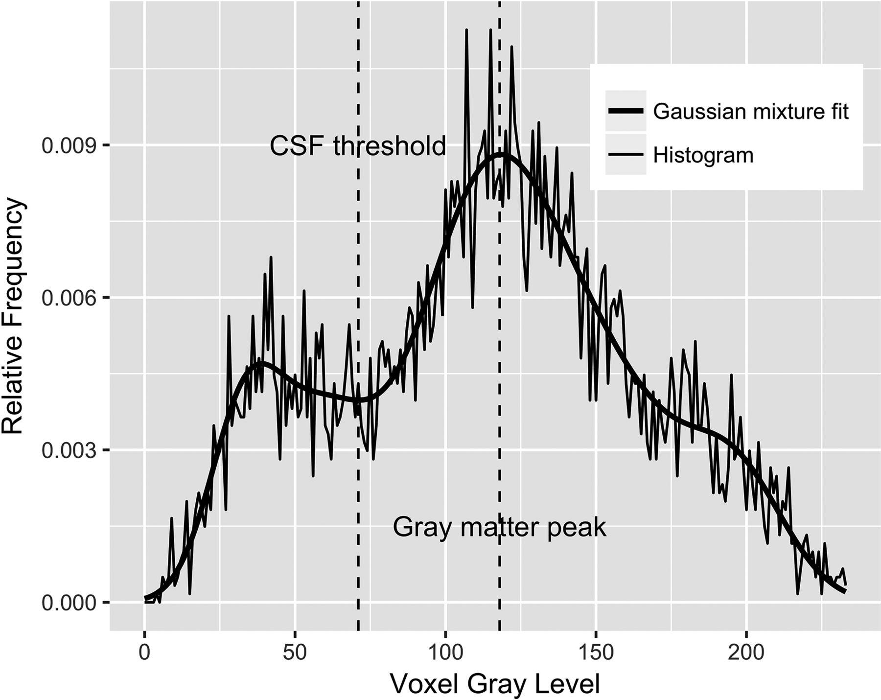
  • Fig 2.
    • Download figure
    • Open in new tab
    • Download powerpoint
    Fig 2.

    Histogram of the voxels with nonzero probabilities on the VOI in Fig 1 (thin line) along with a fitted Gaussian mixture model (thick line) using the expectation-maximization algorithm. The automatically determined CSF threshold is shown as a vertical line approximately located at the intensity value of 70. The HPF is defined as the fraction of voxels in the VOI whose intensities are greater than the CSF threshold.

  • Fig 3.
    • Download figure
    • Open in new tab
    • Download powerpoint
    Fig 3.

    Predicted marginal means of the asymmetry index in different diagnostic groups. AI was found to be significantly different between the CN versus MCI (P < .001), CN versus AD (P < .001), and MCI versus AD (P = .03) groups. AI increased with increasing dementia severity. Error bars indicate 95% CI.
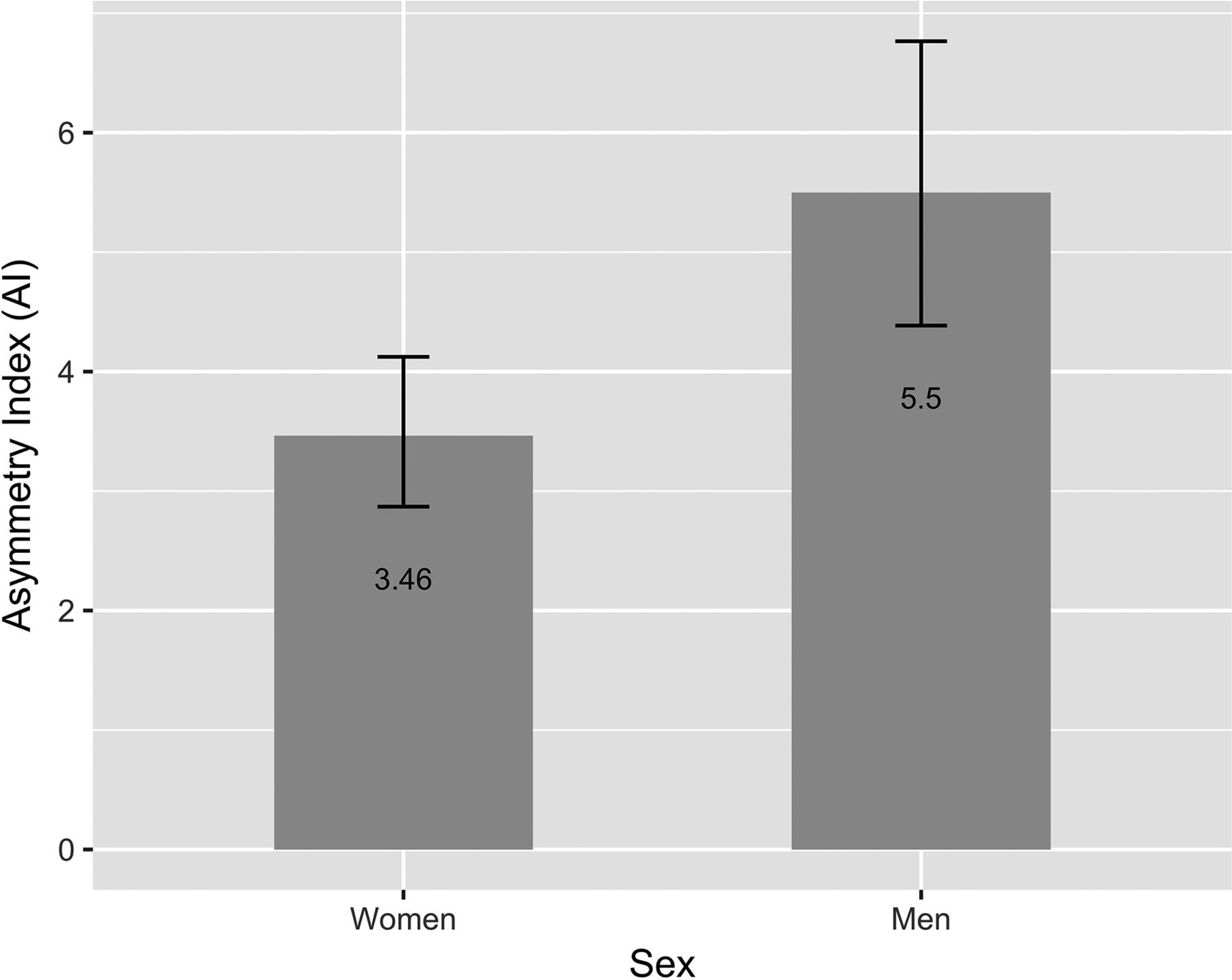
  • Fig 4.
    • Download figure
    • Open in new tab
    • Download powerpoint
    Fig 4.

    Predicted marginal means of the asymmetry index in men and women. The AI was found to be significantly higher in men (P < .01) after controlling for age, intracranial volume, and diagnostic group. Error bars indicate 95% CI.

  • Fig 5.
    • Download figure
    • Open in new tab
    • Download powerpoint
    Fig 5.

    Predicted marginal means of the HPF (averaged across hemispheres) in different diagnostic groups. The HPF was found to be significantly different between the CN versus MCI (P < .001), CN versus AD (P < .001), and MCI versus AD (P < .001) groups. The HPF decreased with increasing dementia severity. Error bars indicate 95% CI.

Tables

  • Figures
    • View popup
    Table 1:

    Cohort demographicsa

    GroupCNMCIAD
    CDR00.51 (n = 28), 2 (n = 2)
    No. of subjects987030
    Sex (female:male)72:2639:3120:10
    Age (yr)75.9 ± 9.0 (60–94)76.2 ± 7.2 (62–92)78.0 ± 6.9 (65–96)
    Education (yr)14.5 ± 2.9 (8–23)13.8 ± 3.2 (6–20)12.8 ± 3.2 (7–20)
    ICV (cm3)1439 ± 150 (1132–1818)1485 ± 187 (1171–1992)1480 ± 118 (1274–1732)
    MMSE29.0 ± 1.2 (25–30)25.6 ± 3.5 (14–30)21.2 ± 4.0 (15–29)
    • Note:—CDR indicates Clinical Dementia Rating; MMSE, Mini-Mental State Examination.

    • ↵a Values given are means and range.

    • View popup
    Table 2:

    Definitions of all mathematic terms used in the mixed-effects model equation (Equation 4)

    iVolume index i = 1, 2, ′, 775
    jSubject identification index j = 1, 2, ′, 198
    yilogit(HPF) or log(AI) for volume i as the response variable
    IDiSubject index of volume i where IDi ∈ (1, 2, ′, 198)
    AgeiAge of subject IDi minus 76.34, the average age of all 198 subjects
    ICViEstimated total intracranial volume of subject IDi minus 1461.5, the average ICV of all 198 subjects
    DXiDiagnostic group of subject IDi, a factor with 3 levels {CN, MCI, AD}
    SexiSex of subject IDi, a factor with 2 levels {Male, Female}
    I{statement}Indicator function that takes on values of 1 or 0 depending on whether the statement is true or false
    β0 ′ β7Model fixed-effects parameters to be estimated and tested against null hypotheses of type Σakβk = 0
    bjModel random effects assumed to be independent and identically distributed Gaussian with mean 0 and variance σb2
    eiRandom residuals assumed to be independent and identically distributed Gaussian with mean 0 and variance σe2
PreviousNext
Back to top

In this issue

American Journal of Neuroradiology: 40 (2)
American Journal of Neuroradiology
Vol. 40, Issue 2
1 Feb 2019
  • Table of Contents
  • Index by author
  • Complete Issue (PDF)
Advertisement
Print
Download PDF
Email Article

Thank you for your interest in spreading the word on American Journal of Neuroradiology.

NOTE: We only request your email address so that the person you are recommending the page to knows that you wanted them to see it, and that it is not junk mail. We do not capture any email address.

Enter multiple addresses on separate lines or separate them with commas.
Sexual Dimorphism and Hemispheric Asymmetry of Hippocampal Volumetric Integrity in Normal Aging and Alzheimer Disease
(Your Name) has sent you a message from American Journal of Neuroradiology
(Your Name) thought you would like to see the American Journal of Neuroradiology web site.
CAPTCHA
This question is for testing whether or not you are a human visitor and to prevent automated spam submissions.
Cite this article
B.A. Ardekani, S.A. Hadid, E. Blessing, A.H. Bachman
Sexual Dimorphism and Hemispheric Asymmetry of Hippocampal Volumetric Integrity in Normal Aging and Alzheimer Disease
American Journal of Neuroradiology Feb 2019, 40 (2) 276-282; DOI: 10.3174/ajnr.A5943

Citation Manager Formats

  • BibTeX
  • Bookends
  • EasyBib
  • EndNote (tagged)
  • EndNote 8 (xml)
  • Medlars
  • Mendeley
  • Papers
  • RefWorks Tagged
  • Ref Manager
  • RIS
  • Zotero
0 Responses
Respond to this article
Share
Bookmark this article
Sexual Dimorphism and Hemispheric Asymmetry of Hippocampal Volumetric Integrity in Normal Aging and Alzheimer Disease
B.A. Ardekani, S.A. Hadid, E. Blessing, A.H. Bachman
American Journal of Neuroradiology Feb 2019, 40 (2) 276-282; DOI: 10.3174/ajnr.A5943
del.icio.us logo Twitter logo Facebook logo Mendeley logo
  • Tweet Widget
  • Facebook Like
  • Google Plus One
Purchase

Jump to section

  • Article
    • Abstract
    • ABBREVIATIONS:
    • Materials and Methods
    • Results
    • Discussion
    • Conclusions
    • References
  • Figures & Data
  • Info & Metrics
  • Responses
  • References
  • PDF

Related Articles

  • No related articles found.
  • PubMed
  • Google Scholar

Cited By...

  • Alzheimers subtypes A supervised, unsupervised, multimodal, multilayered embedded recursive (SUMMER) AI study
  • Dynamic Individual Prediction of Conversion from Mild Cognitive Impairment to Probable Alzheimers Disease using Joint Modeling
  • Crossref
  • Google Scholar

This article has not yet been cited by articles in journals that are participating in Crossref Cited-by Linking.

More in this TOC Section

  • Diagnostic Neuroradiology of Monoclonal Antibodies
  • Clinical Outcomes After Chiari I Decompression
  • Segmentation of Brain Metastases with BLAST
Show more Adult Brain

Similar Articles

Advertisement

Indexed Content

  • Current Issue
  • Accepted Manuscripts
  • Article Preview
  • Past Issues
  • Editorials
  • Editor's Choice
  • Fellows' Journal Club
  • Letters to the Editor
  • Video Articles

Cases

  • Case Collection
  • Archive - Case of the Week
  • Archive - Case of the Month
  • Archive - Classic Case

More from AJNR

  • Trainee Corner
  • Imaging Protocols
  • MRI Safety Corner
  • Book Reviews

Multimedia

  • AJNR Podcasts
  • AJNR Scantastics

Resources

  • Turnaround Time
  • Submit a Manuscript
  • Submit a Video Article
  • Submit an eLetter to the Editor/Response
  • Manuscript Submission Guidelines
  • Statistical Tips
  • Fast Publishing of Accepted Manuscripts
  • Graphical Abstract Preparation
  • Imaging Protocol Submission
  • Evidence-Based Medicine Level Guide
  • Publishing Checklists
  • Author Policies
  • Become a Reviewer/Academy of Reviewers
  • News and Updates

About Us

  • About AJNR
  • Editorial Board
  • Editorial Board Alumni
  • Alerts
  • Permissions
  • Not an AJNR Subscriber? Join Now
  • Advertise with Us
  • Librarian Resources
  • Feedback
  • Terms and Conditions
  • AJNR Editorial Board Alumni

American Society of Neuroradiology

  • Not an ASNR Member? Join Now

© 2025 by the American Society of Neuroradiology All rights, including for text and data mining, AI training, and similar technologies, are reserved.
Print ISSN: 0195-6108 Online ISSN: 1936-959X

Powered by HighWire