Advertisement

Main menu

  • Home
  • Content
    • Current Issue
    • Accepted Manuscripts
    • Article Preview
    • Past Issue Archive
    • Video Articles
    • AJNR Case Collection
    • Case of the Week Archive
    • Case of the Month Archive
    • Classic Case Archive
  • Special Collections
    • AJNR Awards
    • Low-Field MRI
    • Alzheimer Disease
    • ASNR Foundation Special Collection
    • Photon-Counting CT
    • View All
  • Multimedia
    • AJNR Podcasts
    • AJNR SCANtastic
    • Trainee Corner
    • MRI Safety Corner
    • Imaging Protocols
  • For Authors
    • Submit a Manuscript
    • Submit a Video Article
    • Submit an eLetter to the Editor/Response
    • Manuscript Submission Guidelines
    • Statistical Tips
    • Fast Publishing of Accepted Manuscripts
    • Graphical Abstract Preparation
    • Imaging Protocol Submission
    • Author Policies
  • About Us
    • About AJNR
    • Editorial Board
    • Editorial Board Alumni
  • More
    • Become a Reviewer/Academy of Reviewers
    • Subscribers
    • Permissions
    • Alerts
    • Feedback
    • Advertisers
    • ASNR Home

User menu

  • Alerts
  • Log in

Search

  • Advanced search
American Journal of Neuroradiology

American Journal of Neuroradiology

ASHNR American Society of Functional Neuroradiology ASHNR American Society of Pediatric Neuroradiology ASSR
  • Alerts
  • Log in

Advanced Search

  • Home
  • Content
    • Current Issue
    • Accepted Manuscripts
    • Article Preview
    • Past Issue Archive
    • Video Articles
    • AJNR Case Collection
    • Case of the Week Archive
    • Case of the Month Archive
    • Classic Case Archive
  • Special Collections
    • AJNR Awards
    • Low-Field MRI
    • Alzheimer Disease
    • ASNR Foundation Special Collection
    • Photon-Counting CT
    • View All
  • Multimedia
    • AJNR Podcasts
    • AJNR SCANtastic
    • Trainee Corner
    • MRI Safety Corner
    • Imaging Protocols
  • For Authors
    • Submit a Manuscript
    • Submit a Video Article
    • Submit an eLetter to the Editor/Response
    • Manuscript Submission Guidelines
    • Statistical Tips
    • Fast Publishing of Accepted Manuscripts
    • Graphical Abstract Preparation
    • Imaging Protocol Submission
    • Author Policies
  • About Us
    • About AJNR
    • Editorial Board
    • Editorial Board Alumni
  • More
    • Become a Reviewer/Academy of Reviewers
    • Subscribers
    • Permissions
    • Alerts
    • Feedback
    • Advertisers
    • ASNR Home
  • Follow AJNR on Twitter
  • Visit AJNR on Facebook
  • Follow AJNR on Instagram
  • Join AJNR on LinkedIn
  • RSS Feeds

AJNR Awards, New Junior Editors, and more. Read the latest AJNR updates

Research ArticleAdult Brain

Multinodular and Vacuolating Neuronal Tumor of the Cerebrum: A New “Leave Me Alone” Lesion with a Characteristic Imaging Pattern

R.H. Nunes, C.C. Hsu, A.J. da Rocha, L.L.F. do Amaral, L.F.S. Godoy, T.W. Watkins, V.H. Marussi, M. Warmuth-Metz, H.C. Alves, F.G. Goncalves, B.K. Kleinschmidt-DeMasters and A.G. Osborn
American Journal of Neuroradiology October 2017, 38 (10) 1899-1904; DOI: https://doi.org/10.3174/ajnr.A5281
R.H. Nunes
aFrom the Division of Neuroradiology (R.H.N., A.J.d.R., L.L.F.d.A., H.C.A.), Santa Casa de Sao Paulo School of Medical Sciences, Sao Paulo, Brazil
bDivision of Neuroradiology (R.H.N., A.J.d.R.), Diagnosticos da America SA, Sao Paulo, Brazil
  • Find this author on Google Scholar
  • Find this author on PubMed
  • Search for this author on this site
  • ORCID record for R.H. Nunes
C.C. Hsu
cDepartment of Medical Imaging (C.C.H., T.W.W.), Princess Alexandra Hospital, Brisbane, Queensland, Australia
  • Find this author on Google Scholar
  • Find this author on PubMed
  • Search for this author on this site
  • ORCID record for C.C. Hsu
A.J. da Rocha
aFrom the Division of Neuroradiology (R.H.N., A.J.d.R., L.L.F.d.A., H.C.A.), Santa Casa de Sao Paulo School of Medical Sciences, Sao Paulo, Brazil
bDivision of Neuroradiology (R.H.N., A.J.d.R.), Diagnosticos da America SA, Sao Paulo, Brazil
  • Find this author on Google Scholar
  • Find this author on PubMed
  • Search for this author on this site
  • ORCID record for A.J. da Rocha
L.L.F. do Amaral
aFrom the Division of Neuroradiology (R.H.N., A.J.d.R., L.L.F.d.A., H.C.A.), Santa Casa de Sao Paulo School of Medical Sciences, Sao Paulo, Brazil
dDivision of Neuroradiology (L.L.F.d.A., V.H.M.), Medimagem, Hospital da Beneficencia Portuguesa de Sao Paulo, Sao Paulo, Brazil
  • Find this author on Google Scholar
  • Find this author on PubMed
  • Search for this author on this site
  • ORCID record for L.L.F. do Amaral
L.F.S. Godoy
eDepartment of Radiology (L.F.S.G.), Hospital Sirio Libanes, Sao Paulo, Brazil
fDepartment of Radiology (L.F.S.G.), Medical School, Universidade de Sao Paulo, Sao Paulo, Brazil
  • Find this author on Google Scholar
  • Find this author on PubMed
  • Search for this author on this site
  • ORCID record for L.F.S. Godoy
T.W. Watkins
cDepartment of Medical Imaging (C.C.H., T.W.W.), Princess Alexandra Hospital, Brisbane, Queensland, Australia
  • Find this author on Google Scholar
  • Find this author on PubMed
  • Search for this author on this site
  • ORCID record for T.W. Watkins
V.H. Marussi
dDivision of Neuroradiology (L.L.F.d.A., V.H.M.), Medimagem, Hospital da Beneficencia Portuguesa de Sao Paulo, Sao Paulo, Brazil
  • Find this author on Google Scholar
  • Find this author on PubMed
  • Search for this author on this site
  • ORCID record for V.H. Marussi
M. Warmuth-Metz
gDepartment of Neuroradiology (M.W.-M.), University Hospital Würzburg, Würzburg, Germany
  • Find this author on Google Scholar
  • Find this author on PubMed
  • Search for this author on this site
  • ORCID record for M. Warmuth-Metz
H.C. Alves
aFrom the Division of Neuroradiology (R.H.N., A.J.d.R., L.L.F.d.A., H.C.A.), Santa Casa de Sao Paulo School of Medical Sciences, Sao Paulo, Brazil
  • Find this author on Google Scholar
  • Find this author on PubMed
  • Search for this author on this site
  • ORCID record for H.C. Alves
F.G. Goncalves
hDepartment of Radiology (F.G.G.), Children's Hospital of Brasilia, Brasilia, Brazil
  • Find this author on Google Scholar
  • Find this author on PubMed
  • Search for this author on this site
  • ORCID record for F.G. Goncalves
B.K. Kleinschmidt-DeMasters
iDepartment of Pathology (B.K.K.-D.), University of Colorado, Aurora, Colorado
  • Find this author on Google Scholar
  • Find this author on PubMed
  • Search for this author on this site
  • ORCID record for B.K. Kleinschmidt-DeMasters
A.G. Osborn
jDepartment of Radiology (A.G.O.), University of Utah, Salt Lake City, Utah.
  • Find this author on Google Scholar
  • Find this author on PubMed
  • Search for this author on this site
  • ORCID record for A.G. Osborn
  • Article
  • Figures & Data
  • Supplemental
  • Info & Metrics
  • Responses
  • References
  • PDF
Loading

Article Figures & Data

Figures

  • Tables
  • Fig 1.
    • Download figure
    • Open in new tab
    • Download powerpoint
    Fig 1.

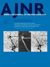
    Typical imaging presentation on FLAIR. Selected axial (A–C) and coronal (D) FLAIR images of presumed MVNT demonstrate supratentorial hyperintense intra-axial lesions consisting of groups of multiple nodules, located on the subcortical ribbon and superficial subcortical white matter following the gyral contour. The images illustrate the variability in the size and location of the lesions and the degree of surrounding signal change (A, Case 21. B, Case 23. C, Case 7. D, Case 19).

  • Fig 2.
    • Download figure
    • Open in new tab
    • Download powerpoint
    Fig 2.

    A 47-year-old woman who presented with nonfocal headache (case 1). Axial FLAIR (A) and coronal T2-weighted I (B) MR images demonstrate a left temporoparietal hyperintense subcortical lesion (arrows). The lesion had ill-defined borders, and the surrounding white matter appeared slightly hyperintense on T2/FLAIR. After 60 months of clinical stability, the study was performed on a 3T scanner. Coronal FLAIR (C), FLAIR 3D reconstruction (D), selected coronal T2WI (E), and selected axial thin-section T2WI (F) show the unchanged lesion located on the inner surface of otherwise normal-appearing left temporoparietal cortex surrounding a sulcus. The groups of multiple coalescent intralesional and perilesional nodules are better depicted. The images illustrate that the signal changes previously demonstrated in the surrounding white matter consist of innumerable very tiny nodules clustered together, sparing the overlying cortex.

  • Fig 3.
    • Download figure
    • Open in new tab
    • Download powerpoint
    Fig 3.

    Most prevalent MR imaging presentations. A 50-year-old asymptomatic man with a history of skin melanoma (case 15). A–E, Selected axial MR images show a right frontal subcortical lesion consisting of groups of multiple nodules, hyperintense on FLAIR/T2WI (A and B). Selected axial thin-section T2WI (C) depicts mild hypointensity in comparison with the CSF. High signal is seen on an axial DWI (D) with high ADC values (not shown) due to the T2-shinethrough effect. No abnormal enhancement is depicted (E). No changes were detected on imaging follow-up after 25 months (not shown). A 41-year-old man who presented with nonfocal headache (case 8). F–J, Selected axial MR images reveal a left opercular subcortical lesion consisting of groups of multiple nodules, hyperintense on FLAIR/T2WI (F and G). High signal is seen on an axial DWI (H) with high ADC values (I) due to the T2-shinethrough effect. No abnormal enhancement is depicted (J). No changes were detected on imaging follow-up after 24 months (not shown).

  • Fig 4.
    • Download figure
    • Open in new tab
    • Download powerpoint
    Fig 4.

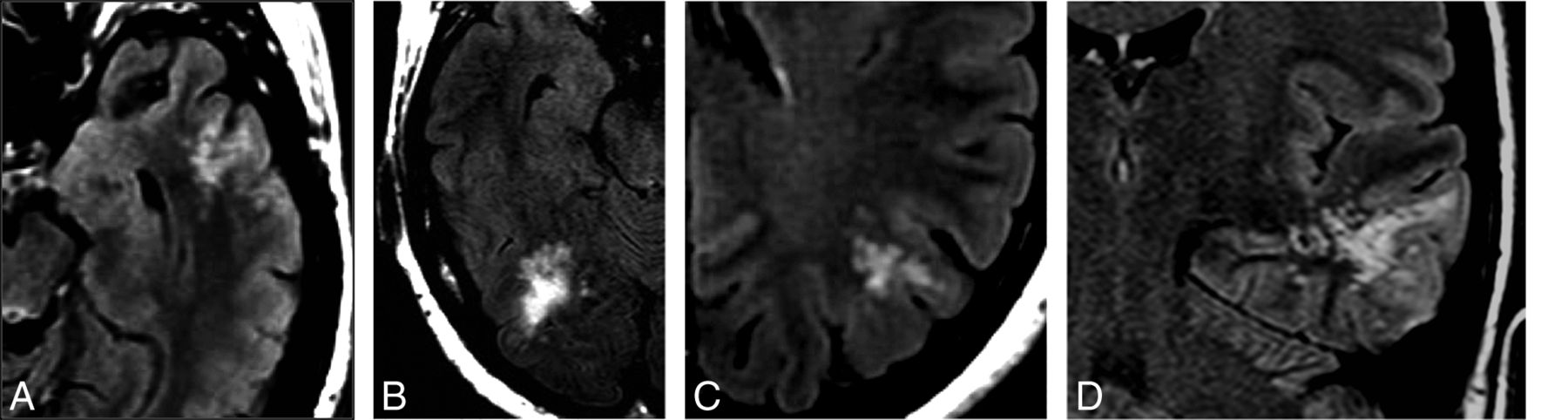
    A 53-year-old male patient with temporal lobe epilepsy (case 31). A–D, MR imaging demonstrates a probable MVNT in the left parahippocampal gyrus (arrows). E, FDG-PET MR brain imaging shows focal hypometabolism in the left medial temporal lobe corresponding to the site of suspected lesion. F and G, Hematoxylin-eosin-stained histopathology slides demonstrate the abnormal clustering and vacuolation of the neuronal cells. Images were obtained with 20× magnification (F) and 200× magnification (G).

  • Fig 5.
    • Download figure
    • Open in new tab
    • Download powerpoint
    Fig 5.

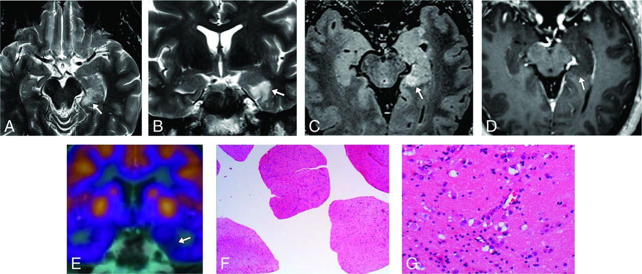
    A 43-year-old male patient with headaches (case 33). A, Axial FLAIR image demonstrates a left frontal lobe lesion (arrow) consisting of multiple coalescent T2 and FLAIR hyperintense nodules located in the deep cortical ribbon and without mass effect. B, Low-power photomicrograph with immunohistochemistry staining for neurofibrillary protein shows positive staining between variably sized nonstaining vacuolating nodules.

  • Fig 6.
    • Download figure
    • Open in new tab
    • Download powerpoint
    Fig 6.

    MVNT typical presentation graphic illustration. Schematic coronal view depicts a nodular “bubbly-appearing” lesion located on the inner surface of otherwise normal-appearing left temporal lobe cortex within the deep cortical ribbon and superficial subcortical white matter. Reprinted from Osborn's Brain9 with permission from Elsevier.

Tables

  • Figures
  • Summary of findings of patients with presumed MVNT

    Demographics33 patients; mean age, 39 yr; female/male ratio (1.4:1)
    Clinical dataNonfocal headache (n = 16), possible seizure (n = 8), other reasons (n = 9)
    Follow-up scan 24–144 mo (mean, 36.8 mo)
    LocationParietal lobe (n = 9), frontal lobe (n = 8), temporal lobe (n = 6), occipital lobe (n = 2), >1 lobe (n = 8)
    MRI findings (total)
        Hyperintense lesions on T2WI in comparison with the normal white matter33 (100%)
        Hyperintense lesions on FLAIR in comparison with the CSF33 (100%)
        Hypointense lesions on thin-section T2WI in comparison with the CSF21 (100%)
        Presence of abnormal white matter signal surrounding the bubble-like lesions15 (45%)
        Contrast enhancement1 (3%)
        Restricted diffusion or blooming on susceptibility-weighted imaging0 (0%)
PreviousNext
Back to top

In this issue

American Journal of Neuroradiology: 38 (10)
American Journal of Neuroradiology
Vol. 38, Issue 10
1 Oct 2017
  • Table of Contents
  • Index by author
  • Complete Issue (PDF)
Advertisement
Print
Download PDF
Email Article

Thank you for your interest in spreading the word on American Journal of Neuroradiology.

NOTE: We only request your email address so that the person you are recommending the page to knows that you wanted them to see it, and that it is not junk mail. We do not capture any email address.

Enter multiple addresses on separate lines or separate them with commas.
Multinodular and Vacuolating Neuronal Tumor of the Cerebrum: A New “Leave Me Alone” Lesion with a Characteristic Imaging Pattern
(Your Name) has sent you a message from American Journal of Neuroradiology
(Your Name) thought you would like to see the American Journal of Neuroradiology web site.
CAPTCHA
This question is for testing whether or not you are a human visitor and to prevent automated spam submissions.
Cite this article
R.H. Nunes, C.C. Hsu, A.J. da Rocha, L.L.F. do Amaral, L.F.S. Godoy, T.W. Watkins, V.H. Marussi, M. Warmuth-Metz, H.C. Alves, F.G. Goncalves, B.K. Kleinschmidt-DeMasters, A.G. Osborn
Multinodular and Vacuolating Neuronal Tumor of the Cerebrum: A New “Leave Me Alone” Lesion with a Characteristic Imaging Pattern
American Journal of Neuroradiology Oct 2017, 38 (10) 1899-1904; DOI: 10.3174/ajnr.A5281

Citation Manager Formats

  • BibTeX
  • Bookends
  • EasyBib
  • EndNote (tagged)
  • EndNote 8 (xml)
  • Medlars
  • Mendeley
  • Papers
  • RefWorks Tagged
  • Ref Manager
  • RIS
  • Zotero
0 Responses
Respond to this article
Share
Bookmark this article
Multinodular and Vacuolating Neuronal Tumor of the Cerebrum: A New “Leave Me Alone” Lesion with a Characteristic Imaging Pattern
R.H. Nunes, C.C. Hsu, A.J. da Rocha, L.L.F. do Amaral, L.F.S. Godoy, T.W. Watkins, V.H. Marussi, M. Warmuth-Metz, H.C. Alves, F.G. Goncalves, B.K. Kleinschmidt-DeMasters, A.G. Osborn
American Journal of Neuroradiology Oct 2017, 38 (10) 1899-1904; DOI: 10.3174/ajnr.A5281
del.icio.us logo Twitter logo Facebook logo Mendeley logo
  • Tweet Widget
  • Facebook Like
  • Google Plus One
Purchase

Jump to section

  • Article
    • Abstract
    • ABBREVIATIONS:
    • Case Series
    • Results
    • Discussion
    • Conclusions
    • Acknowledgments
    • Footnotes
    • References
  • Figures & Data
  • Supplemental
  • Info & Metrics
  • Responses
  • References
  • PDF

Related Articles

  • PubMed
  • Google Scholar

Cited By...

  • Multinodular and vacuolating neuronal tumour: emphasis on expression of early and late neuronal immunomarkers
  • Cortically Based Brain Tumors in Children: A Decision-Tree Approach in the Radiology Reading Room
  • Multinodular and Vacuolating Neuronal Tumor-like Lesion of the Spinal Cord: Two Case Reports
  • Long-Term Follow-up of Multinodular and Vacuolating Neuronal Tumors and Implications for Surveillance Imaging
  • Newly Recognized CNS Tumors in the 2021 World Health Organization Classification: Imaging Overview with Histopathologic and Genetic Correlation
  • Uncommon Glioneuronal Tumors: A Radiologic and Pathologic Synopsis
  • The 2021 World Health Organization Classification of Tumors of the Central Nervous System: What Neuroradiologists Need to Know
  • Multinodular and Vacuolating Neuronal Tumor of the Cerebrum: Does the Name Require Review?
  • Multinodular and Vacuolating Posterior Fossa Lesions of Unknown Significance
  • Multiparametric Imaging Improves Confidence in the Diagnosis of Multinodular and Vacuolating Neuronal Tumor of the Cerebrum
  • Reply:
  • Crossref
  • Google Scholar

This article has not yet been cited by articles in journals that are participating in Crossref Cited-by Linking.

More in this TOC Section

  • Diagnostic Neuroradiology of Monoclonal Antibodies
  • Cerebral ADC Changes in Fabry Disease
  • ML for Glioma Molecular Subtype Prediction
Show more Adult Brain

Similar Articles

Advertisement

Indexed Content

  • Current Issue
  • Accepted Manuscripts
  • Article Preview
  • Past Issues
  • Editorials
  • Editor's Choice
  • Fellows' Journal Club
  • Letters to the Editor
  • Video Articles

Cases

  • Case Collection
  • Archive - Case of the Week
  • Archive - Case of the Month
  • Archive - Classic Case

More from AJNR

  • Trainee Corner
  • Imaging Protocols
  • MRI Safety Corner
  • Book Reviews

Multimedia

  • AJNR Podcasts
  • AJNR Scantastics

Resources

  • Turnaround Time
  • Submit a Manuscript
  • Submit a Video Article
  • Submit an eLetter to the Editor/Response
  • Manuscript Submission Guidelines
  • Statistical Tips
  • Fast Publishing of Accepted Manuscripts
  • Graphical Abstract Preparation
  • Imaging Protocol Submission
  • Evidence-Based Medicine Level Guide
  • Publishing Checklists
  • Author Policies
  • Become a Reviewer/Academy of Reviewers
  • News and Updates

About Us

  • About AJNR
  • Editorial Board
  • Editorial Board Alumni
  • Alerts
  • Permissions
  • Not an AJNR Subscriber? Join Now
  • Advertise with Us
  • Librarian Resources
  • Feedback
  • Terms and Conditions
  • AJNR Editorial Board Alumni

American Society of Neuroradiology

  • Not an ASNR Member? Join Now

© 2025 by the American Society of Neuroradiology All rights, including for text and data mining, AI training, and similar technologies, are reserved.
Print ISSN: 0195-6108 Online ISSN: 1936-959X

Powered by HighWire