
Article Figures & Data
Figures
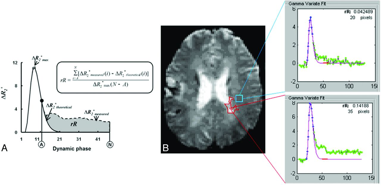
- Fig 1.
A, Estimation of rR by use of the ΔR2* versus time curve measured from the DSC regions of interest (ΔR2*measured) as well as its γ-variate fit (ΔR2*fit). ΔR2*max is the maximum of ΔR2*theoretical. A is the dynamic phase corresponding to the onset of the recirculation phase measured at half-height of the descending aspect of the ΔR2* curve, and N is the final DSC phase. B, Example of rR measurement: case 733, section 11, perfusion scan. MS lesion (red region of interest) and NAWM (blue region of interest) and rR calculation results. rR for WM lesion is 0.142; rR for NAWM region is 0.042.
- Fig 2.
rR values of NAWM and WM lesion.
- Fig 3.
rR values of NAWM are significantly lower in impaired than in nonimpaired groups by use of A, the Symbol Digit Modalities Test (P = .007), and B, the Paced Auditory Serial Addition Test (P = .024).


Tables
Outcomes of multivariate logistic regression analyses for NAWM rR after controlling for potential confounders
Cognitive Test Odds Ratio (5–95% CI) P Value SDMT 0.177 (0.040–0.785) .023 COWAT 0.662 (0.002–2.226) .130 PASAT 1.6 0.053 (0.003–0.961) .047 Note:—SDMT indicates Symbol Digit Modalities Test; COWAT, Controlled Word Association Test; PASAT, Paced Auditory Serial Addition Test.
Confounders for Symbol Digit Modalities Test: brain parenchymal fraction, WM fraction, WM lesion fraction; Controlled Word Association Test: WM fraction, WM lesion fraction, gray matter fraction; and Paced Auditory Serial Addition Test: brain parenchymal fraction, WM fraction, WM lesion fraction.
NAWM rR continues to be significantly lower in the impaired group for both the Symbol Digit Modalities Test and the Paced Auditory Serial Addition Test.


Jump to section
Related Articles
Cited By...
- No citing articles found.