
Article Figures & Data
Figures
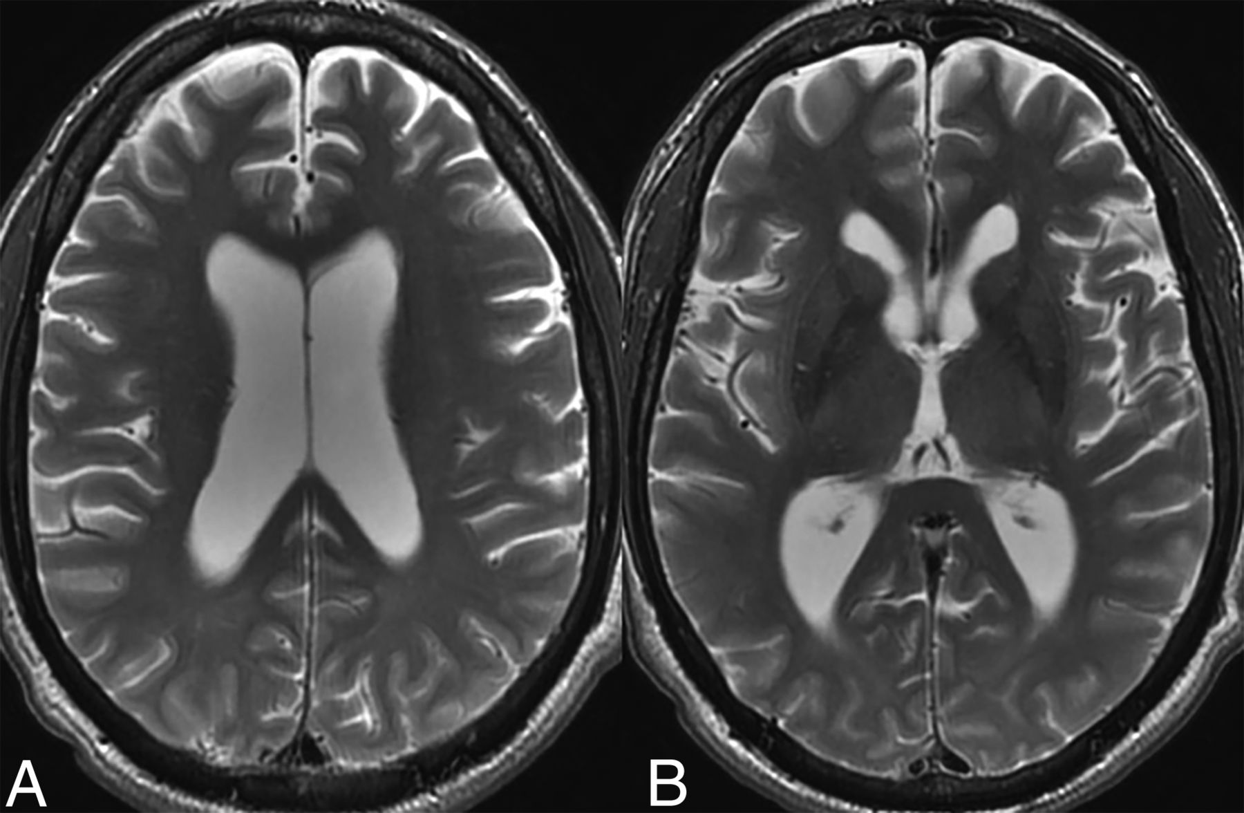
- FIG 1.
Parenchymal atrophy. Axial T2-weighted images in a 55-year-old patient with ECD demonstrate diffuse cortical atrophy with mild ex-vacuo dilation of the lateral ventricles. There were no focal lesions.
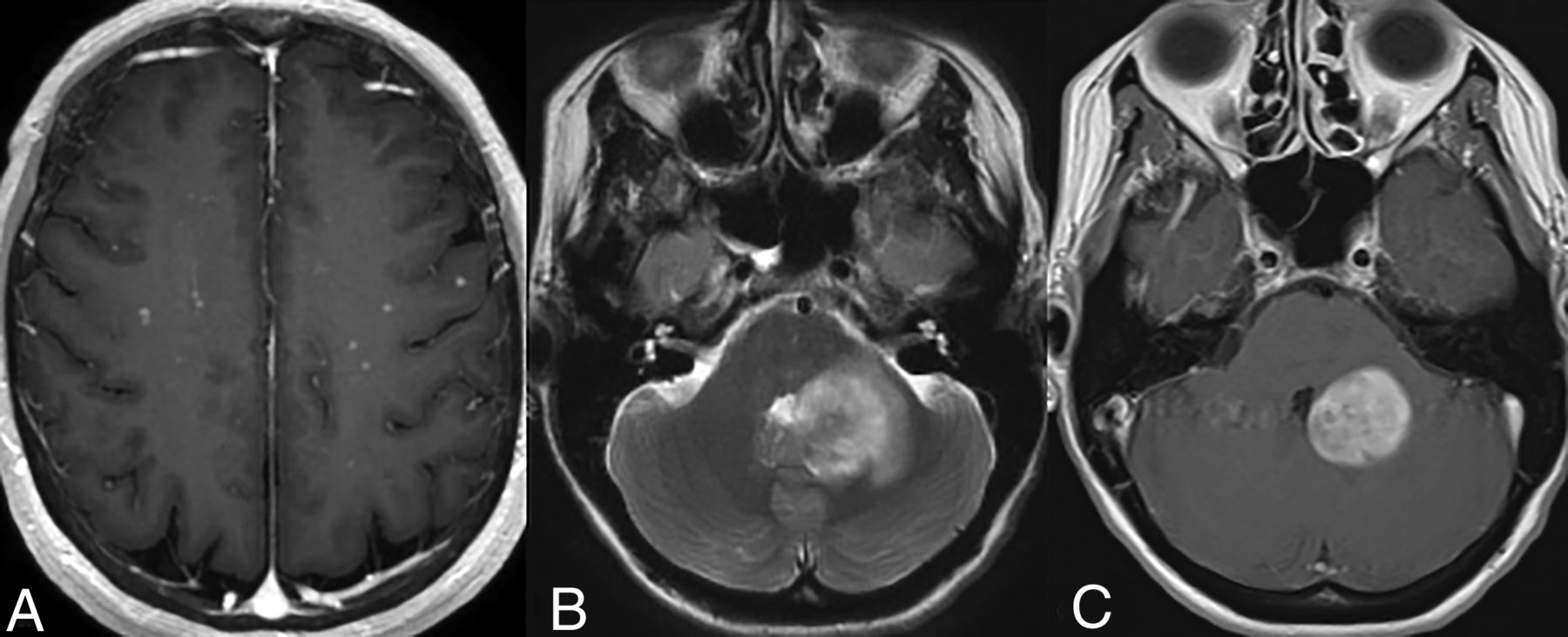
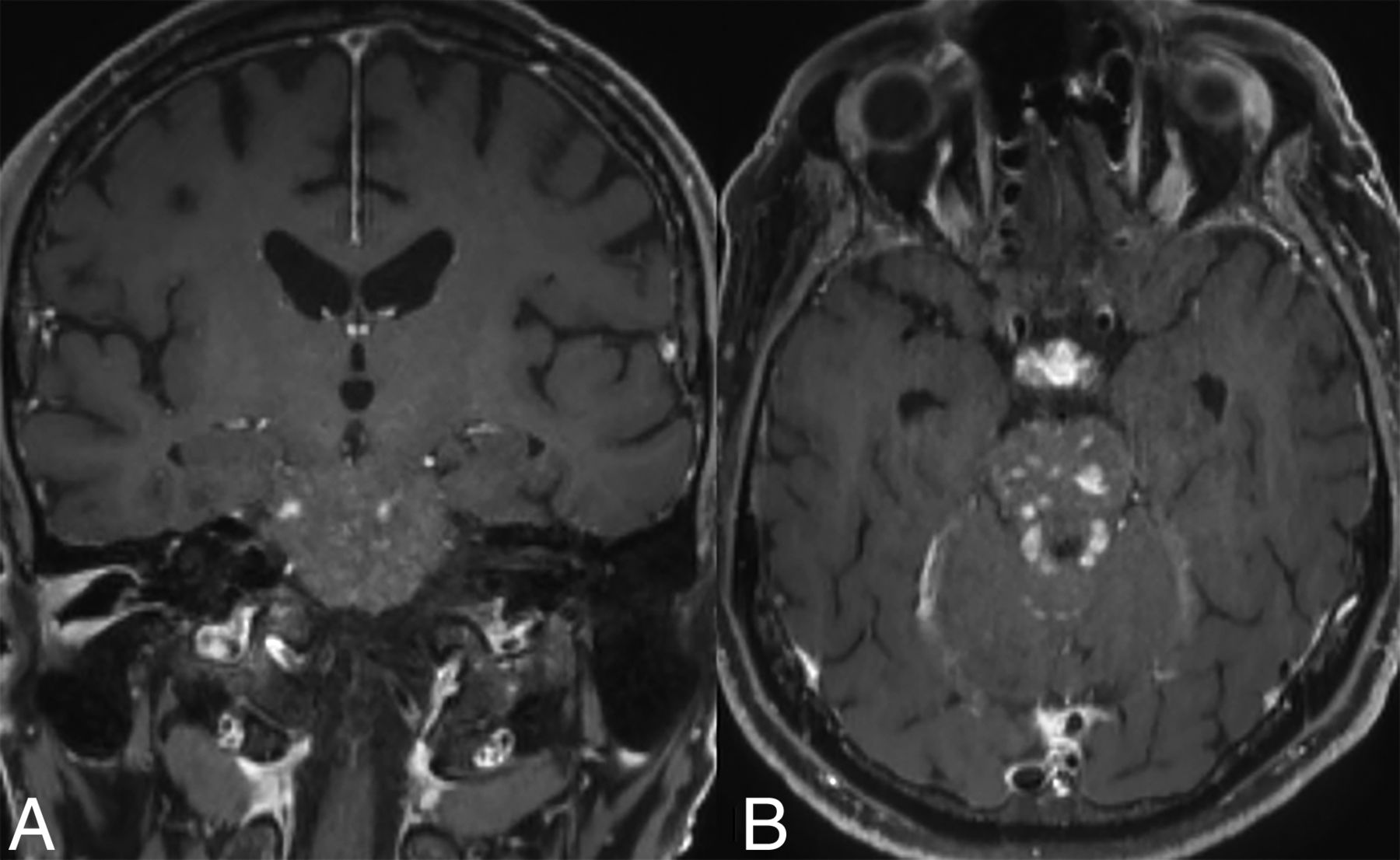
- FIG 2.
Parenchymal lesions. T1-CE maximum intensity projection images in a patient (A) reveal scattered micronodular enhancing lesions bilaterally. Axial T2 WI (B) and T1-CE images (C) in a different patient demonstrate a masslike T2 hyperintense periventricular lesion with homogeneous enhancement and mild surrounding edema.
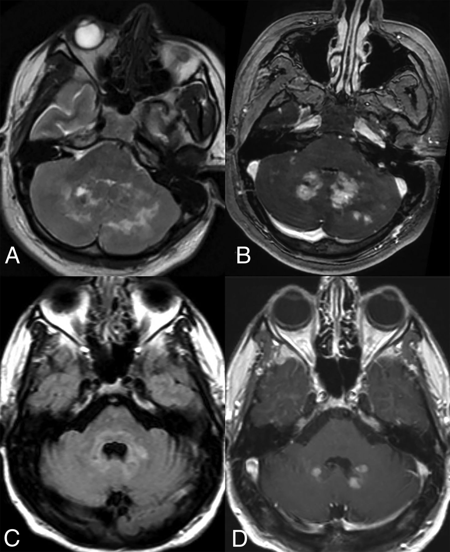
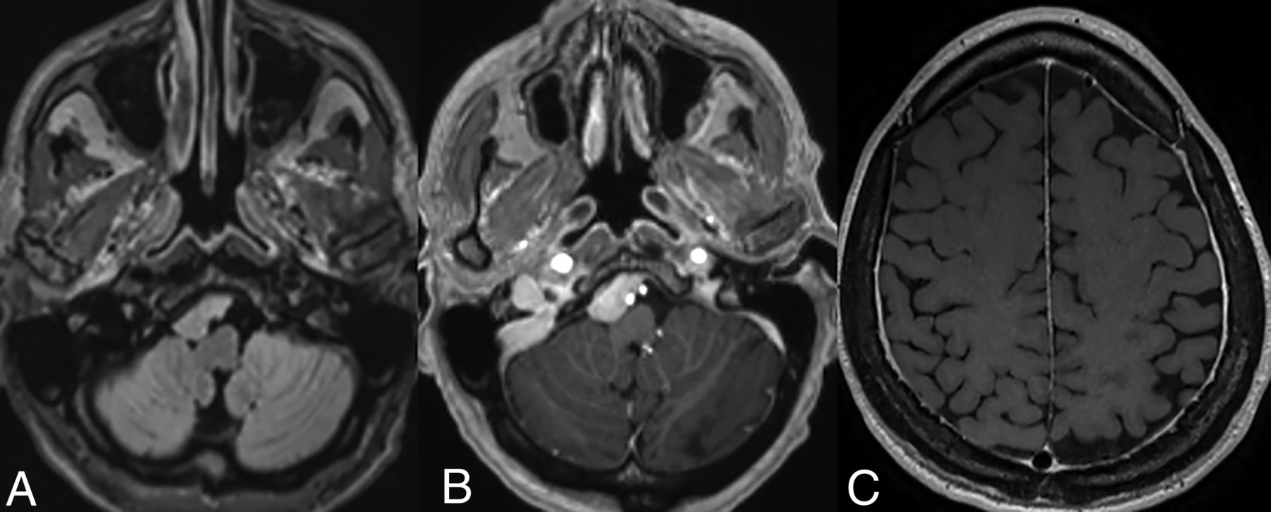
- FIG 3.
Cerebellar involvement. Axial T2-WI (A) and T1-CE (B) images in a patient demonstrate ill-defined T2-isointense lesions in bilateral cerebellar hemispheres, most prominently along the dentate nuclei. Axial FLAIR (C) and T1-CE (D) images in a different patient reveal more focal involvement of the bilateral dentate nuclei.
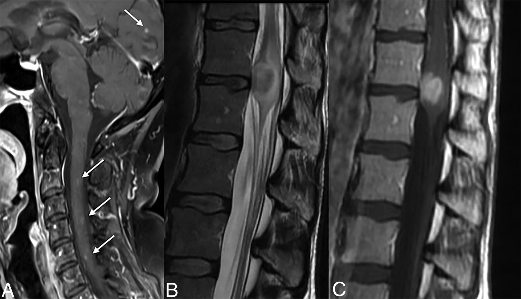
- FIG 4.
Spinal involvement. Sagittal T1-CE image (A) reveals tiny discrete enhancing foci involving the cervical cord and the occipital lobe. Sagittal T2 WI (B) and T1-CE images (C) in a different patient demonstrate a masslike involvement of the conus.
- FIG 5.
Coronal (A) and axial (B) T1-CE images in a patient with ECD demonstrate punctate areas of enhancement relatively confined to the pons, mimicking CLIPPERS. Note the thickening and enhancement within the infundibulum in (B) suggesting involvement of the HPA axis by the disease.
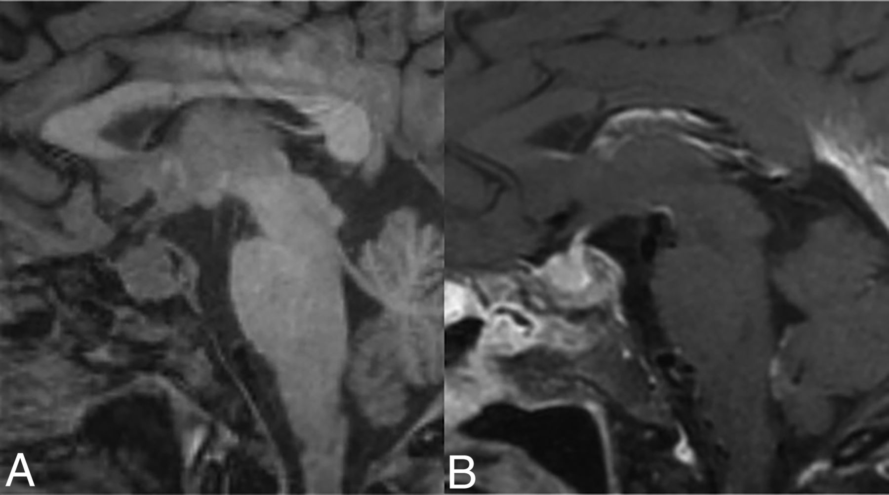
- FIG 6.
HPA axis involvement. Sagittal T1 noncontrast image (A) demonstrates an enlarged pituitary gland with loss of the posterior pituitary bright spot. Also, note abnormal marrow signal in clivus from bony involvement. Sagittal T1-CE image (B) demonstrates heterogeneous enhancement.
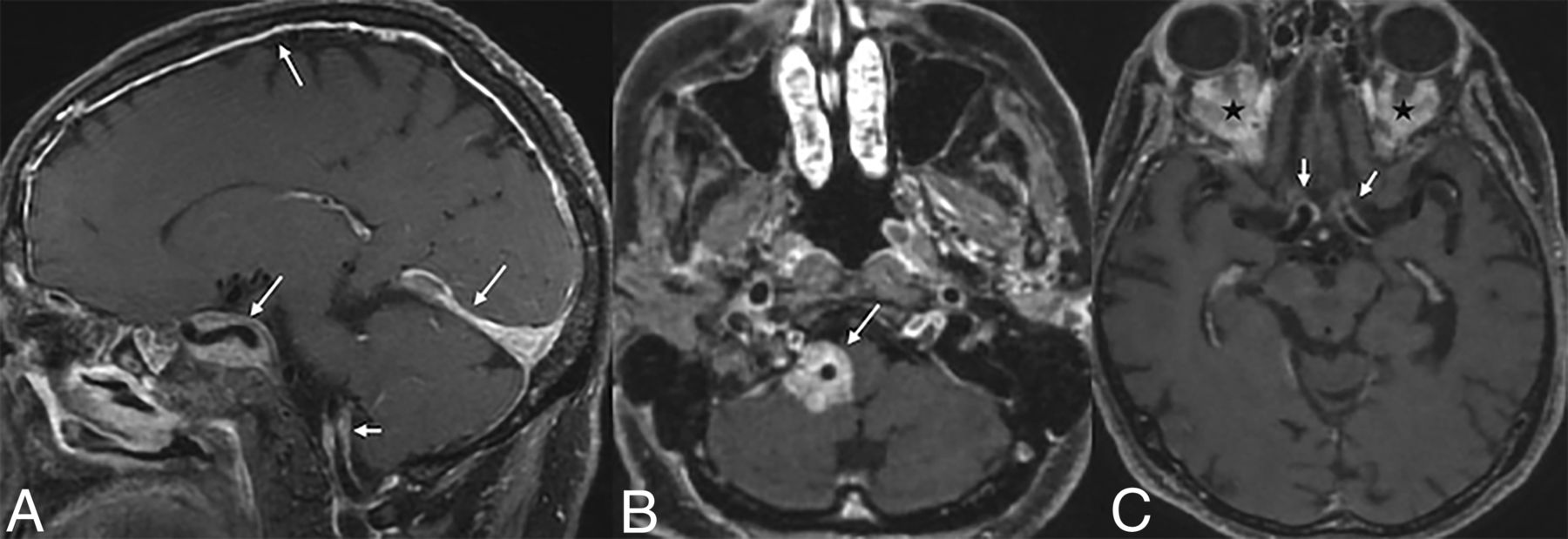
- FIG 7.
Vascular involvement. Sagittal (A) and axial (B and C) T1-CE in 3 different patients with ECD showing vascular involvement. There is vertebral artery involvement (short arrow, A) along with dural, tentorial, and cavernous sinus involvement (long arrows, A). In the second patient (B), there is vascular sheathing and enhancement around the right vertebral artery (arrow, B). The third image (C) shows similar involvement of the ICA vessels (arrows) along with orbital infiltrative lesions (black stars).
- FIG 8.
Axial FLAIR (A) and T1-CE (B) images demonstrate an enhancing dural-based lesion along the clivus. A T1-CE (C) image of another patient reveals diffuse pachymeningeal enhancement.
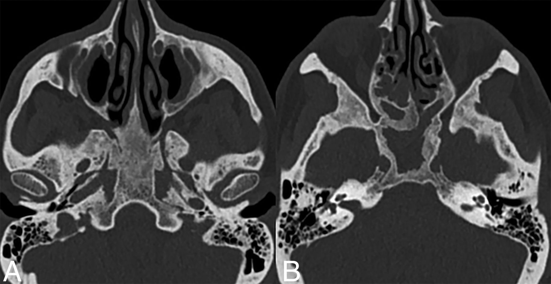
- FIG 9.
Sinonasal ECD. Axial CT images (bone kernel) in a patient with ECD demonstrate skull base and sinus involvement characterized by thickening and osteosclerosis of the basisphenoid and paranasal sinuses.
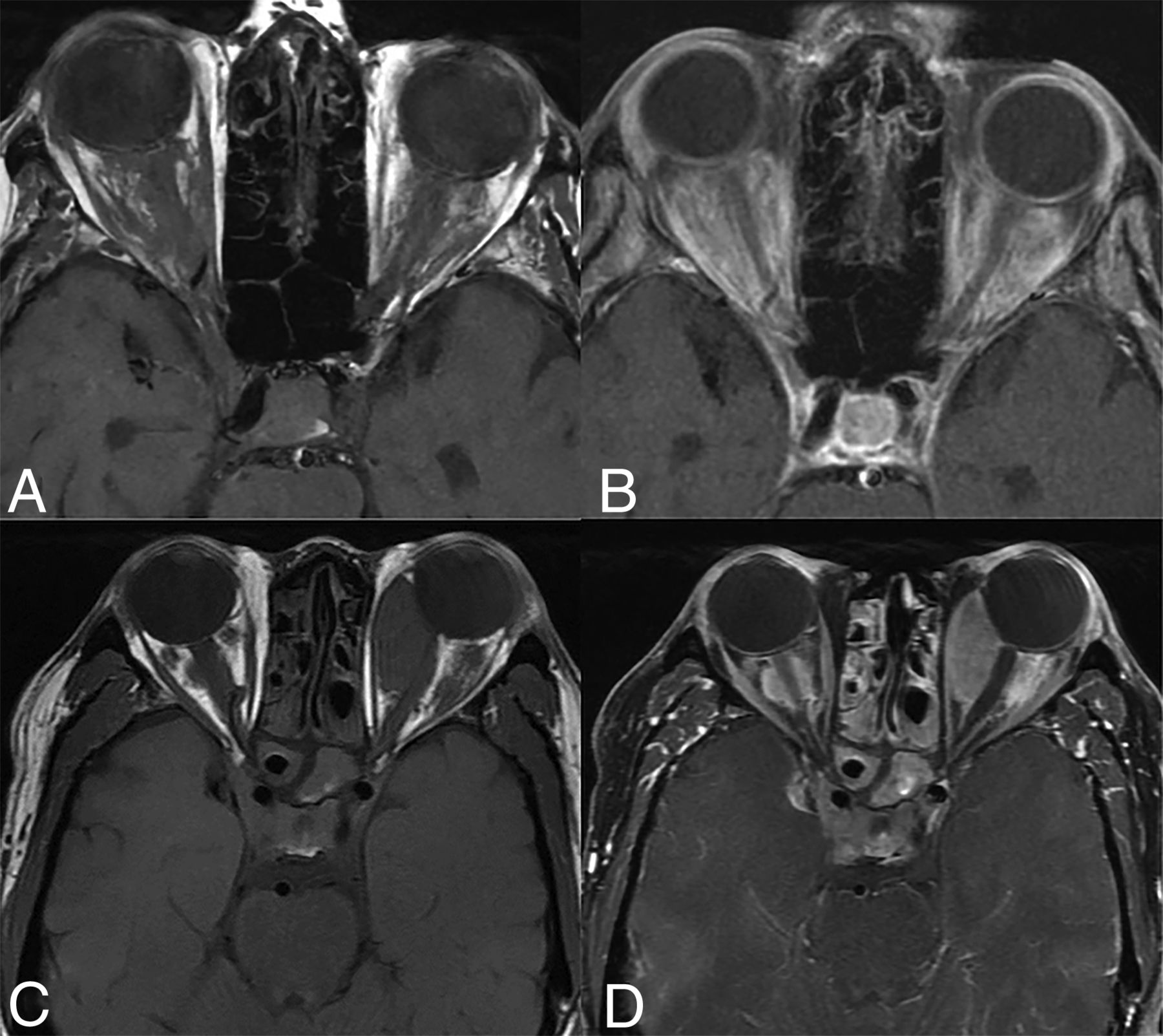
- FIG 10.
Orbital involvement in ECD. Axial T1 WI precontrast (A and C) and T1-CE (B and D) images of 2 different patients showing infiltrative (A and B) and masslike (C and D) orbital involvement. There is involvement of the bilateral cavernous sinuses in both cases along with paranasal sinus involvement in the bottom row (C and D).


Tables
Erdheim-Chester Disease Langerhans Cell Histiocytosis Rosai-Dorfman Disease Demographics M > F, sixth decade of life M > F, first 2 decades of life M > F, second to third decade of life BRAFV00E mutation Positive (more than LCD) Positive N/A Histopathology Foamy histiocytes, CD68+, CD1a- Mononucleated dendritic cells, CD1a+, Birbeck granules Multinucleated histiocytes, CD68+, S-100+, CD1a-; may be associated with immunoglobulin G4-related disease Neurodegeneration and atrophy + ++ + Intraparenchymal lesions Present (often multiple, more edema) Present (often solitary, lesser edema) Present (often multiple, more edema) HPA involvement + +++ Rare Extra-axial lesions ++ + + Craniofacial involvement Sclerotic lesions (calvarial involvement is less common) Lytic lesions (calvarial involvement is more common) Rare Orbital involvement ++ + + Vascular involvement + − − Note:—M indicates male; F, female; N/A, not applicable.
- Table 2:
Summary of the CNS imaging findings in Erdheim-Chester disease (reported prevalence in parentheses)
Compartment Imaging Finding CNS involvement (25%–76%) Parenchymal volume loss (cortical and cerebellar atrophy), increased radial diffusivity of water molecules Parenchyma (supratentorial [approx. 46%]; posterior fossa [20%–46%]; white matter [60%–87%]; basal ganglia [7%]) Scattered lesions (micronodular, nodular, masslike), T2 prolongation, heterogeneous enhancement, minimal edema, increased Ktrans values T2/FLAIR hyperintense lesions of indeterminate significance May have signal drop-out on SWI in basal ganglia Prolonged gadolinium contrast retention HPA (17%–44%) Thickening of pituitary stalk, hypothalamic T2 hyperintensity Loss of posterior pituitary bright spot, nodular or micronodular lesions, pituitary atrophy Vascular (10%–17%) Periadventitial vessel inflammation, vascular sheathing Dural venous sinus thrombosis or infiltration Extra-axial (dural [30%–50%], leptomeningeal [6%–7%]) Dura-based solitary or multiple lesions (enhancing, isointense), pachymeningeal thickening, spinal extradural involvement Leptomeningeal disease Craniofacial (40%–50%) Osteosclerotic lesions (calvaria, skull base, paranasal sinuses), bone thickening Orbital (18%–30%) Intraconal or extraconal masses Optic nerve sheath enlargement Choroidal involvement (intraorbital mass)


Jump to section
Related Articles
Cited By...
- No citing articles found.