Advertisement

Main menu

  • Home
  • Content
    • Current Issue
    • Accepted Manuscripts
    • Article Preview
    • Past Issue Archive
    • Video Articles
    • AJNR Case Collection
    • Case of the Week Archive
    • Case of the Month Archive
    • Classic Case Archive
  • Special Collections
    • AJNR Awards
    • Low-Field MRI
    • Alzheimer Disease
    • ASNR Foundation Special Collection
    • Photon-Counting CT
    • View All
  • Multimedia
    • AJNR Podcasts
    • AJNR SCANtastic
    • Trainee Corner
    • MRI Safety Corner
    • Imaging Protocols
  • For Authors
    • Submit a Manuscript
    • Submit a Video Article
    • Submit an eLetter to the Editor/Response
    • Manuscript Submission Guidelines
    • Statistical Tips
    • Fast Publishing of Accepted Manuscripts
    • Graphical Abstract Preparation
    • Imaging Protocol Submission
    • Author Policies
  • About Us
    • About AJNR
    • Editorial Board
    • Editorial Board Alumni
  • More
    • Become a Reviewer/Academy of Reviewers
    • Subscribers
    • Permissions
    • Alerts
    • Feedback
    • Advertisers
    • ASNR Home

User menu

  • Alerts
  • Log in

Search

  • Advanced search
American Journal of Neuroradiology

American Journal of Neuroradiology

ASHNR American Society of Functional Neuroradiology ASHNR American Society of Pediatric Neuroradiology ASSR
  • Alerts
  • Log in

Advanced Search

  • Home
  • Content
    • Current Issue
    • Accepted Manuscripts
    • Article Preview
    • Past Issue Archive
    • Video Articles
    • AJNR Case Collection
    • Case of the Week Archive
    • Case of the Month Archive
    • Classic Case Archive
  • Special Collections
    • AJNR Awards
    • Low-Field MRI
    • Alzheimer Disease
    • ASNR Foundation Special Collection
    • Photon-Counting CT
    • View All
  • Multimedia
    • AJNR Podcasts
    • AJNR SCANtastic
    • Trainee Corner
    • MRI Safety Corner
    • Imaging Protocols
  • For Authors
    • Submit a Manuscript
    • Submit a Video Article
    • Submit an eLetter to the Editor/Response
    • Manuscript Submission Guidelines
    • Statistical Tips
    • Fast Publishing of Accepted Manuscripts
    • Graphical Abstract Preparation
    • Imaging Protocol Submission
    • Author Policies
  • About Us
    • About AJNR
    • Editorial Board
    • Editorial Board Alumni
  • More
    • Become a Reviewer/Academy of Reviewers
    • Subscribers
    • Permissions
    • Alerts
    • Feedback
    • Advertisers
    • ASNR Home
  • Follow AJNR on Twitter
  • Visit AJNR on Facebook
  • Follow AJNR on Instagram
  • Join AJNR on LinkedIn
  • RSS Feeds

AJNR Awards, New Junior Editors, and more. Read the latest AJNR updates

Research ArticleNEUROVASCULAR/STROKE IMAGING

A Comparative Study of CT Perfusion Postprocessing Tools in Medium/Distal Vessel Occlusion Stroke

Helene Corbaz, Nikolaos Ntoulias, Alex Brehm, Julia Wolleb, Philippe Claude Cattin, Florentin Bieder, Victor Schulze-Zachau and Marios-Nikos Psychogios
American Journal of Neuroradiology April 2025, DOI: https://doi.org/10.3174/ajnr.A8616
Helene Corbaz
aFrom the Department of Biomedical Engineering (H.C., J.W., P.C.C., F.B.), University Basel, Basel, Switzerland
  • Find this author on Google Scholar
  • Find this author on PubMed
  • Search for this author on this site
Nikolaos Ntoulias
bDepartment of Diagnostic & Interventional Neuroradiology (N.N., A.B., V.S.-Z., M.-N.P.), Radiology & Nuclear Medicine Clinic, University Hospital Basel, Basel, Switzerland
  • Find this author on Google Scholar
  • Find this author on PubMed
  • Search for this author on this site
Alex Brehm
bDepartment of Diagnostic & Interventional Neuroradiology (N.N., A.B., V.S.-Z., M.-N.P.), Radiology & Nuclear Medicine Clinic, University Hospital Basel, Basel, Switzerland
  • Find this author on Google Scholar
  • Find this author on PubMed
  • Search for this author on this site
  • ORCID record for Alex Brehm
Julia Wolleb
aFrom the Department of Biomedical Engineering (H.C., J.W., P.C.C., F.B.), University Basel, Basel, Switzerland
  • Find this author on Google Scholar
  • Find this author on PubMed
  • Search for this author on this site
Philippe Claude Cattin
aFrom the Department of Biomedical Engineering (H.C., J.W., P.C.C., F.B.), University Basel, Basel, Switzerland
  • Find this author on Google Scholar
  • Find this author on PubMed
  • Search for this author on this site
  • ORCID record for Philippe Claude Cattin
Florentin Bieder
aFrom the Department of Biomedical Engineering (H.C., J.W., P.C.C., F.B.), University Basel, Basel, Switzerland
  • Find this author on Google Scholar
  • Find this author on PubMed
  • Search for this author on this site
Victor Schulze-Zachau
bDepartment of Diagnostic & Interventional Neuroradiology (N.N., A.B., V.S.-Z., M.-N.P.), Radiology & Nuclear Medicine Clinic, University Hospital Basel, Basel, Switzerland
  • Find this author on Google Scholar
  • Find this author on PubMed
  • Search for this author on this site
  • ORCID record for Victor Schulze-Zachau
Marios-Nikos Psychogios
bDepartment of Diagnostic & Interventional Neuroradiology (N.N., A.B., V.S.-Z., M.-N.P.), Radiology & Nuclear Medicine Clinic, University Hospital Basel, Basel, Switzerland
cClinical Research Department(M.-N.P.), University Basel, Basel, Switzerland
  • Find this author on Google Scholar
  • Find this author on PubMed
  • Search for this author on this site
  • Article
  • Figures & Data
  • Info & Metrics
  • Responses
  • References
  • PDF
Loading

Article Figures & Data

Figures

  • Tables
  • Figure
    • Download figure
    • Open in new tab
    • Download powerpoint
  • FIG 1.
    • Download figure
    • Open in new tab
    • Download powerpoint
    FIG 1.

    Patient flow chart. *The full-text search was conducted in the local PACS and included the descriptors “acute stroke,” “cerebrovascular insult,” and “National Institutes of Health Stroke Scale” as well as corresponding abbreviations.
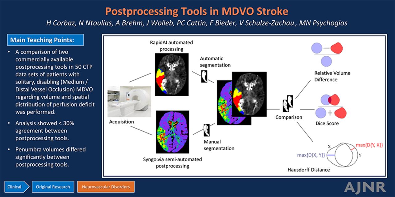
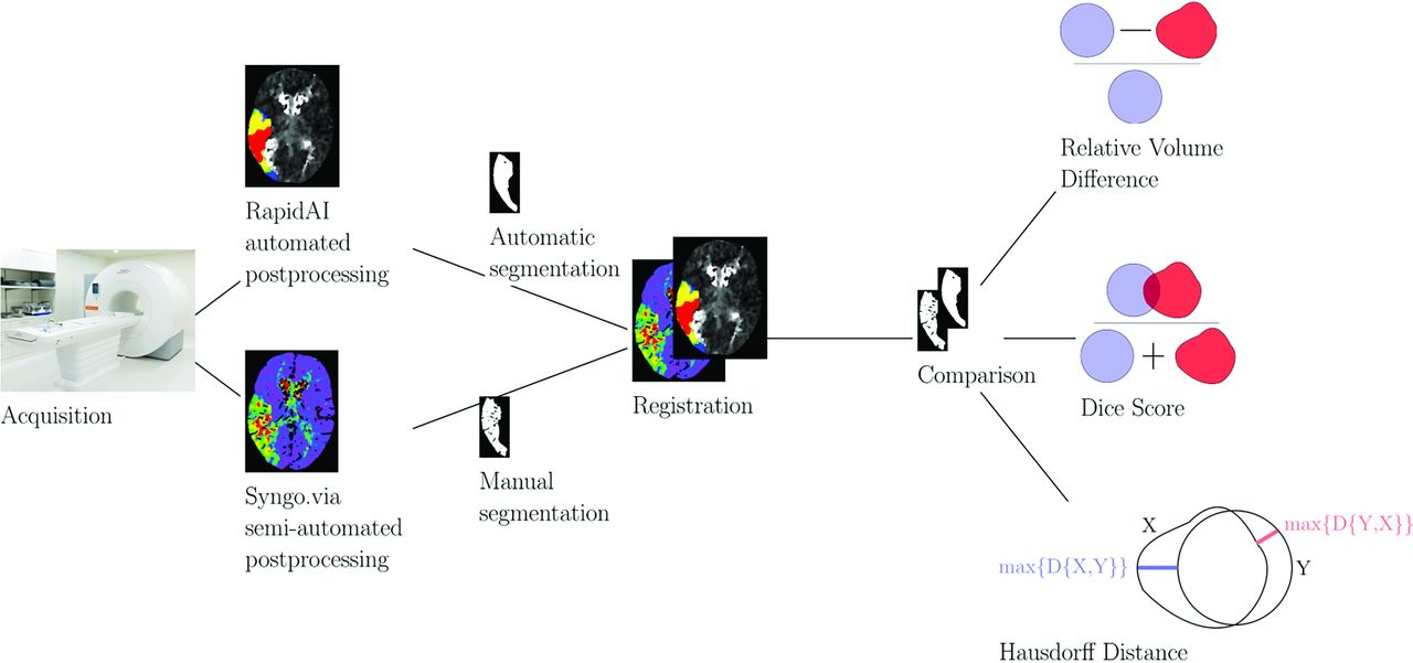
  • FIG 2.
    • Download figure
    • Open in new tab
    • Download powerpoint
    FIG 2.

    Study workflow.

  • FIG 3.
    • Download figure
    • Open in new tab
    • Download powerpoint
    FIG 3.

    Classification scheme.
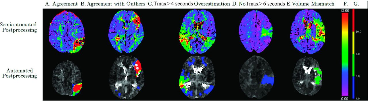
  • FIG 4.
    • Download figure
    • Open in new tab
    • Download powerpoint
    FIG 4.

    Examples of different categories of agreement or disagreement. Figure shows Tmax maps for semiautomated (upper row) and automated postprocessing. A, Agreement. B, Agreement with outliers. Please note contralateral areas of Tmax > 4 seconds indicated in blue in the RapidAI map. C, Tmax > 4 seconds overestimation. Please note how the Tmax >4 seconds lesion of RapidAI extends beyond the lesion of perfusion delay in the Syngo.via Tmax map. D, No Tmax > 6 seconds lesion. E, Volume mismatch. F, Color reference for perfusion maps derived from Syngo.via. G, Color reference for perfusion maps derived from RapidAI.

  • FIG 5.
    • Download figure
    • Open in new tab
    • Download powerpoint
    FIG 5.

    Patterns of agreement and disagreement between semiautomated and automated postprocessing.

  • FIG 6.
    • Download figure
    • Open in new tab
    • Download powerpoint
    FIG 6.

    Bland-Altman plots for the comparison of mismatch estimates of semiautomated and automated postprocessing. Mismatch volume derived from semiautomated postprocessing (mL) as compared with mismatch volume derived from automated postprocessing (mL). A, In the upper graphic, automated postprocessing mismatch volume was defined as volume of Tmax >4 seconds – volume of CBF <30%. B, In the lower graphic, automated postprocessing mismatch volume was defined as volume of Tmax > 6 seconds – volume of CBF <30%. In both graphics, semiautomated postprocessing mismatch volume was defined as volume of Tmax lesion – volume of CBV lesion. For both comparisons, the discrepancy of mismatch volume was larger for patients with larger average volume of the perfusion lesion. SD indicates standard deviation.

Tables

  • Figures
    • View popup
    Table 1:

    Patient baseline characteristics

    All Patients (n = 49)
    Age (years, median [IQR])76 (15)
    Sex (male patients)33 (67.3%)
    Median NIHSS at admission (IQR)6 (5)
    • Note:—One patient experienced 2 distinct MDVO strokes with a time lag of several months and CTP data sets from both events were included.

    • View popup
    Table 2:

    Patient baseline characteristics

    Occlusion LocationAll Data Sets (n = 50)
    M222 (44%)
    M318 (36%)
    M42 (4%)
    A10
    A20
    A31 (2%)
    P10
    P26 (12%)
    P31 (2%)
    • Note:—One patient experienced 2 distinct MDVO strokes with a time lag of several months and CTP data sets from both events were included.

    • View popup
    Table 3:

    Distribution of the perfusion lesion volumes

    Median Volume (mL)IQRRange
    Tissue at riska42.833.82–161.8
    Tmax > 4 secondsb67.467.74.2–295.4
    Tmax > 6 secondsb25.421.40–96
    Infarct corea7.325.20–56.8
    CBF <30%b2.89.00–49.1
    Tmax > 10 secondsb7.417.80–63.7
    • Note:—Volumes were estimated by multiplying the computed volume with pixel size.

    • ↵a Results of semiautomated postprocessing.

    • ↵b Results of automated postprocessing.

    • View popup
    Table 4:

    Median of the DS, HD, and the RVD for the different comparisons


    Median DSMedian HD [mm]Median RVD
    Tissue at riska/Tmax > 6 secondsb0.5942.310.79
    Tissue at riska/Tmax > 4 secondsb0.56105.02−0.30
    Infarct corea/CBF <30%b0.0917.381.40
    Infarct corea/Tmax > 10 secondsb0.3959.662.28
    • ↵a Results of semiautomated postprocessing.

    • ↵b Results of automated postprocessing.

PreviousNext
Back to top
Advertisement
Print
Download PDF
Email Article

Thank you for your interest in spreading the word on American Journal of Neuroradiology.

NOTE: We only request your email address so that the person you are recommending the page to knows that you wanted them to see it, and that it is not junk mail. We do not capture any email address.

Enter multiple addresses on separate lines or separate them with commas.
A Comparative Study of CT Perfusion Postprocessing Tools in Medium/Distal Vessel Occlusion Stroke
(Your Name) has sent you a message from American Journal of Neuroradiology
(Your Name) thought you would like to see the American Journal of Neuroradiology web site.
CAPTCHA
This question is for testing whether or not you are a human visitor and to prevent automated spam submissions.
Cite this article
Helene Corbaz, Nikolaos Ntoulias, Alex Brehm, Julia Wolleb, Philippe Claude Cattin, Florentin Bieder, Victor Schulze-Zachau, Marios-Nikos Psychogios
A Comparative Study of CT Perfusion Postprocessing Tools in Medium/Distal Vessel Occlusion Stroke
American Journal of Neuroradiology Apr 2025, DOI: 10.3174/ajnr.A8616

Citation Manager Formats

  • BibTeX
  • Bookends
  • EasyBib
  • EndNote (tagged)
  • EndNote 8 (xml)
  • Medlars
  • Mendeley
  • Papers
  • RefWorks Tagged
  • Ref Manager
  • RIS
  • Zotero
0 Responses
Respond to this article
Share
Bookmark this article
CTP Postprocessing Tools in MDVO Stroke
Helene Corbaz, Nikolaos Ntoulias, Alex Brehm, Julia Wolleb, Philippe Claude Cattin, Florentin Bieder, Victor Schulze-Zachau, Marios-Nikos Psychogios
American Journal of Neuroradiology Apr 2025, DOI: 10.3174/ajnr.A8616
del.icio.us logo Twitter logo Facebook logo Mendeley logo
  • Tweet Widget
  • Facebook Like
  • Google Plus One
Purchase

Jump to section

  • Article
    • Graphical Abstract
    • Abstract
    • ABBREVIATIONS:
    • MATERIALS AND METHODS
    • RESULTS
    • DISCUSSION
    • CONCLUSIONS
    • Acknowledgments
    • Footnotes
    • References
  • Figures & Data
  • Info & Metrics
  • Responses
  • References
  • PDF

Related Articles

  • No related articles found.
  • PubMed
  • Google Scholar

Cited By...

  • No citing articles found.
  • Crossref
  • Google Scholar

This article has not yet been cited by articles in journals that are participating in Crossref Cited-by Linking.

More in this TOC Section

  • The insula and malignant cerebral edema
  • ICH Shape & Density Predict Rebleeding after MICE
  • Circle of Willis Variants and Stroke Outcomes
Show more Neurovascular/Stroke Imaging

Similar Articles

Advertisement

Indexed Content

  • Current Issue
  • Accepted Manuscripts
  • Article Preview
  • Past Issues
  • Editorials
  • Editor's Choice
  • Fellows' Journal Club
  • Letters to the Editor
  • Video Articles

Cases

  • Case Collection
  • Archive - Case of the Week
  • Archive - Case of the Month
  • Archive - Classic Case

More from AJNR

  • Trainee Corner
  • Imaging Protocols
  • MRI Safety Corner
  • Book Reviews

Multimedia

  • AJNR Podcasts
  • AJNR Scantastics

Resources

  • Turnaround Time
  • Submit a Manuscript
  • Submit a Video Article
  • Submit an eLetter to the Editor/Response
  • Manuscript Submission Guidelines
  • Statistical Tips
  • Fast Publishing of Accepted Manuscripts
  • Graphical Abstract Preparation
  • Imaging Protocol Submission
  • Evidence-Based Medicine Level Guide
  • Publishing Checklists
  • Author Policies
  • Become a Reviewer/Academy of Reviewers
  • News and Updates

About Us

  • About AJNR
  • Editorial Board
  • Editorial Board Alumni
  • Alerts
  • Permissions
  • Not an AJNR Subscriber? Join Now
  • Advertise with Us
  • Librarian Resources
  • Feedback
  • Terms and Conditions
  • AJNR Editorial Board Alumni

American Society of Neuroradiology

  • Not an ASNR Member? Join Now

© 2025 by the American Society of Neuroradiology All rights, including for text and data mining, AI training, and similar technologies, are reserved.
Print ISSN: 0195-6108 Online ISSN: 1936-959X

Powered by HighWire