Advertisement

Main menu

  • Home
  • Content
    • Current Issue
    • Accepted Manuscripts
    • Article Preview
    • Past Issue Archive
    • Video Articles
    • AJNR Case Collection
    • Case of the Week Archive
    • Case of the Month Archive
    • Classic Case Archive
  • Special Collections
    • AJNR Awards
    • Low-Field MRI
    • Alzheimer Disease
    • ASNR Foundation Special Collection
    • Photon-Counting CT
    • View All
  • Multimedia
    • AJNR Podcasts
    • AJNR SCANtastic
    • Trainee Corner
    • MRI Safety Corner
    • Imaging Protocols
  • For Authors
    • Submit a Manuscript
    • Submit a Video Article
    • Submit an eLetter to the Editor/Response
    • Manuscript Submission Guidelines
    • Statistical Tips
    • Fast Publishing of Accepted Manuscripts
    • Graphical Abstract Preparation
    • Imaging Protocol Submission
    • Author Policies
  • About Us
    • About AJNR
    • Editorial Board
    • Editorial Board Alumni
  • More
    • Become a Reviewer/Academy of Reviewers
    • Subscribers
    • Permissions
    • Alerts
    • Feedback
    • Advertisers
    • ASNR Home

User menu

  • Alerts
  • Log in

Search

  • Advanced search
American Journal of Neuroradiology

American Journal of Neuroradiology

ASHNR American Society of Functional Neuroradiology ASHNR American Society of Pediatric Neuroradiology ASSR
  • Alerts
  • Log in

Advanced Search

  • Home
  • Content
    • Current Issue
    • Accepted Manuscripts
    • Article Preview
    • Past Issue Archive
    • Video Articles
    • AJNR Case Collection
    • Case of the Week Archive
    • Case of the Month Archive
    • Classic Case Archive
  • Special Collections
    • AJNR Awards
    • Low-Field MRI
    • Alzheimer Disease
    • ASNR Foundation Special Collection
    • Photon-Counting CT
    • View All
  • Multimedia
    • AJNR Podcasts
    • AJNR SCANtastic
    • Trainee Corner
    • MRI Safety Corner
    • Imaging Protocols
  • For Authors
    • Submit a Manuscript
    • Submit a Video Article
    • Submit an eLetter to the Editor/Response
    • Manuscript Submission Guidelines
    • Statistical Tips
    • Fast Publishing of Accepted Manuscripts
    • Graphical Abstract Preparation
    • Imaging Protocol Submission
    • Author Policies
  • About Us
    • About AJNR
    • Editorial Board
    • Editorial Board Alumni
  • More
    • Become a Reviewer/Academy of Reviewers
    • Subscribers
    • Permissions
    • Alerts
    • Feedback
    • Advertisers
    • ASNR Home
  • Follow AJNR on Twitter
  • Visit AJNR on Facebook
  • Follow AJNR on Instagram
  • Join AJNR on LinkedIn
  • RSS Feeds

AJNR Awards, New Junior Editors, and more. Read the latest AJNR updates

Research ArticleHEAD AND NECK IMAGING

The “Outline Sign”: Thin Hyperenhancing Perimeter as an MR Imaging Feature of Meningioma. A Useful Tool in the Temporal Bone Region for Differentiating Meningiomas from Schwannomas and Paragangliomas

Anil K. Vasireddi, Katherine L. Reinshagen, Donghoon Shin, Laura V. Romo and Amy F. Juliano
American Journal of Neuroradiology January 2025, DOI: https://doi.org/10.3174/ajnr.A8452
Anil K. Vasireddi
aFrom the Department of Radiology (A.K.V., D.S.), Massachusetts General Hospital, Harvard Medical School, Boston, Massachusetts
  • Find this author on Google Scholar
  • Find this author on PubMed
  • Search for this author on this site
  • ORCID record for Anil K. Vasireddi
Katherine L. Reinshagen
bDepartment of Radiology (K.L.R, L.V.R., A.F.J.), Massachusetts Eye and Ear, Harvard Medical School, Boston, Massachusetts
  • Find this author on Google Scholar
  • Find this author on PubMed
  • Search for this author on this site
  • ORCID record for Katherine L. Reinshagen
Donghoon Shin
aFrom the Department of Radiology (A.K.V., D.S.), Massachusetts General Hospital, Harvard Medical School, Boston, Massachusetts
  • Find this author on Google Scholar
  • Find this author on PubMed
  • Search for this author on this site
Laura V. Romo
bDepartment of Radiology (K.L.R, L.V.R., A.F.J.), Massachusetts Eye and Ear, Harvard Medical School, Boston, Massachusetts
  • Find this author on Google Scholar
  • Find this author on PubMed
  • Search for this author on this site
  • ORCID record for Laura V. Romo
Amy F. Juliano
bDepartment of Radiology (K.L.R, L.V.R., A.F.J.), Massachusetts Eye and Ear, Harvard Medical School, Boston, Massachusetts
  • Find this author on Google Scholar
  • Find this author on PubMed
  • Search for this author on this site
  • ORCID record for Amy F. Juliano
  • Article
  • Figures & Data
  • Info & Metrics
  • Responses
  • References
  • PDF
Loading

Article Figures & Data

Figures

  • Tables
  • FIG 1.
    • Download figure
    • Open in new tab
    • Download powerpoint
    FIG 1.

    Study design.

  • FIG 2.
    • Download figure
    • Open in new tab
    • Download powerpoint
    FIG 2.

    A, Contrast-enhanced axial T1-weighted image in a 46-year-old man demonstrates a large mass in the anterior cranial fossa continuing to the olfactory groove, which was subsequently resected and confirmed to be a meningioma. The mass demonstrates a thin rim of hyperenhancement (“outline sign”) that is especially prominent along its right posterolateral margin (arrow). B, A magnified view of this meningioma shows areas of rim enhancement (arrowheads) with a jagged appearance (“serrated edges”). Axial (C) and coronal (D) contrast-enhanced T1-weighted images in a 47-year-old with pathologically proven meningioma show an outline sign around the periphery of the lesion (arrows). Note the serrated pattern of this enhancing edge.

  • FIG 3.
    • Download figure
    • Open in new tab
    • Download powerpoint
    FIG 3.

    Potential pitfalls of the outline sign. A, Axial contrast-enhanced T1-weighted image in a 34-year-old with pathologically proven vestibular schwannoma shows small focal short/discrete (left arrow) or punctate/rounded/nodular (right arrow) areas of enhancement including some near the periphery (arrows). The short/discrete or punctate/nodular rather than thin curvilinear appearance is not consistent with the outline sign. B, Axial contrast-enhanced T1-weighted image in a 46-year-old with pathologically proven vestibular schwannoma. There is linear enhancement along the margin of the internal auditory canal (arrows), and it is difficult to determine whether this is intrinsic to the dural lining or if it is along the periphery of the tumor that may have cystic components at its lateral aspect. However, as this linear enhancement reaches the cerebellopontine angle cistern (arrowheads), it appears to continue along the posterior petrous ridge away from the tumor, favoring the former theory of being related to the dura. As such, this is equivocal for an outline sign. C, Axial contrast-enhanced T1-weighted image shows a pathologically proven left vestibular schwannoma in a 49-year-old patient. The central portion of the tumor shows diminished enhancement within areas of focal cystic change, falsely exaggerating the degree of mural enhancement (arrows). This enhancing periphery is thicker than 1 mm and does not have the thin appearance of the outline sign.
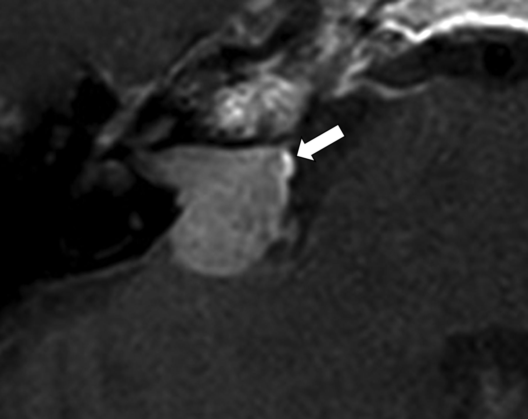
  • FIG 4.
    • Download figure
    • Open in new tab
    • Download powerpoint
    FIG 4.

    Contrast-enhanced axial T1-weighted image in a 40-year-old shows an enhancing lesion in the right cerebellopontine angle extending into the right internal auditory canal. The lesion was reported as a vestibular schwannoma by the interpreting radiologist. Histopathology upon resection revealed a meningioma. In retrospect, an incomplete rim of enhancement in keeping with an outline sign (arrow) is visible.

Tables

  • Figures
    • View popup
    Table 1:

    Clinical features of meningiomas, schwannomas, and paragangliomas

    Meningiomas (n = 14)Schwannomas (n = 22)Paragangliomas (n = 3)
    Mean age (standard deviation) in years49.5 (19.1)50.5 (13.0)45.6 (14.8)
    SexWoman (n = 9)Man (n = 5)Woman (n = 14)Man (n = 8)Woman (n = 2)Man (n = 1)
    Tumor location
    • Sphenoid wing (n = 6)

    • Olfactory groove (n = 2)

    • Tentorial leaflet (n = 2)

    • Frontal convexity (n = 1)

    • Cerebellopontine angle (n = 1)

    • Foramen magnum (n = 1)

    • Parafalcine (n = 1)

    • Internal auditory canal/cerebellopontine angle (n = 18)

    • Cochlea (n = 1)

    • Meckel cave (n = 1)

    • Orbit (n = 1)

    • Submandibular region (n = 1)

    • Carotid body tumor (n = 2)

    • Jugulotympanic paraganglioma (n = 1)

    • View popup
    Table 2:

    Reader assessments on presence of outline sign performed independently and as a consensus opinion classified by tumor histopathology

    Tumor Histopathology# Positive Outline Sign–Reader A (%)# Positive Outline Sign–Reader B (%)# of Cases in Which There was Interreader Agreement, of the Total Number of Cases (%)
    Meningioma (n = 14)12 (86%)13 (93%)13/14 (93%)
    Schwannoma (n = 22)3 (14%)4 (18%)19/22 (86%)
    Paraganglioma (n = 3)1 (33%)0 (0%)2/3 (67%)
    • View popup
    Table 3:

    Presence of outline sign and/or serrated edges (meningioma versus other tumor types) following consensus read

    Meningioma (n=14)Schwannoma (n=22)Paraganglioma (n=3)P
    Outline sign12/14 (86%)3/22 (14%)1/3 (33%)<.001
    Serrated edges8/14 (57%)2/22 (9%)1/3 (33%).0158
PreviousNext
Back to top
Advertisement
Print
Download PDF
Email Article

Thank you for your interest in spreading the word on American Journal of Neuroradiology.

NOTE: We only request your email address so that the person you are recommending the page to knows that you wanted them to see it, and that it is not junk mail. We do not capture any email address.

Enter multiple addresses on separate lines or separate them with commas.
The “Outline Sign”: Thin Hyperenhancing Perimeter as an MR Imaging Feature of Meningioma. A Useful Tool in the Temporal Bone Region for Differentiating Meningiomas from Schwannomas and Paragangliomas
(Your Name) has sent you a message from American Journal of Neuroradiology
(Your Name) thought you would like to see the American Journal of Neuroradiology web site.
CAPTCHA
This question is for testing whether or not you are a human visitor and to prevent automated spam submissions.
Cite this article
Anil K. Vasireddi, Katherine L. Reinshagen, Donghoon Shin, Laura V. Romo, Amy F. Juliano
The “Outline Sign”: Thin Hyperenhancing Perimeter as an MR Imaging Feature of Meningioma. A Useful Tool in the Temporal Bone Region for Differentiating Meningiomas from Schwannomas and Paragangliomas
American Journal of Neuroradiology Jan 2025, DOI: 10.3174/ajnr.A8452

Citation Manager Formats

  • BibTeX
  • Bookends
  • EasyBib
  • EndNote (tagged)
  • EndNote 8 (xml)
  • Medlars
  • Mendeley
  • Papers
  • RefWorks Tagged
  • Ref Manager
  • RIS
  • Zotero
0 Responses
Respond to this article
Share
Bookmark this article
The “Outline Sign”: Thin Hyperenhancing Perimeter as an MR Imaging Feature of Meningioma. A Useful Tool in the Temporal Bone Region for Differentiating Meningiomas from Schwannomas and Paragangliomas
Anil K. Vasireddi, Katherine L. Reinshagen, Donghoon Shin, Laura V. Romo, Amy F. Juliano
American Journal of Neuroradiology Jan 2025, DOI: 10.3174/ajnr.A8452
del.icio.us logo Twitter logo Facebook logo Mendeley logo
  • Tweet Widget
  • Facebook Like
  • Google Plus One
Purchase

Jump to section

  • Article
    • Abstract
    • MATERIALS AND METHODS
    • RESULTS
    • DISCUSSION
    • CONCLUSIONS
    • Footnotes
    • References
  • Figures & Data
  • Info & Metrics
  • Responses
  • References
  • PDF

Related Articles

  • No related articles found.
  • PubMed
  • Google Scholar

Cited By...

  • No citing articles found.
  • Crossref
  • Google Scholar

This article has not yet been cited by articles in journals that are participating in Crossref Cited-by Linking.

More in this TOC Section

  • MRI of the Parasellar Ligaments
  • ASL Sensitivity for Head and Neck Paraganglioma
  • Post SRS Peritumoral Hyperintense Signal of VSs
Show more Head and Neck Imaging

Similar Articles

Advertisement

Indexed Content

  • Current Issue
  • Accepted Manuscripts
  • Article Preview
  • Past Issues
  • Editorials
  • Editor's Choice
  • Fellows' Journal Club
  • Letters to the Editor
  • Video Articles

Cases

  • Case Collection
  • Archive - Case of the Week
  • Archive - Case of the Month
  • Archive - Classic Case

More from AJNR

  • Trainee Corner
  • Imaging Protocols
  • MRI Safety Corner
  • Book Reviews

Multimedia

  • AJNR Podcasts
  • AJNR Scantastics

Resources

  • Turnaround Time
  • Submit a Manuscript
  • Submit a Video Article
  • Submit an eLetter to the Editor/Response
  • Manuscript Submission Guidelines
  • Statistical Tips
  • Fast Publishing of Accepted Manuscripts
  • Graphical Abstract Preparation
  • Imaging Protocol Submission
  • Evidence-Based Medicine Level Guide
  • Publishing Checklists
  • Author Policies
  • Become a Reviewer/Academy of Reviewers
  • News and Updates

About Us

  • About AJNR
  • Editorial Board
  • Editorial Board Alumni
  • Alerts
  • Permissions
  • Not an AJNR Subscriber? Join Now
  • Advertise with Us
  • Librarian Resources
  • Feedback
  • Terms and Conditions
  • AJNR Editorial Board Alumni

American Society of Neuroradiology

  • Not an ASNR Member? Join Now

© 2025 by the American Society of Neuroradiology All rights, including for text and data mining, AI training, and similar technologies, are reserved.
Print ISSN: 0195-6108 Online ISSN: 1936-959X

Powered by HighWire