
Article Figures & Data
Figures
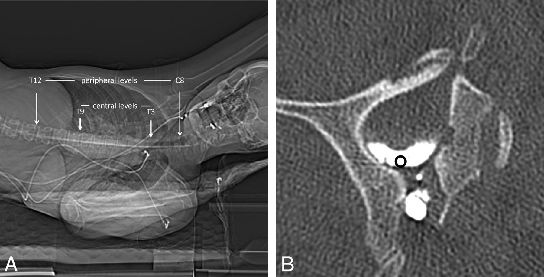
- FIG 1.
Technique for measurement of attenuation in the lateral thecal sac. A, CT scout image indicating the levels assessed on each scan. B, Axial CT image shows representative placement of an ROI immediately caudal to the dural emergence of the nerve root.
- FIG 2.
Box-and-whisker plots summarizing the technical performance of the 10-mL control and 20-mL study groups. The distribution of average attenuation within the lateral-most part of the thecal sac on the total (A), central (B), and peripheral (C) portions of the thoracic spine is shown. The number of levels adequately opacified by contrast (D) is also shown. Boxplots highlighted using bold lines, Asterisks in A, B, and D indicate groups showing statistically significant differences.
- FIG 3.
Cases in which a CVF was found on the second side examined. A, Axial CT image shows a left T8 CVF in a patient who had received 20 mL of contrast. B, Axial CT MIP image shows a right T7 CVF in a patient who had received 10 mL of contrast. C, Axial CT image shows a right T9 CVF in a patient who had received 20 mL of contrast. Arrows in all images point to opacified draining veins from the CVFs.
- FIG 4.
Coronal CT reformats showing the cranial and caudal margins of an IOCM bolus on the first side examined (A) and on the second side examined (B) of a patient who had received 20 mL of IOCM. Notice that a peripheral level may have very poor opacification even if a large and dense bolus of contrast is still present adjacently.
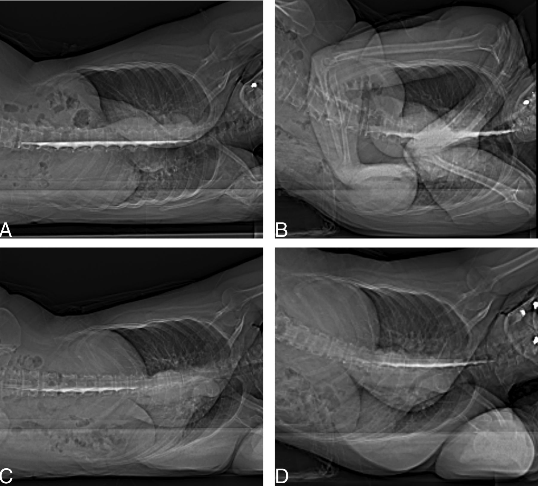
- FIG 5.
Anterior-posterior scout images of a patient who had received 20 mL of IOCM, demonstrating that the residual contrast bolus stays dense, even after the patient is repositioned a few times. Patient positioning is shown as follows: right decubitus, hips neutral (A); right decubitus, hips elevated (B); left decubitus, hips neutral (C); and left decubitus, hips elevated (D).


Tables
General characteristics of the population under study
Age Sex Opening Pressure Contrast Volume Contrast Type Iodine Dose First Decubitus CVF Bern Score 1 41 F 13 20 Iodixanol 320 6.4 Left L T8 4 2 59 F 5 20 Iodixanol 320 6.4 Right L T8 7 3 64 F 11 20 Iodixanol 320 6.4 Right R T2 8 4 20 M 25 20 Iodixanol 320 6.4 Right None 4 5 34 M 18 20 Iodixanol 320 6.4 Right None 0 6 52 F 16 20 Iodixanol 320 6.4 Right None 0 7 42 F – 20 Iodixanol 320 6.4 Right None 0 8 40 F 18 20 Iodixanol 320 6.4 Left None 0 9 15 M 14 20 Iodixanol 320 6.4 Right None 2 10 64 F – 20 Iodixanol 320 6.4 Left R T9 6 11 63 M 8 20 Iodixanol 320 6.4 Right None 0 12 31 M 27 20 Iodixanol 320 6.4 Right None 0 13 41 F 11 20 Iodixanol 320 6.4 Left None 0 14 51 M 7 20 Iodixanol 320 6.4 Right R T7 6 15 51 F 10 20 Iodixanol 320 6.4 Right R T10 4 16 68 M 1 20 Iodixanol 320 6.4 Right None 0 17 70 M 7 20 Iodixanol 320 6.4 Right R T6 7 18 46 F 11 20 Iodixanol 320 6.4 Right R T11 6 19 65 F 28 10 Iohexol 300 3.0 Right None 1 20 44 F 10 10 Iohexol 300 3.0 Left None 2 21 40 F 12 10 Iodixanol 320 3.2 Left R T7 9 22 30 M 16 10 Iohexol 300 3.0 Right R C8 9 23 29 F 15 10 Iohexol 300 3.0 Right None 0 24 61 M 13 10 Iodixanol 320 3.2 Right None 0 Note:—R indicates right; L, left; en dash, information not available or not measured; M, male; F, female.


