Advertisement

Main menu

  • Home
  • Content
    • Current Issue
    • Accepted Manuscripts
    • Article Preview
    • Past Issue Archive
    • Video Articles
    • AJNR Case Collection
    • Case of the Week Archive
    • Case of the Month Archive
    • Classic Case Archive
  • Special Collections
    • AJNR Awards
    • Low-Field MRI
    • Alzheimer Disease
    • ASNR Foundation Special Collection
    • Photon-Counting CT
    • View All
  • Multimedia
    • AJNR Podcasts
    • AJNR SCANtastic
    • Trainee Corner
    • MRI Safety Corner
    • Imaging Protocols
  • For Authors
    • Submit a Manuscript
    • Submit a Video Article
    • Submit an eLetter to the Editor/Response
    • Manuscript Submission Guidelines
    • Statistical Tips
    • Fast Publishing of Accepted Manuscripts
    • Graphical Abstract Preparation
    • Imaging Protocol Submission
    • Author Policies
  • About Us
    • About AJNR
    • Editorial Board
    • Editorial Board Alumni
  • More
    • Become a Reviewer/Academy of Reviewers
    • Subscribers
    • Permissions
    • Alerts
    • Feedback
    • Advertisers
    • ASNR Home

User menu

  • Alerts
  • Log in

Search

  • Advanced search
American Journal of Neuroradiology

American Journal of Neuroradiology

ASHNR American Society of Functional Neuroradiology ASHNR American Society of Pediatric Neuroradiology ASSR
  • Alerts
  • Log in

Advanced Search

  • Home
  • Content
    • Current Issue
    • Accepted Manuscripts
    • Article Preview
    • Past Issue Archive
    • Video Articles
    • AJNR Case Collection
    • Case of the Week Archive
    • Case of the Month Archive
    • Classic Case Archive
  • Special Collections
    • AJNR Awards
    • Low-Field MRI
    • Alzheimer Disease
    • ASNR Foundation Special Collection
    • Photon-Counting CT
    • View All
  • Multimedia
    • AJNR Podcasts
    • AJNR SCANtastic
    • Trainee Corner
    • MRI Safety Corner
    • Imaging Protocols
  • For Authors
    • Submit a Manuscript
    • Submit a Video Article
    • Submit an eLetter to the Editor/Response
    • Manuscript Submission Guidelines
    • Statistical Tips
    • Fast Publishing of Accepted Manuscripts
    • Graphical Abstract Preparation
    • Imaging Protocol Submission
    • Author Policies
  • About Us
    • About AJNR
    • Editorial Board
    • Editorial Board Alumni
  • More
    • Become a Reviewer/Academy of Reviewers
    • Subscribers
    • Permissions
    • Alerts
    • Feedback
    • Advertisers
    • ASNR Home
  • Follow AJNR on Twitter
  • Visit AJNR on Facebook
  • Follow AJNR on Instagram
  • Join AJNR on LinkedIn
  • RSS Feeds

AJNR Awards, New Junior Editors, and more. Read the latest AJNR updates

Review ArticleReview Articles

A Neuroradiologist’s Guide to Operationalizing the Response Assessment in Neuro-Oncology (RANO) Criteria Version 2.0 for Gliomas in Adults

Benjamin M. Ellingson, Francesco Sanvito, Timothy F. Cloughesy, Raymond Y. Huang, Javier E. Villanueva-Meyer, Whitney B. Pope, Daniel P. Barboriak, Lalitha K. Shankar, Marion Smits, Timothy J. Kaufmann, Jerrold L. Boxerman, Michael Weller, Evanthia Galanis, John de Groot, Mark R. Gilbert, Andrew B. Lassman, Mark S. Shiroishi, Ali Nabavizadeh, Minesh Mehta, Roger Stupp, Wolfgang Wick, David A. Reardon, Michael A. Vogelbaum, Martin van den Bent, Susan M. Chang and Patrick Y. Wen
American Journal of Neuroradiology September 2024, DOI: https://doi.org/10.3174/ajnr.A8396
Benjamin M. Ellingson
aFrom the UCLA Brain Tumor Imaging Laboratory, Department of Radiological Sciences (B.M.E., F.S.), David Geffen School of Medicine, University of California Los Angeles, Los Angeles, CA
  • Find this author on Google Scholar
  • Find this author on PubMed
  • Search for this author on this site
  • ORCID record for Benjamin M. Ellingson
Francesco Sanvito
aFrom the UCLA Brain Tumor Imaging Laboratory, Department of Radiological Sciences (B.M.E., F.S.), David Geffen School of Medicine, University of California Los Angeles, Los Angeles, CA
  • Find this author on Google Scholar
  • Find this author on PubMed
  • Search for this author on this site
  • ORCID record for Francesco Sanvito
Timothy F. Cloughesy
bUCLA Brain Tumor Program, Department of Neurology (T.F.C.), David Geffen School of Medicine, University of California Los Angeles, Los Angeles, CA
  • Find this author on Google Scholar
  • Find this author on PubMed
  • Search for this author on this site
  • ORCID record for Timothy F. Cloughesy
Raymond Y. Huang
cDepartment of Radiology, Brigham and Women's Hospital (R.Y.H.), Harvard Medical School, Boston, MA
  • Find this author on Google Scholar
  • Find this author on PubMed
  • Search for this author on this site
  • ORCID record for Raymond Y. Huang
Javier E. Villanueva-Meyer
dDepartments of Radiology and Neurosurgery (J.E.V.-M.), University of California San Francisco, CA
  • Find this author on Google Scholar
  • Find this author on PubMed
  • Search for this author on this site
  • ORCID record for Javier E. Villanueva-Meyer
Whitney B. Pope
wDepartment of Radiological Sciences (W.B.P.), David Geffen School of Medicine, University of California Los Angeles, Los Angeles, CA
  • Find this author on Google Scholar
  • Find this author on PubMed
  • Search for this author on this site
  • ORCID record for Whitney B. Pope
Daniel P. Barboriak
eDepartment of Radiology (D.P.B.), Duke University Medical Center, Durham, NC
  • Find this author on Google Scholar
  • Find this author on PubMed
  • Search for this author on this site
  • ORCID record for Daniel P. Barboriak
Lalitha K. Shankar
fClinical Trials Branch, Cancer Imaging Program (L.K.S.), National Cancer Institute, National Institutes of Health, Bethesda, MD
  • Find this author on Google Scholar
  • Find this author on PubMed
  • Search for this author on this site
Marion Smits
gDepartment of Radiology & Nuclear Medicine (M.S.), Erasmus MC - University Medical Centre Rotterdam, Rotterdam, The Netherlands
  • Find this author on Google Scholar
  • Find this author on PubMed
  • Search for this author on this site
  • ORCID record for Marion Smits
Timothy J. Kaufmann
hDepartment of Radiology (T.J.K.), Mayo Clinic, Rochester, MN
  • Find this author on Google Scholar
  • Find this author on PubMed
  • Search for this author on this site
  • ORCID record for Timothy J. Kaufmann
Jerrold L. Boxerman
iDepartment of Diagnostic Imaging (J.L.B.), Rhode Island Hospital and Alpert Medical School of Brown University, Providence, RI
  • Find this author on Google Scholar
  • Find this author on PubMed
  • Search for this author on this site
  • ORCID record for Jerrold L. Boxerman
Michael Weller
jDepartment of Neurology (M.W.), University Hospital and University of Zurich, Zurich, Switzerland
  • Find this author on Google Scholar
  • Find this author on PubMed
  • Search for this author on this site
Evanthia Galanis
kDepartment of Oncology (E.G.), Mayo Clinic, Rochester, MN
  • Find this author on Google Scholar
  • Find this author on PubMed
  • Search for this author on this site
John de Groot
lDivision of Neuro-Oncology, Department of Neurosurgery (J.d.G., S.M.C.), University of California, San Francisco, CA
  • Find this author on Google Scholar
  • Find this author on PubMed
  • Search for this author on this site
Mark R. Gilbert
mNeuro-Oncology Branch, Center for Cancer Research (M.R.G.), National Cancer Institute, National Institutes of Health, Bethesda, MD
  • Find this author on Google Scholar
  • Find this author on PubMed
  • Search for this author on this site
Andrew B. Lassman
nDivision of Neuro-Oncology, Department of Neurology (A.B.L.), Herbert Irving Comprehensive Cancer Center and Irving Institute for Clinical and Translational Research, Columbia University Vagelos College of Physicians and Surgeons and New York-Presbyterian Hospital, New York, NY
  • Find this author on Google Scholar
  • Find this author on PubMed
  • Search for this author on this site
Mark S. Shiroishi
oDepartment of Radiology (M.S.S.), Keck School of Medicine of the University of Southern California (USC), Los Angeles, CA
  • Find this author on Google Scholar
  • Find this author on PubMed
  • Search for this author on this site
  • ORCID record for Mark S. Shiroishi
Ali Nabavizadeh
pDepartment of Radiology (A.N.), Perelman School of Medicine, University of Pennsylvania, Philadelphia, PA
  • Find this author on Google Scholar
  • Find this author on PubMed
  • Search for this author on this site
  • ORCID record for Ali Nabavizadeh
Minesh Mehta
qMiami Cancer Institute (M.M.), Miami, FL
  • Find this author on Google Scholar
  • Find this author on PubMed
  • Search for this author on this site
Roger Stupp
rMalnati Brain Tumor Institute, Lurie Comprehensive Cancer Center and Departments of Neurological Surgery, Neurology and Division of Hematology/Oncology (R.S.), Northwestern University, Chicago, IL
  • Find this author on Google Scholar
  • Find this author on PubMed
  • Search for this author on this site
  • ORCID record for Roger Stupp
Wolfgang Wick
sDepartment of Neurology Heidelberg University Hospital & Clinical Cooperation Unit Neurooncology (W.W.), German Cancer Consortium (DKTK), German Cancer Research Center (DKFZ), Heidelberg, Germany
  • Find this author on Google Scholar
  • Find this author on PubMed
  • Search for this author on this site
David A. Reardon
tCenter For Neuro-Oncology, Dana-Farber Cancer Institute and Harvard Medical School(D.A.R., P.Y.W.), Boston, MA
  • Find this author on Google Scholar
  • Find this author on PubMed
  • Search for this author on this site
  • ORCID record for David A. Reardon
Michael A. Vogelbaum
uDepartment of Neuro-Oncology (M.A.V.), Moffitt Cancer Center, Tampa, FL
  • Find this author on Google Scholar
  • Find this author on PubMed
  • Search for this author on this site
Martin van den Bent
vDepartment Neuro-Oncology (M.v.d.B.), Erasmus MC Cancer Institute, Rotterdam, The Netherlands
  • Find this author on Google Scholar
  • Find this author on PubMed
  • Search for this author on this site
Susan M. Chang
lDivision of Neuro-Oncology, Department of Neurosurgery (J.d.G., S.M.C.), University of California, San Francisco, CA
  • Find this author on Google Scholar
  • Find this author on PubMed
  • Search for this author on this site
Patrick Y. Wen
tCenter For Neuro-Oncology, Dana-Farber Cancer Institute and Harvard Medical School(D.A.R., P.Y.W.), Boston, MA
  • Find this author on Google Scholar
  • Find this author on PubMed
  • Search for this author on this site
  • Article
  • Figures & Data
  • Supplemental
  • Info & Metrics
  • Responses
  • References
  • PDF
Loading

Article Figures & Data

Figures

  • Tables
  • FIG 1.
    • Download figure
    • Open in new tab
    • Download powerpoint
    FIG 1.

    Flow-chart to identify target lesions for CE (A), non-CE (B), and “mixed” (C) tumors. * the size condition for measurable disease is: ≥10 mm on 3 ⊥ planes for 3D images with thin slice (more often applied to CE disease), or ≥ 10 mm on in-plane ⊥ diameters and visible on ≥2 slices for 2D images with thicker slice (more often applied to non-CE disease). Of note, these conditions apply to trials employing bidimensional measurements for treatment response assessment, while for trials employing volumetric segmentations various strategies can be applied for the identification of target lesions (please refer to the dedicated paragraph in the main text). ° “mixed” tumors is short for “tumors with both CE and non-CE components”. † in mixed tumors, up to a total of 4 target lesions is allowed (i.e., x + y ≤ 4).

  • FIG 2.
    • Download figure
    • Open in new tab
    • Download powerpoint
    FIG 2.

    Guidelines and tips for bidirectional “2D” measurements. The measurement protocol for target lesions with 3D images and 2D imaging (A) is used both to verify that the lesion meets the criteria for “measurable disease” and to obtain measurements to calculate the tumor burden. Measurements in CE tumors (B) should not include the surgical cavity or cystic areas, and should avoid areas where the lesion shows necrotic features or is not distinguishable from post-surgical meningeal thickening. In non-CE tumors (C), the presence of infiltrative disease with unclear boundaries can pose a serious challenge when performing measurements. If possible, slices where the tumor shows more defined margins should be preferred for target lesion measurements. In “mixed” tumors (D), the CE lesions lie within the boundaries of non-CE lesions. CE and non-CE target lesions should be measured separately and not necessarily on the same plane/slice. Then, longitudinal changes in CE- and non-CE tumor burden are tracked in parallel to define the response category.

  • FIG. 3
    • Download figure
    • Open in new tab
    • Download powerpoint
    FIG. 3

    Schema for determining radiographic treatment response assessment as per RANO 2.0. To assess MR/PR, the change in tumor burden should be compared to the baseline MRI, while for PD it should be compared to the nadir. In clinical trials applying the “mixed” tumor criteria, the whole evaluation should be performed in parallel for both the CE and the non-CE tumor burden at each timepoint in order to assign the response category (e.g., PD, SD, PR, …), then the overall response category is assigned based on both CE and non-CE categories: PD+SD/MR/PR/CR=PD; MR/PR+SD=MR/PR; CR+SD/MR/PR=SD/MR/PR; SD+SD=SD (see text for details). ° the additional categories preliminary and durable “minor response” (MR: > –50% and < –25% 2D or > –65% and < –40% 3D) should be taken into consideration, only for non-CE disease. * confirmation scans obtained after ≥4 weeks to confirm PD can be waived in the following scenarios: in CE tumors ≥3 months after RT completion and if the treatment does not include agents highly associated with PsP; in the evaluation of non-CE progression in non-CE tumors or mixed tumors (as PsP is typically contrast-enhancing). Confirmation scans after ≥4 weeks to confirm durable MR/PR/CR are always required. If a patient is lost to follow-up (censored) before obtaining a confirmation scan (when required), the last timepoint showing preliminary CR/PR/MR/PD is classified as SD.

  • FIG 4.
    • Download figure
    • Open in new tab
    • Download powerpoint
    FIG 4.

    RANO 2.0 evaluations in example cases of newly-diagnosed CE glioblastoma. In case A, radiographic findings are stable (stable disease, SD) for 13.1 months from baseline, in the absence of measurable disease around the resection cavity. The appearance of a new measurable CE nodule after ≥12 weeks after RT completion (at 16.8 months in this case) classifies PD generally without the need of a confirmatory scan (unless clearly required by the trial), and the study treatment is stopped. In case B, the first follow-up scan shows early progressive disease (PD) <12 weeks after RT completion, “preliminary PD” (prel. PD) is annotated, the patient is kept on treatment, and a confirmation scan is obtained after ≥4 weeks. After confirmation of PD, the treatment is stopped and the confirmed PD (conf. PD) event is backdated to the day of preliminary PD. The time interval between the post-RT baseline scan and the date of confirmed PD corresponds to the time of progression (TTP). In case C, the first follow-up scan shows early PD <12 weeks after RT completion, annotated as preliminary PD. The confirmation scan after >4 weeks shows substantially stable findings, with no further size increase of the target lesion, therefore the preliminary PD timepoint is reclassified as pseudoprogression (PsP), and the patient is kept on treatment.
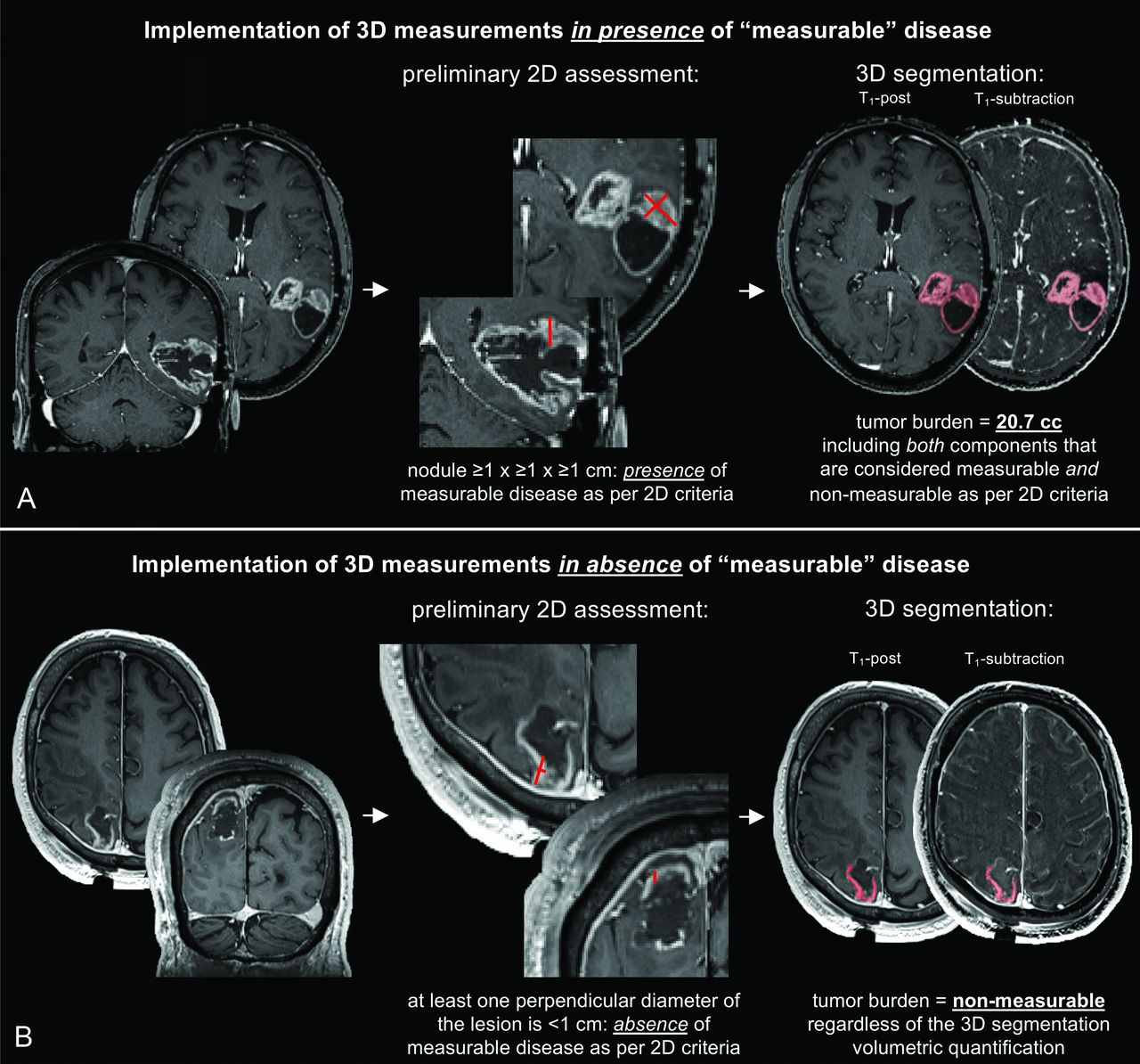
  • FIG 5.
    • Download figure
    • Open in new tab
    • Download powerpoint
    FIG 5.

    Proposed implementation of volumetric (3D) assessments. A preliminary evaluation should be performed to confirm the presence of measurable disease according to the classic bidimensional criteria. If 2D-defined measurable disease is present, the volumetric tumor burden may be computed by including the whole segmentation of the appropriate tissue (e.g., only CE tissue for CE tumors) which comprises both 2D-defined measurable components and 2D-defined non-measurable components (A). A quality check should always be performed by the reader to correct errors in the segmentation, for instance to exclude vascular structures or healthy meningeal tissue. If 2D-defined measurable disease is absent, the volumetric tumor burden should be set to “non-measurable”, regardless of the 3D segmentation (B). In the cases shown, T1-subtraction maps were used to only include in the segmentation voxels that showed increased T1-weighted signal when comparing post- to pre-contrast T1-weighted normalized images.

Tables

  • Figures
    • View popup
    Table 1:

    Main features of RANO 2.0

    RANO 2.0 overview
    Baseline
     Newly diagnosed tumorsPost-RT scan
     Recurrent tumorsPre-Tx scan
    Tumor components to evaluate
     CE tissueIn all tumors
     Non-CE tissue- In non-CE tumors- In “mixed” tumors with CE + non-CE components- Optional in CE tumors for trials using agents that affectvascular permeability (e.g., antiangiogenic)
    Measurement technique
     Sum of bidimensional products or volumetric segmentations
    Thresholds applied to target lesions
     CRcomplete disappearance of all lesions
     PD≥ +25% in sum of bidimensional productsor ≥ +40% in volume
     PR≤ –50% in sum of bidimensional productsor ≤ –65% in volume
     MRa≤ –25% and > –50% in sum of bidimensional productsor ≤ –40% and > –65% in volume
     SDcriteria for CR / PR / MR / PD are not met
    Confirmatory scans (performed after >4 weeks)
     For PD- Mandatory for CE tumors ≤12 weeks from RT- Optional for CE tumors >12 weeks from RT (recommended for agentsb highly associated with PsP) - Unnecessary for non-CE tumors
     For CR/PRMandatory for all tumors at all timepoints
    • Note:—CE indicates contrast-enhancing; CR, complete response; MR, minor response; PD, progressive disease; PsP, pseudoprogression; PR, partial response; RT, radiotherapy; SD, stable disease; Tx, treatment.

    • ↵a MR only applies to non-CE disease.

    • ↵b e.g., immunotherapies.

    • View popup
    Table 2:

    Summary of RANO 2.0 criteria

    RANO 2.0 categories
    Complete Response (CR) – compare to baseline
     disappearance of target and non-target lesions sustained for ≥4 weeksa
     and no new lesionsb
     and clinical status is stable or improved
     and off corticosteroids (or on physiologic replacement dose)
    Partial Response (PR) – compare to baseline
     at least 50% decrease in tumor burden with 2D measurements, or 65% with 3D measurements, sustained for ≥4 weeksa,e
     and no newly-measurable lesionsb,c,d
     and clinical status is stable or improved
     and corticosteroid dose is stable compared to baseline (or on physiologic replacement dose)
    Minor Response (MR, only applicable to non-CE disease) – compare to baseline
     25% to 50% decrease in tumor burden with 2D measurements, or 40% to 65% with 3D measurements, sustained for ≥4 weeksa
     and no newly-measurable lesionsb,c,d
     and clinical status is stable or improved
     and corticosteroid dose is stable compared to baseline (or on physiologic replacement)
    Progressive Disease (PD)f – compare to nadir
     at least 25% increase in tumor burden with 2D measurements, or 40% with 3D measurements, with or without confirmation scan after ≥4 weeksg
     or appearance of newly-measurable lesionsb,c,d
     or appearance of leptomeningeal disease
     or clinical deterioration not ascribable to steroid dose reduction or other causes apart from the tumor
     or failure to return for evaluation as a result of death or clinical deterioration
    Stable Disease (SD)
     all scenarios that do not meet criteria for CR, PR, MR, or PD.
     e.g., stable radiographic findings without clinical deterioration.
    • Note:—CE indicates contrast-enhancing; CR, complete response; MR, minor response; PD, progressive disease; PsP, pseudoprogression; PR, partial response; SD, stable disease.

    • ↵a Confirmation scans after ≥4 weeks to confirm durable MR/PR/CR are always required. If confirmed, MR/PR/CR is backdated to the date of preliminary MR/PR/CR. If a patient is lost to follow-up (censored) before confirmation, preliminary CR/PR/MR is considered SD.

    • ↵b Disregard new non-CE lesions unequivocally ascribable to post-RT.

    • ↵c Either new measurable lesions or previously non-measurable lesions that became measurable and grew ≥5x≥5 mm.

    • ↵d For CE-tumors, only CE lesions should be considered.

    • ↵e In mixed tumors, the assessment should be performed in-parallel for both CE and non-CE components, then combined.

    • ↵f Corticosteroid dose increase alone does not define PD.

    • ↵g Confirmation scans for PD can be waived: in CE tumors >3 months after RT completion not treated with agents highly associated with PsP; in the evaluation of non-CE progression in non-CE tumors or mixed tumors.

PreviousNext
Back to top
Advertisement
Print
Download PDF
Email Article

Thank you for your interest in spreading the word on American Journal of Neuroradiology.

NOTE: We only request your email address so that the person you are recommending the page to knows that you wanted them to see it, and that it is not junk mail. We do not capture any email address.

Enter multiple addresses on separate lines or separate them with commas.
A Neuroradiologist’s Guide to Operationalizing the Response Assessment in Neuro-Oncology (RANO) Criteria Version 2.0 for Gliomas in Adults
(Your Name) has sent you a message from American Journal of Neuroradiology
(Your Name) thought you would like to see the American Journal of Neuroradiology web site.
CAPTCHA
This question is for testing whether or not you are a human visitor and to prevent automated spam submissions.
Cite this article
Benjamin M. Ellingson, Francesco Sanvito, Timothy F. Cloughesy, Raymond Y. Huang, Javier E. Villanueva-Meyer, Whitney B. Pope, Daniel P. Barboriak, Lalitha K. Shankar, Marion Smits, Timothy J. Kaufmann, Jerrold L. Boxerman, Michael Weller, Evanthia Galanis, John de Groot, Mark R. Gilbert, Andrew B. Lassman, Mark S. Shiroishi, Ali Nabavizadeh, Minesh Mehta, Roger Stupp, Wolfgang Wick, David A. Reardon, Michael A. Vogelbaum, Martin van den Bent, Susan M. Chang, Patrick Y. Wen
A Neuroradiologist’s Guide to Operationalizing the Response Assessment in Neuro-Oncology (RANO) Criteria Version 2.0 for Gliomas in Adults
American Journal of Neuroradiology Sep 2024, DOI: 10.3174/ajnr.A8396

Citation Manager Formats

  • BibTeX
  • Bookends
  • EasyBib
  • EndNote (tagged)
  • EndNote 8 (xml)
  • Medlars
  • Mendeley
  • Papers
  • RefWorks Tagged
  • Ref Manager
  • RIS
  • Zotero
0 Responses
Respond to this article
Share
Bookmark this article
A Neuroradiologist’s Guide to Operationalizing the Response Assessment in Neuro-Oncology (RANO) Criteria Version 2.0 for Gliomas in Adults
Benjamin M. Ellingson, Francesco Sanvito, Timothy F. Cloughesy, Raymond Y. Huang, Javier E. Villanueva-Meyer, Whitney B. Pope, Daniel P. Barboriak, Lalitha K. Shankar, Marion Smits, Timothy J. Kaufmann, Jerrold L. Boxerman, Michael Weller, Evanthia Galanis, John de Groot, Mark R. Gilbert, Andrew B. Lassman, Mark S. Shiroishi, Ali Nabavizadeh, Minesh Mehta, Roger Stupp, Wolfgang Wick, David A. Reardon, Michael A. Vogelbaum, Martin van den Bent, Susan M. Chang, Patrick Y. Wen
American Journal of Neuroradiology Sep 2024, DOI: 10.3174/ajnr.A8396
del.icio.us logo Twitter logo Facebook logo Mendeley logo
  • Tweet Widget
  • Facebook Like
  • Google Plus One
Purchase

Jump to section

  • Article
    • Abstract
    • ABBREVIATIONS:
    • STANDARDIZED BRAIN TUMOR IMAGING PROTOCOL (BTIP) AND RADIOGRAPHIC READ PARADIGMS
    • OVERVIEW OF UPDATES IN RANO 2.0
    • DEFINITIONS OF MEASURABLE, NON-MEASURABLE, AND TARGET LESIONS (FIGURE 1)
    • METHODOLOGY FOR 2D MEASUREMENTS (FIGURE 2)
    • STEP-BY-STEP GUIDE TO RANO 2.0 RADIOGRAPHIC READS
    • ADDITIONAL NOTES ON THE EVALUATION OF CE AND NON-CE DISEASE
    • CORRESPONDENCE BETWEEN 2D AND 3D MEASUREMENTS
    • CLINICAL DETERIORATION AND CORTICOSTEROID USE
    • CONCLUSIONS
    • Footnotes
    • REFERENCES
  • Figures & Data
  • Supplemental
  • Info & Metrics
  • Responses
  • References
  • PDF

Related Articles

  • PubMed
  • Google Scholar

Cited By...

  • Reply:
  • Crossref
  • Google Scholar

This article has not yet been cited by articles in journals that are participating in Crossref Cited-by Linking.

More in this TOC Section

  • An Atlas of Neonatal Neurovascular Imaging Anatomy as Depicted with Microvascular Imaging: The Intracranial Arteries
  • An Atlas of Neonatal Neurovascular Imaging Anatomy as Depicted with Microvascular Imaging: The Intracranial Veins
  • Clinical Translation of Hyperpolarized 13C Metabolic Probes for Glioma Imaging
Show more Review Articles

Similar Articles

Advertisement

Indexed Content

  • Current Issue
  • Accepted Manuscripts
  • Article Preview
  • Past Issues
  • Editorials
  • Editor's Choice
  • Fellows' Journal Club
  • Letters to the Editor
  • Video Articles

Cases

  • Case Collection
  • Archive - Case of the Week
  • Archive - Case of the Month
  • Archive - Classic Case

More from AJNR

  • Trainee Corner
  • Imaging Protocols
  • MRI Safety Corner
  • Book Reviews

Multimedia

  • AJNR Podcasts
  • AJNR Scantastics

Resources

  • Turnaround Time
  • Submit a Manuscript
  • Submit a Video Article
  • Submit an eLetter to the Editor/Response
  • Manuscript Submission Guidelines
  • Statistical Tips
  • Fast Publishing of Accepted Manuscripts
  • Graphical Abstract Preparation
  • Imaging Protocol Submission
  • Evidence-Based Medicine Level Guide
  • Publishing Checklists
  • Author Policies
  • Become a Reviewer/Academy of Reviewers
  • News and Updates

About Us

  • About AJNR
  • Editorial Board
  • Editorial Board Alumni
  • Alerts
  • Permissions
  • Not an AJNR Subscriber? Join Now
  • Advertise with Us
  • Librarian Resources
  • Feedback
  • Terms and Conditions
  • AJNR Editorial Board Alumni

American Society of Neuroradiology

  • Not an ASNR Member? Join Now

© 2025 by the American Society of Neuroradiology All rights, including for text and data mining, AI training, and similar technologies, are reserved.
Print ISSN: 0195-6108 Online ISSN: 1936-959X

Powered by HighWire