
Article Figures & Data
Figures
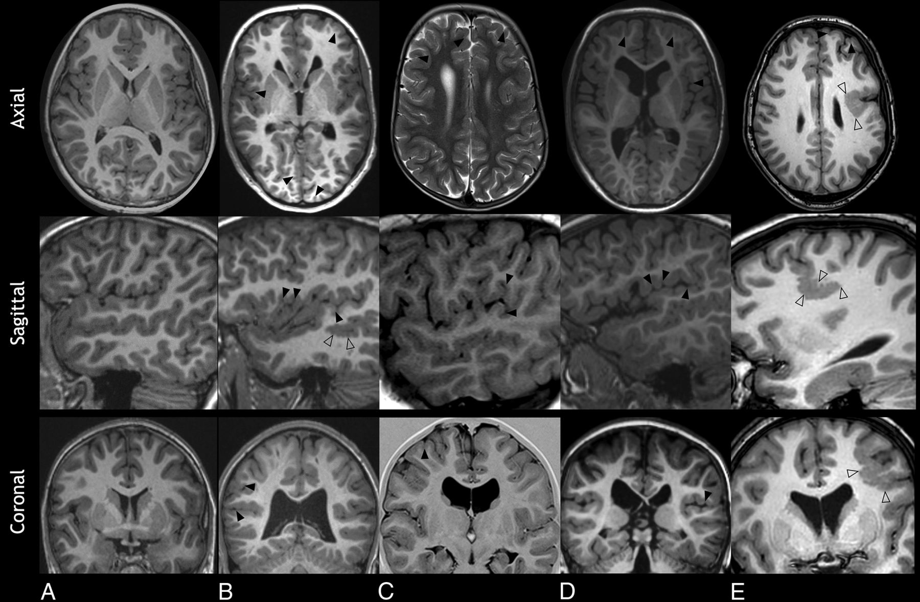
- FIG 1.
Malformations of cortical development in 4 patients with Sotos syndrome (B–E). A, An 11-year-old healthy control. Axial, sagittal, and coronal T1WIs show a normal appearance of the cortex and sulcation pattern. B, A 15-year-old boy. Axial and coronal T1WIs show diffuse dysgyria (arrowheads), and sagittal T1WI shows undulating gyri in keeping with polymicrogyria (empty arrowheads). C, A 5.5-year-old girl. Axial T2WI and coronal T1 inversion recovery show frontal dysgyria with shallow sulci in disorganized orientation (arrowheads). Sagittal T1WI shows perisylvian dysgyria. D, A 3-year-old boy. Axial and coronal T1WI shows asymmetric, left-sided, frontal dysgyria (arrowheads). Sagittal T1WI shows perisylvian dysgyria (arrowheads). E, A 13-year-old girl. Axial, sagittal, and coronal T1WI shows polymicrogyria with regionally increased gyral/sulcal frequency and greater corticomedullary junction irregularity compared with dysgyria (empty arrowheads), accompanied by frontal dysgyria (arrowheads).
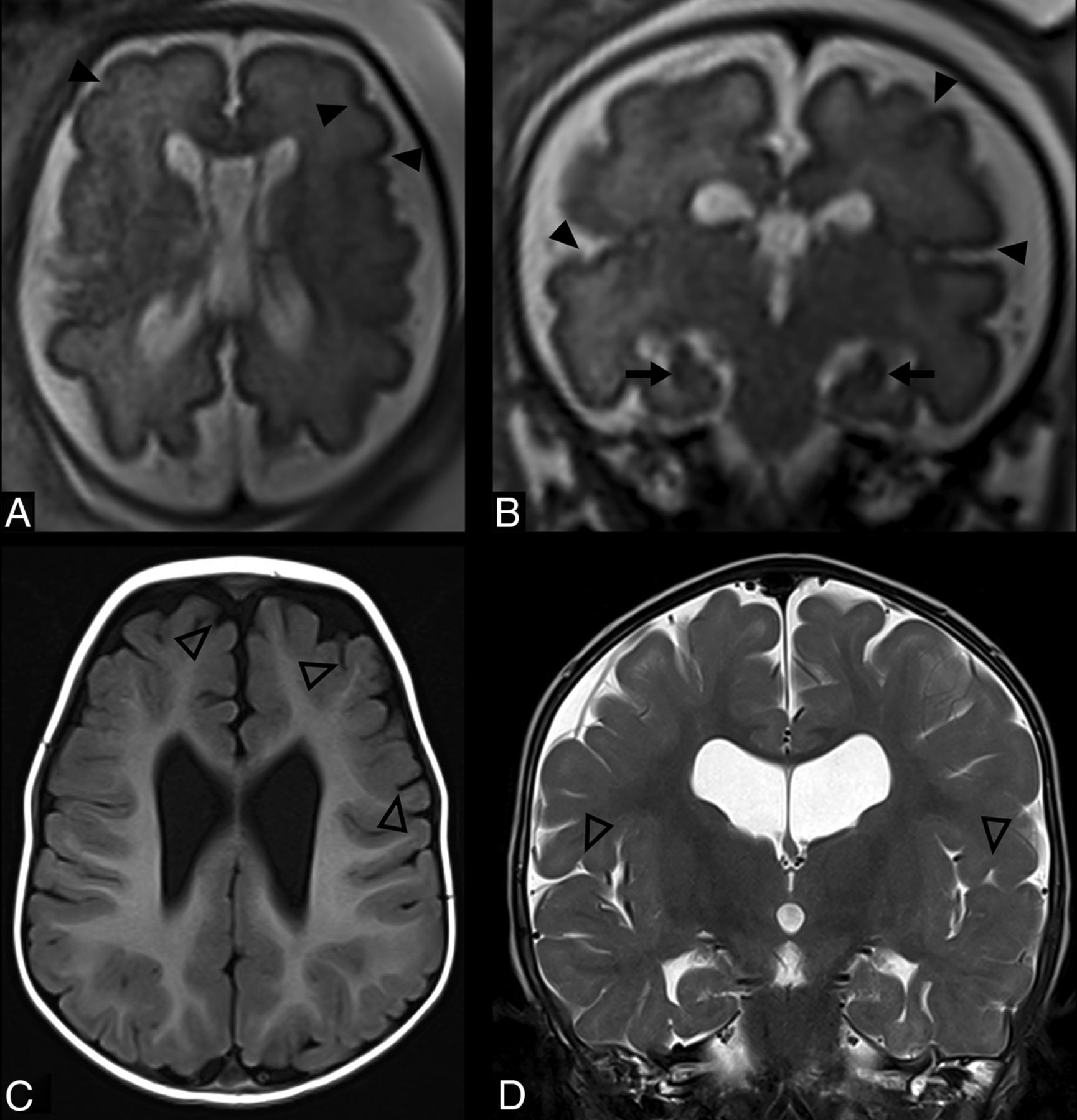
- FIG 2.
Imaging characteristics of Sotos syndrome in 3 patients (A, C, and D). A, A 16-year-old girl. Sagittal T1WI shows midline abnormalities including a thin corpus callosum (white dashed arrow), a thin anterior commissure (white empty arrow), and brainstem dysmorphism, a shallow pontomedullary sulcus (white empty arrowhead). B, A 16-year-old girl, an age-matched healthy control. Sagittal T1WI shows a normal midline appearance of the corpus callosum (white dashed arrow), anterior commissure (white arrow), and a pontomedullary sulcus (white arrowhead). C, A 10-month-old boy. Axial T2WI shows reduced white matter volume in the posterior cerebrum with enlargement of the ventricular atria and occipital horns and enlarged perivascular spaces (black arrows). D, A 3-year-old boy. Coronal T2WI demonstrates enlarged CSF spaces (asterisks), bilateral incomplete hippocampal rotation (black dashed arrows), and ventriculomegaly (black arrowheads).
- FIG 3.
A patient with Sotos syndrome. Fetal MR imaging at 32 weeks’ gestational age (A and B) and postnatal MR imaging at 1 year of age (C and D). A and B, Axial and coronal T2WI shows mild asymmetry of sulcation (arrowheads), mild ventriculomegaly, enlargement of the cavum septum pellucidum/vergae, and taller-than-wide hippocampal formations consistent with incomplete hippocampal rotation bilaterally (arrows). C and D, Axial T1WI and coronal T2WI show mild dysgyria in the frontal and perisylvian regions (empty arrowheads), ventriculomegaly, incomplete hippocampal rotation, and thinning of the corpus callosum. Distention of the cavum septum pellucidum/vergae has intervally resolved.


Tables
Feature No. (%) Overgrowth (height and weight) 46 (59.7%) Macrocephaly 52 (67.5%) Developmental delay 65 (88.3%) Intellectual disability 45 (58%) Autism spectrum disorder 9 (11.7%) Attention deficithyperactivity disorder 6 (7.8%) Seizures 24 (31.2%) Electroencephalogram abnormality 9 (37.5%) Neurologic findings 55 (71.4%) Hypotonia 48 (62.3%) Nystagmus 11 (14.3%) Increased intracranial pressure 5 (6.5%) Ophthalmologic findings 29 (37.7%) Dysphagia 13 (16.9%) Scoliosis 27 (35.1%) Joint hypermobility 23 (29.9%) Hearing loss 9 (11.7%) Craniosynostosis 6 (7.8%) Feature No. (%) Malformations of cortical development 73 (94.8%) Dysgyria 71 (92.2%) Frontal 59 (83.1%) Perisylvian 33 (45.2%) Opercular 8 (11.3%) Shallow sulci 30 (42.3%) Polygyria 36 (50.7%) Polymicrogyria 17 (22.1%) Perisylvian 12 (70.6%) Periventricular nodular heterotopia 2 (2.7%) Focal cortical dysplasia 1 (1.4%) Incomplete hippocampal rotation 39 (50.6%) Unilateral 10 (25.6%) Bilateral 29 (74.4%) Feature Hippocampi Appearance P Value Incomplete Rotation (n = 39) No. (%) Normal (n = 38) No. (%) Malformations of cortical development 39 (100%) 34 (89.5%) .055 Dysgyria 39 (100%) 32 (84.2%) .012 Polymicrogyria 9 (23.1%) 8 (21.1%) .83 Periventricular nodular heterotopia 2 (100%) 0 (0%) .49 Ventriculomegaly 33 (84.6%) 34 (89.5%) .74 Septum pellucidum abnormality 16 (41%) 10 (26.3%) .17 Thin anterior commissure 20 (87%) 18 (90%) >.999 Missing massa intermedia 9 (40.9%) 9 (47.4%) .68 White matter signal change 10 (25.6%) 1 (2.6%) .004


Jump to section
Related Articles
Cited By...
- No citing articles found.