
Article Figures & Data
Figures
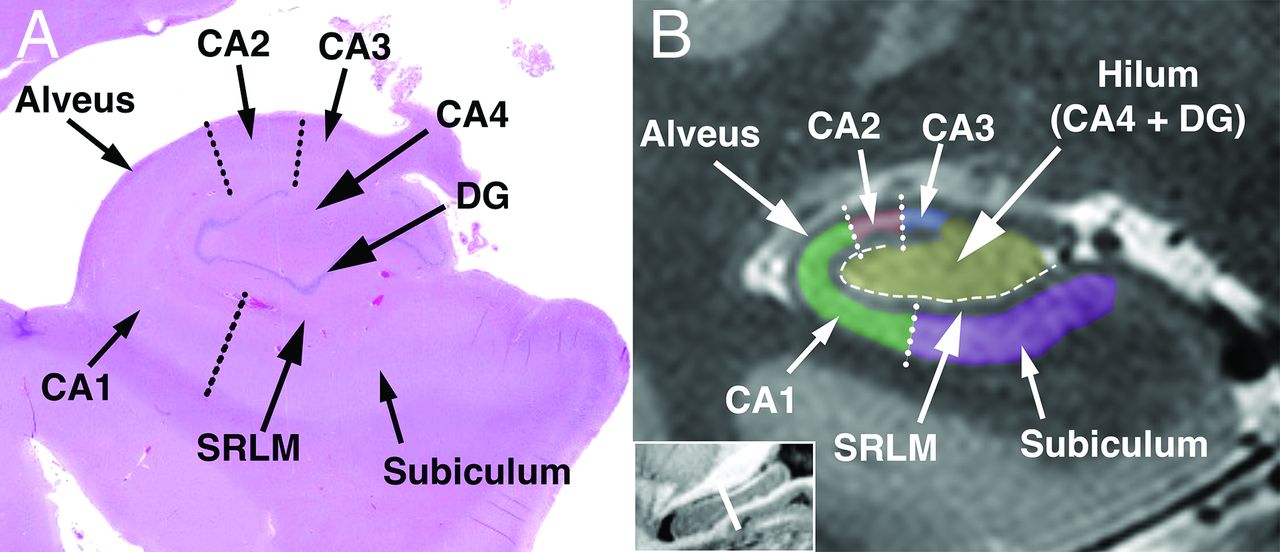
- FIG 1.
Normal hippocampal anatomy. A, Coronal histologic section through the body of the hippocampus compared with (B) coronal 7T T2-weighted 2D turbo-spin-echo MRI. Dotted lines represent approximate subfield borders, and dashed line represents the location of the hippocampal sulcus.
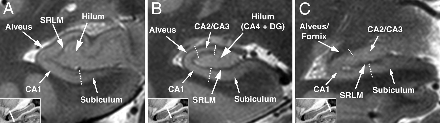
- FIG 2.
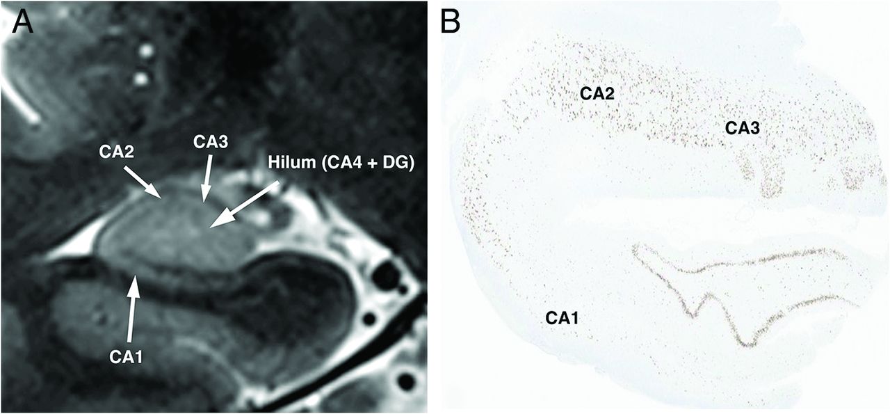
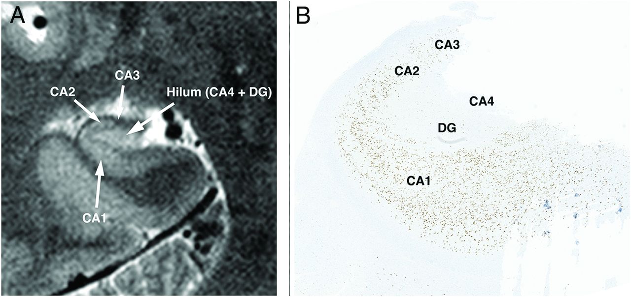
Normal hippocampal anatomy. Oblique coronal 7T T2-weighted 2D turbo-spin-echo through the hippocampus shows the normal hippocampal head (A), body (B), and tail (C).
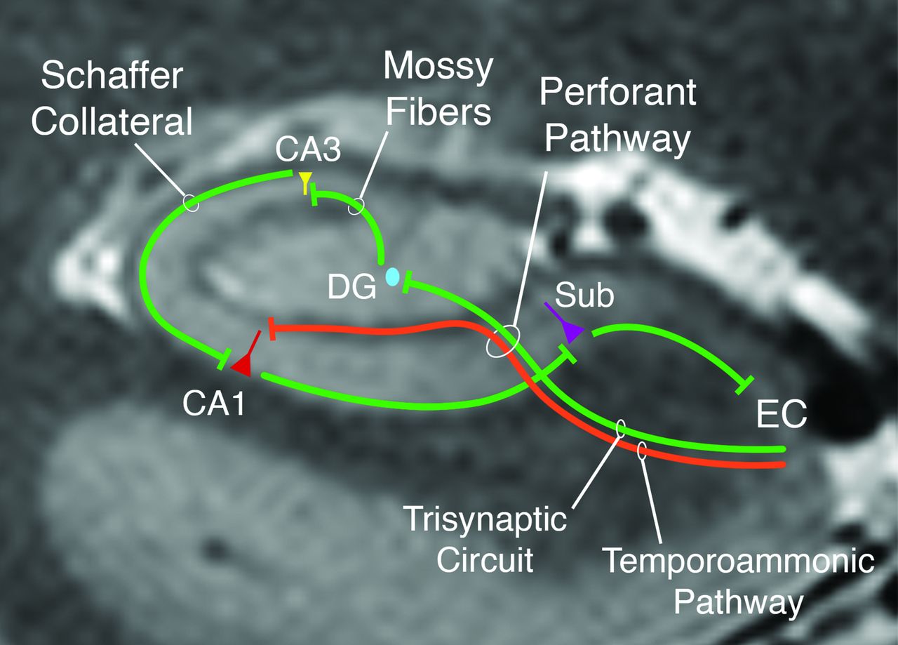
- FIG 3.
Illustration of the 2 primary hippocampal circuits implicated in hippocampal damage from temporal lobe epilepsy, the trisynaptic and temporoammonic pathways. The classic trisynaptic pathway projects from layer II of the entorhinal cortex (EC) to the DG via the perforant pathway for the first synapse. The second synapse is from the granule cells in the DG to the pyramidal layer of the CA3 region. The third synapse is formed by CA3 neurons projecting to CA1 neurons through Schaffer collaterals. Meanwhile, the temporoammonic pathway consists of monosynaptic connections from layer III of the EC projecting directly to the pyramidal layers of CA1. Sub = subiculum.
- FIG 4.
Patterns of reactive astrogliosis on glial fibrillary acidic protein (GFAP) staining of hippocampal tissue. Hippocampal tissue in a patient with no HS shows a single GFAP staining reactive astrocyte (A, arrow). Moderate number of GFAP reactive astrogliosis is noted in a different patient with long-standing epilepsy (B, arrows) with decreased neuronal attenuation. Severe reactive astrogliosis characterized by attenuated meshwork of GFAP-labeled fine processes (C) in a hippocampal specimen of a patient with advanced ILAE type 1 HS.
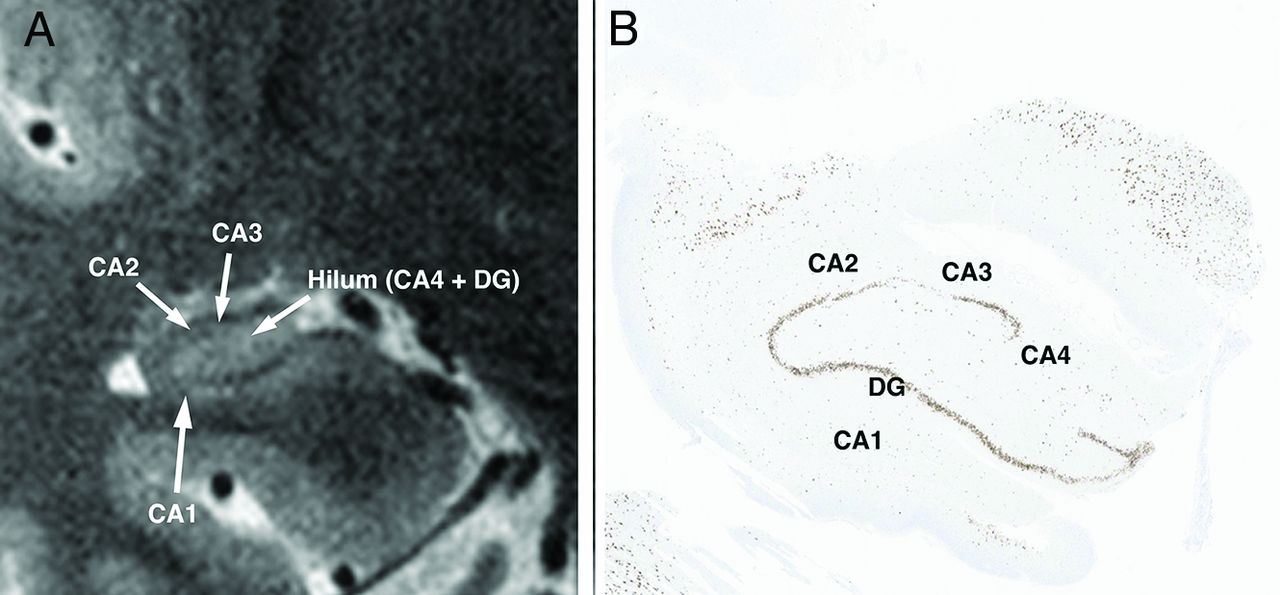
- FIG 5.
Type 1 ILAE HS. A, Oblique coronal T2-weighted image shows volume loss affecting CA1–CA3 and the hippocampal hilum. B, Microscopy by using neuronal nuclear (NeuN), a marker of neuronal cells, confirms the MRI findings with severe neuronal loss throughout CA1–CA4 along with neuronal loss in the DG.
- FIG 6.
HS ILAE type 2 (CA1-predominant neuronal cell loss and gliosis). A, Oblique coronal T2-weighted image shows atrophy of the CA1 segment of hippocampus on coronal T2-weighted MRI with loss of the normal tapering of CA1 from the CA1-subiculum junction to the CA1–CA2 junction. There is relative sparing of the hilum and other CA segments. B, Microscopy by using NeuN marker shows moderate neuronal loss limited to the CA1 sector with preserved neuronal attenuation in other CA sectors.
- FIG 7.
HS ILAE type 3 (hilar predominant neuronal cell loss and gliosis). A, Oblique coronal T2-weighted image shows atrophy of the hilum of hippocampus on coronal T2-weighted MRI, as evidenced by overall decrease in volume and height of the hilum. There is relative sparing of CA1–CA3, which show no substantial volume loss or loss of tapering of CA1–CA3. B, Microscopy by using NeuN marker shows moderate neuronal loss limited to the hilum region with preserved neuronal attenuation in CA1–CA3.
- FIG 8.
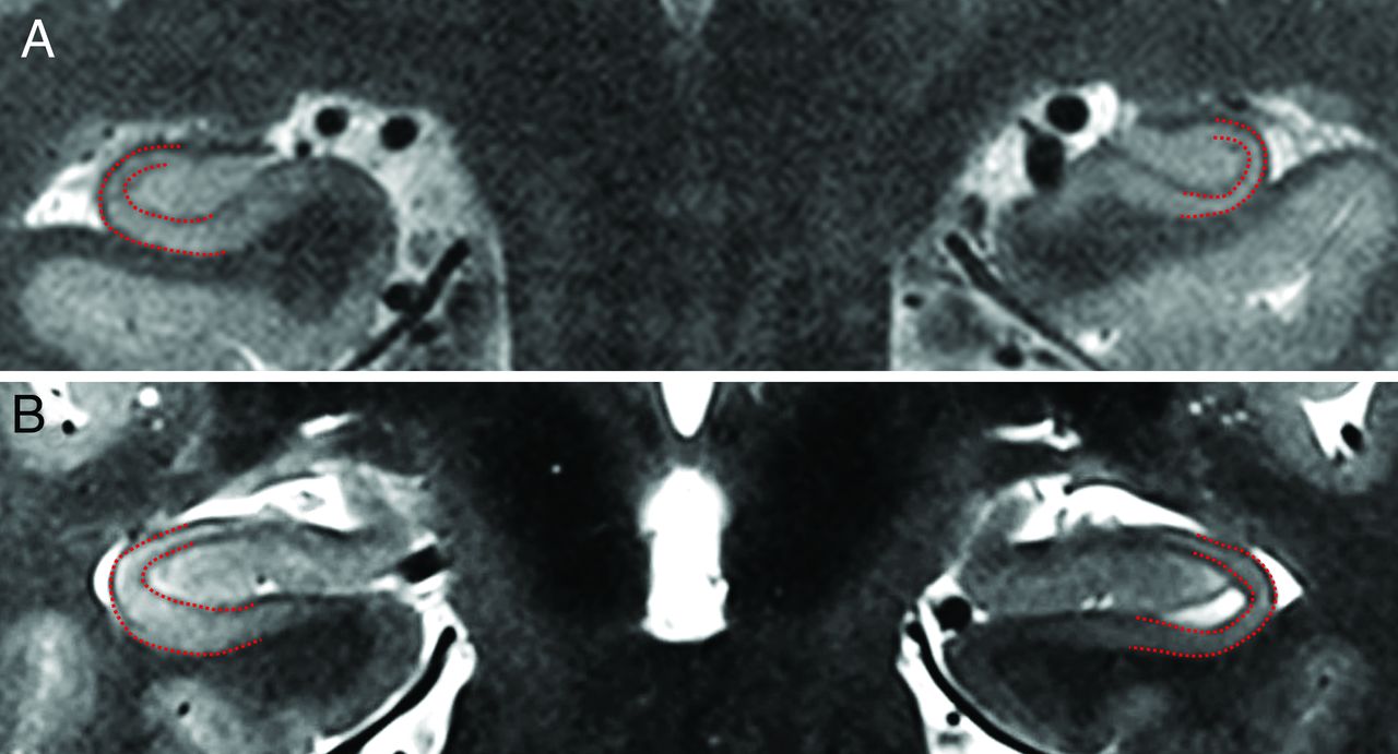
ILAE type 2 HS with predominant CA1 cell loss and gliosis. A, Oblique coronal T2-weighted image shows the normal tapering of the right CA1 (red outline) as it extends from the subiculum toward CA2. In the left hippocampus, there is loss of this normal tapering (red outline) with a flat appearance of CA1 having similar width at its inferior junction with the subiculum and its superior junction at CA2. B, Oblique coronal T2-weighted image in another patient shows the normal tapering of CA1 (red outline) in the right hippocampus compared with the abnormal left hippocampus showing a flat appearance and loss of tapering in CA1 (red outline).


Tables
Proposed radiologic HS grading
Subfield Type 1 (Classic) Type 2 (CA1-Predominant) Type 3 (Hilar Predominant) No HS CA1 ++ +/++ – – CA2/CA3 +/++ – – – Hilum (CA4 + DG) +/++ – +/++ – Note:—+ indicates mild volume loss; ++, moderate-to-severe volume loss; −, minimal to no volume loss.


Jump to section
- Article
- SUMMARY:
- ABBREVIATIONS:
- OVERVIEW OF HIPPOCAMPAL ANATOMY
- PATHOPHYSIOLOGY OF HS IN EPILEPSY
- ILAE CLASSIFICATION OF HS IN EPILEPSY
- CLINICAL IMPLICATIONS OF THE ILAE CLASSIFICATION OF HS
- PROPOSED RADIOLOGIC TYPES CORRELATING WITH ILAE PATHOLOGY CLASSIFICATION
- QUALITATIVE ASSESSMENT OF HS
- QUANTITATIVE ASSESSMENT OF HS
- DISCUSSION
- CONCLUSIONS
- Footnotes
- References
- Figures & Data
- Supplemental
- Info & Metrics
- Responses
- References
Related Articles
Cited By...
- No citing articles found.