
Article Figures & Data
Figures
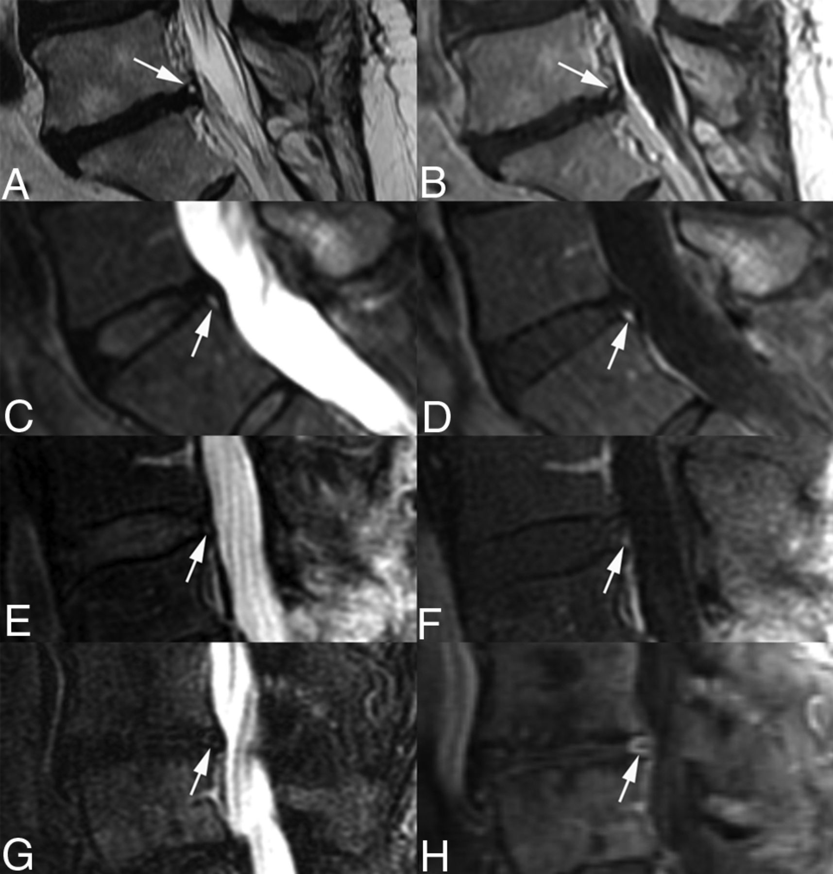
- FIG 1.
T2-weighted sequence grading of annular fissures detected on postcontrast T1-weighted imaging. A, Grade 4 annular fissure: annular fissure signal has high signal intensity (arrow) with signal that is greater than that of adjacent spinal fluid, consistent with an HIZ. B, Enhancement in the Grade 4 annular fissure (arrow). C, Grade 3 annular fissure: annular fissure signal has high signal intensity (arrow) with signal that is less than that of the adjacent spinal fluid, consistent with an HIZ. D, Enhancement in the Grade 3 annular fissure (arrow). E, Grade 2 annular fissure: annular fissure signal is focally abnormal but with mixed intermediate-to-low signal intensity (arrow) appearing close to the signal of the degenerative disc without a definite HIZ. F, Enhancement in the Grade 2 annular fissure (arrow). G, Grade 1 annular fissure: annular fissure signal is abnormal and has low signal intensity (arrow), appearing similar to the signal of other portions of the severely degenerative disc. H, Enhancement in the Grade 1 annular fissure (arrow).
- FIG 2.
Receiver operating curves (ROCs) for predicting concordant pain (A) and significant pain (B) based on HIZ and EAF features. Sens indicates sensitivity; Spec, specificity.
- FIG 3.
A 49-year-old woman with long-standing LBP and some bilateral leg radiation (most severe VAS = 10/10; immediate preprocedural VAS = 5/10) with prior discectomy at L5–S1. Concordant pain was provoked at L2–3, L3–4, and L4–5, with all 3 levels demonstrating partial provoked pain improvement with intradiscal lidocaine, and all 3 levels appearing contained on postdiscogram CT. A, Sagittal postcontrast T1-weighted image demonstrates posterior EAFs at L2–3, L3–4 (thin arrows), and L4–5 (thick arrow). B, Sagittal T2-weighted image. The L2–3 HIZ, considered Grade 4, and L3–4 HIZ, considered Grade 2 by consensus group 1, were identified as HIZs by consensus group 2 (thin arrows). The EAF at L4–5 considered a Grade 1 HIZ by consensus group 1 was not identified as an HIZ by consensus group 2 (thick arrow). C, Sagittal reconstructed CT image demonstrates the posterior annular margin at L4–5, consistent with the EAF (thick arrow). D and E, Axial postdiscogram CT images through the L4–5 disc demonstrates severe annular degeneration (Dallas Grade 3) with the posterior annular margin noted, consistent with the EAF (thick arrow).
- FIG 4.
A 29-year-old woman with complex LBP and leg pain (most severe VAS = 10/10; preprocedural VAS = 5/10) with prior L5–S1 discectomy. Concordant pain was provoked at L4–5 (VAS = 10/10). A, Sagittal T2-weighted sequence demonstrates a small annular fissure along the posterior annular margin (arrow), judged Grade 4 by consensus. B, Sagittal postcontrast T1-weighted sequence demonstrates enhancement in the annular fissure (arrow). C, Axial postdiscogram CT image demonstrates a full-thickness radial annular fissure (arrow) projecting into a small peripheral concentric annular fissure (arrowhead). D, Axial postdiscogram CT image demonstrates the posterior annular margin at the level of the peripheral concentric annular fissure (arrowhead), which corresponds to the HIZ identified on the T2-weighted sequence and EAF region demonstrated on the postcontrast T1-weighted sequence.


Tables
Peripheral Annular Morphology by MR Imaging Discography-Provoked Pain Response Severe Pain Negative for Pain Total Concordant Pain(% of Total) Nonconcordant Pain(% of Total) EAF 22/39 (56.4%) 10/39 (25.6%) 7/39 (18%) 39 (100%) Focal high signal on T2WI only 0 0 1 1 No MR imaging evidence of annular fissure/tear 14/60 (23.3%) 4/60 (6.7%) 42/60 (70.0%) 60 (100%) Total 36/100 (36%) 14/100 (14%) 50/100 (50%) 100 (100%) - Table 2:
Thirty-nine discs positive for EAF—T2WI grade, HIZ detection, and unrecognized fissure/tear pain response
Annular Fissure/Tear CG-1 T2WI Grade EAFs Identified on T1 Enhanced Sequence by CG-1 HIZs Identified on T2-Weighted Sequence by CG-2 (% EAFs) Injection Pain Response of 16 Unrecognized Annular Fissures (HIZs) by CG-2 on T2WI C NC N 4 8 7/8 (87.5%) 1 3 12 10/12 (83.3%) 1 1 2 8 4/8 (50.0%) 1 2 1 1 11 2/11 (18.2%) 7 1 1 Total 39 23/39 (59.0%) 9 4 3 Note:—C indicates concordant levels; NC, nonconcordant levels; N, negative pain response; CG-1, consensus group 1; CG-2, consensus group 2.
- Table 3:
Proportions of discs with EAF for different pain responses by disc levels, sex, and patient age
Disc/Patient Characteristics Discography Pain Response Severe Pain Negative for Pain (EAF %) Overall (EAF %) Concordant Pain (EAF %) Nonconcordant Pain (EAF %) Overall 22/36 (61%) 10/14 (71%) 7/50 (14%) 39/100 (39%) Disc level L1–2 to L2–3a 2/3 (67%) 0/1 (0%) 1/9 (11%) 3/13 (23%) L3–4 4/11 (36%) 4/5 (80%) 4/28 (14%) 12/44 (27%) L4–5 14/15 (93%) 3/3 (100%) 2/10 (20%) 19/28 (68%) L5–S1 2/7 (29%) 3/5 (60%) 0/3 (0%) 5/15 (33%) Sex Female 16/23 (70%) 5/7 (71%) 5/29 (17%) 26/59 (44%) Male 6/13 (46%) 5/7 (71%) 2/21 (10%) 13/41 (32%) Age Younger than 41 yr 16/24 (67%) 7/9 (78%) 6/25 (24%) 29/58 (50%) 41 yr or older 6/12 (50%) 3/5 (60%) 1/25 (4%) 10/42 (24%) ↵a There was a single disc at L1–2 that did not have an EAF or pain on provocation.
- Table 4:
Response to provocative discography by imaging findings of HIZ and EAF (no discs identified with HIZ but without EAF)
Imaging Features Discography Pain Response Severe Pain (n = 50) Negative for Pain(n = 50) Total (n = 100) Concordant (n = 36) Nonconcordant (n = 14) EAF+ HIZ+ 14/36 (39%) 6/14 (43%) 3/50 (6%) 23/100 EAF+ HIZ− 8/36 (22%) 4/14 (29%) 4/50 (8%) 16/100 EAF− HIZ− 14/36 (39%) 4/14 (29%) 43/50 (86%) 61/100 Total 36 (100%) 14 (100%) 50 (100%) 100/100 - Table 6:
EAF T2WI grade by CG-1 versus postdiscogram CT features of internal derangement—39 discs
Annular Fissure T2WI CG-1 Grade Discogram Pain Response and Internal Derangement Features Severe Pain Concordant Pain Nonconcordant Pain Negative for Pain Total RDef DEG RDef DEG RDef DEG RDef DEG 4 2 3 3 0 0 0 5 3 3 4 2 2 1 1 2 7 5 2 1 2 1 2 1 1 3 5 1 2 6 0 1a 1 1 3 8 Total 9 13 6 4a 3 4 18 21 Note:—DEG indicates degenerative change Dallas Grade 3 except for 1 disc; RDef, all radial defects (Dallas Grade 3); CG-1, consensus group 1.
↵a Dallas Grade 1.
- Table 7:
Intradiscal lidocaine response—40 concordant and nonconcordant severely painful discs where anesthetic was injected
Disc State Intradiscal Lidocaine-Provoked Pain Response Total (n = 40) Total Improvement (n = 17) Partial Improvement (n = 7) No Improvement (n = 16) Leaking 14 (82%) 2 (29%) 5 (31%) 21 (53%) Contained 3 (18%) 5 (71%) 11 (69%) 19 (48%) Total 17 (100%) 7 (100%) 16 (100%) 40 (100%)


Jump to section
Related Articles
Cited By...
- No citing articles found.