
Article Figures & Data
Figures
- FIG 1.
A case of MMA origin from basilar artery (BA) pontine perforating branch. In this case, the MMA originates from a pontine branch of the BA, as indicated by the yellow arrow in A, B, and C. A and B, A frontal and lateral view of left vertebral artery (VA) injection, respectively. C, A frontal XperCT (Philips Healthcare) reconstruction with the same MMA origin. D, A distal external carotid artery injection, where the superficial temporal artery (STA) and the IMA are visible, without the typical MMA origin from the IMA.
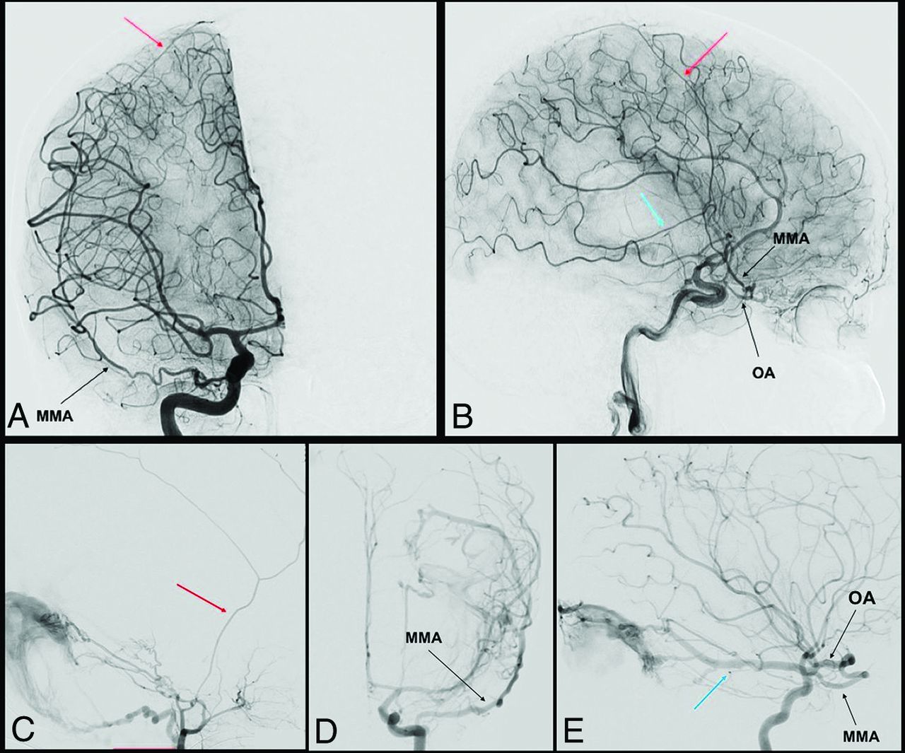
- FIG 2.
Complete and partial MMA origin from OA. The anteroposterior and lateral view angiograms (A and B respectively) show a rare case of a complete MMA origin from the OA. The OA, through the superficial recurrent OA, gives origin to the MMA, that passes through the lateral part of the superior orbital fissure, and bifurcates into its anterior (red arrow) and posterior division (blue arrow). In the angiograms C, D, and E, a rare case of a partial origin of the MMA from the OA is shown. D and E, Angiograms of a left ICA injection in the frontal and lateral views, where the posterior branch of the MMA (blue arrow) originates at the OA and feeds a tentorial AVF. After the external carotid artery injection (C), only the anterior branch of the MMA is enhanced (red arrow).
- FIG 3.
DSA, showing segments and branches of the MMA. A and B, show selective MMA injection from a lateral (A) and frontal (B) view. The MMA bifurcates at the pterional region (green circle) into 2: an anterior (red arrow) and posterior (blue arrow) division. Before its bifurcation, the MMA gives the petrosal branch (Pb), which courses on the petrous apex. The posterior division gives 2 principal branches: the petrosquamosal branch (PSb) and the parieto-occipital branch (POb). The anterior division ends with 2 kinds of terminal branches, visible after common carotid artery injection: the falcine arteries (yellow arrow) (C), which anastomose with branches of the anterior falcine artery from the OA, and contralateral branches (purple arrow) (D) that cross the midline to anastomose with a contralateral MMA.
- FIG 4.
Anastomoses of the MMA. Before the MMA bifurcation (purple circle), the petrous branch (1), from which the superior tympanic artery (2) originates, anastomoses into the middle ear with the caroticotympanic artery (3, from the ICA), and with the inferior tympanic artery (4, from the ascending pharyngeal artery), with the posterior tympanic artery (5, from the occipital artery). The cavernous branch of the MMA (6) on the other side anastomoses with the inferolateral trunk (ILT) (7), which is itself connected to the OA (9) through the deep recurrent OA (8). The ILT, the MMA, and the OA are also linked to each other through the marginal tentorial artery (10), whose origin can vary from the lacrimal artery (11), via superficial recurrent OA (12), to the meningohypophyseal trunk (13). After the MMA bifurcation at the pterion, its frontal division (14) gives a medial branch (15), which can bifurcate intracranially into a lateral meningolacrimal artery (16), and a medial sphenoidal artery (17). Both branches reach the lacrimal artery, even if the meningolacrimal artery more distal than the sphenoidal artery. The anastomoses with the OA and the ILT represent the most dangerous connections in the case of MMA transarterial embolization because of the risk of particle embolism into these arteries. The frontal division of the MMA reaches the convexity, following the coronal suture and anastomoses with the anterior falcine artery (18, OA–anterior ethmoidal artery) and with branches of the contralateral MMA (19). The posterior division of the MMA (20) divides into a petrosquamosal branch (21) and a parieto-occipital branch (22). The former anastomoses with the jugular branch (23) of the ascending pharyngeal artery (24) and with the mastoid branch (25) of the occipital artery (26). The latter is linked to the posterior meningeal artery (27), from the vertebral artery (28) at the border areas.


Tables
- Table 1:
Different origin of the MMA with modifications associated and embryologic explanation
Variations in the Origin of the MMA Embryologic Implications Type Associated Changes Embryologic Explanation Embryo Size (mm) IMA origin Normal anatomy Normal embryology Basilar artery origin Absence foramen spinosum Anastomosis between SA and trigeminal artery; anastomosis between SA and lateral pontine artery 12 Cavernous ICA origin Absence foramen spinosum Anastomosis between inferolateral trunk and SA 16 Partial persistent SA Absence foramen spinosum; enlargement of the facial canal Regression of the proximal part of the maxillomandibular branch; persistence of the intratympanic segment of the SA 24 Complete persistent SA Enlargement of the facial canal Lack of annexation of the maxillomandibular branch by the ventral pharyngeal artery; persistence of the intratympanic segment of the SA 24 Pseudopetrous ICA origin Absence foramen spinosum; enlargement of the facial canal; absence of the exocranial opening of the carotid canal Agenesis of the first and second segments of the ICA; intratympanic anastomosis between inferior tympanic and caroticotympanic arteries; persistence of the intratympanic segment of the SA 4–5; 24 Cervical ICA origin Absence foramen spinosum; enlargement of the facial canal Intratympanic anastomosis between inferior and superior tympanic arteries; regression of the proximal part of the maxillomandibular branch; persistence of the intratympanic segment of the SA 16; 24 Occipital artery origin Absence foramen spinosum; enlargement of the facial canal No clear explanation Distal petrous ICA origin Absence foramen spinosum Lack of annexation of the mandibular artery (first aortic arch) by the SA (second aortic arch) 9 Type Vascular Anatomy Foramen Spinosum I Complete OA origin of the MMA Absence II Partial OA origin of the MMA; anterior division from the OA; posterior division from the IMA Reduced in size III OA origin of the accessory meningeal artery Normal ↵a From Ref.28
MMA Branches Origin from the MMA Territory (Dural and Neural) Possible Anastomosis Petrosal branch Foramen spinosum Trigeminal ganglion and nerves; posteromedial floor of the middle fossa; insertion of tentorium (medial half); superior petrosal sinus Ascending pharyngeal artery (carotid branch); medial and lateral tentorial arteries (ICA) Superior tympanic artery Petrosal branch Greater superficial petrosal nerve; geniculate ganglion; tympanic cavity (superior part) Inferior tympanic artery (ascending pharyngeal artery); caroticotympanic artery (ICA); anterior tympanic artery (IMA); stylomastoid artery (posterior auricular artery) Cavernous branch Petrosal branch Lateral wall of the cavernous sinus Accessory meningeal artery; inferolateral trunk (ICA) Anterior division or frontal branch Pterional region Frontal and anterior parietal convexity; superior sagittal sinus; anterior and middle fossa (lateral part) Anterior and posterior ethmoidal arteries (OA); contralateral MMA Falcine arteries Anterior and posterior division Falx cerebri Anterior falcine artery (OA); anterior cerebral artery; posterior meningeal artery (vertebral artery) Medial branch or sphenoidal branch Anterior division Lesser sphenoid wing; superior orbital fissure; peri-orbital (lateral) Recurrent meningeal branches (OA); inferolateral trunk (ICA) Petrosquamosal branch Posterior division Posterolateral floor of the middle fossa; insertion of tentorium (lateral half); superior petrosal sinus; transverse and sigmoid sinuses; dura of the posterior fossa (superior part) Ascending pharyngeal artery (jugular branch); lateral tentorial artery (ICA); occipital artery (mastoid branch) Parieto-occipital branch Posterior division Temporosquamous dura; parieto-occipital convexity; superior sagittal sinus Posterior meningeal artery (vertebral artery)


Jump to section
Related Articles
Cited By...
- Correspondence on 'Evaluation of ChatGPT in knowledge of newly evolving neurosurgery: middle meningeal artery embolization for subdural hematoma management by Koester et al
- Advancements in super-selective catheterization and drug selection for intra-arterial chemotherapy for retinoblastoma: a 15-year evolution
- Evaluation of an in vivo preclinical model for human middle meningeal artery embolization using the posterior intercostal artery of the swine
- Advancements in super-selective catheterization and drug selection for intra-arterial chemotherapy for retinoblastoma: a 15-year evolution
- Pharyngo-tympano-stapedial middle meningeal artery variant supply to a falcotentorial dural arteriovenous fistula
- Neuroanatomy of cranial dural vessels: implications for subdural hematoma embolization
- Anatomic and Embryologic Analysis of the Dural Branches of the Ophthalmic Artery