
Article Figures & Data
Figures
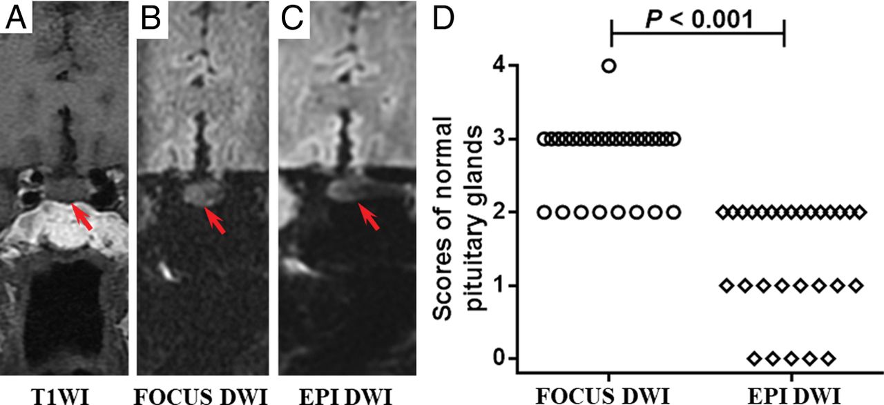
- Fig 1.
A–C, Images of a 68-year-old woman with dizziness for 1 week. The pituitary gland has a score of 3 for FOCUS DWI and 2 for EPI DWI, respectively (red arrow indicates the pituitary gland). The mean ADC value of the FOCUS DWI measurement is 1.19 × 10⁻³ mm2/s. D, The image-quality scores of normal pituitary glands between the 2 methods are statistically significant (P < .001). The number of subjects for each score in FOCUS and EPI DWI sequences is shown in the On-line Table.
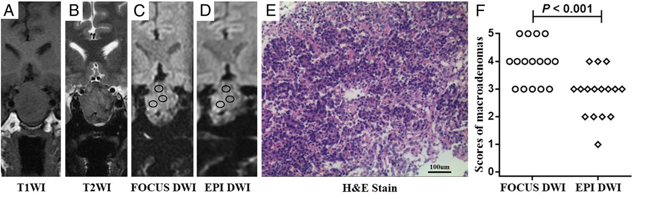
- Fig 2.
A–D, Images of a 51-year-old man with immunohistochemical staining–proved nonfunctional adenoma. The scores of the macroadenoma in FOCUS and EPI DWI are 4 and 3, respectively (black circles indicate the ROIs of ADC measurements). The mean ADC values of the FOCUS and EPI DWI measurements are 0.66 × 10⁻³ mm2/s and 0.80 × 10⁻³ mm2/s, respectively. E, A specimen of the mass at histologic examination (H&E stain; original magnification, ×200) shows plenty of small cells (blue) with scant fibrous stroma (deep pink). F, The image-quality scores of macroadenomas between 2 methods are statistically significant (P < .001). The number of patients for each score in FOCUS and EPI DWI sequences is shown in the On-line Table.


Tables
FOCUS DWI EPI DWI TR (ms) 2200 2200 TE (ms) Minimum Minimum B-value (s/mm2) 0, 500 0, 500 Diffusion directions All All Frequency-encoding direction S/I S/I FOV (cm) 16 × 4.8 24 × 24 Matrix size 128 × 38 160 × 160 NEX for B0 4 4 NEX for b=500 12 12 Slice thickness (mm) 2.0 2.0 Intersection gap (mm) 0 0 Spatial resolution (mm3) 1.25 × 1.26 × 2 1.5 × 1.5 × 2 Acquisition time (min:s) 01:30 01:30 Note:—S/I indicates superior/inferior.
Score Criteria Normal pituitary gland 0 Pronounced artifacts; pituitary stalk and gland cannot be recognized 1 Considerable artifacts; the stalk is visible, but the gland cannot be recognized 2 Pituitary stalk and gland are visible, with moderate-to-obvious image distortion and/or most of the pituitary gland (>50%) exhibiting signal loss 3 Pituitary stalk and gland are distinctly visible, with mild image distortion and/or <50% of the pituitary gland exhibiting signal loss 4 Visualization of pituitary stalk and gland is as clear as that on T1WI Pituitary macroadenoma 0 Pronounced artifacts; adenoma cannot be recognized 1 Considerable artifacts; adenoma is visible, with or without most of the adenoma (>50%) exhibiting signal loss 2 Adenoma is visible, with obvious image distortion and with or without nearly half of the adenoma (25%–50%) exhibiting signal loss 3 Adenoma is distinctly visible; moderate image distortion, with or without <25% of the adenoma exhibiting signal loss 4 Adenoma is distinctly visible, with only mild image distortion 5 Visualization of adenoma is as clear as that on T1WI


Jump to section
Related Articles
Cited By...
- No citing articles found.