
Article Figures & Data
Figures
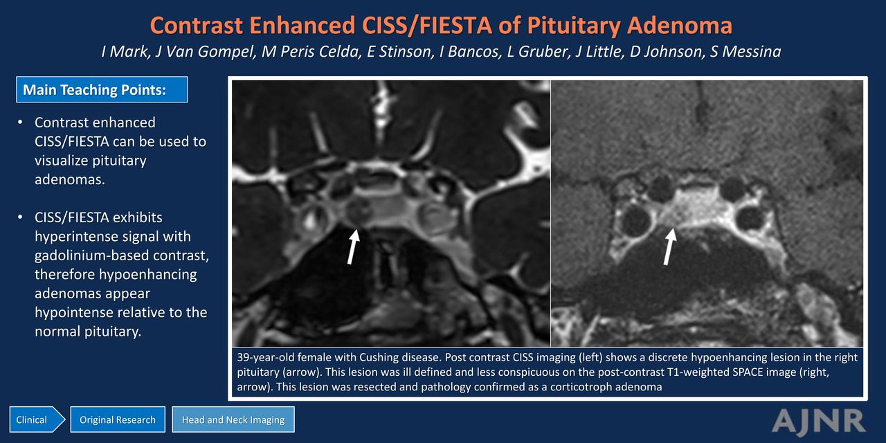
- FIG 1.
A 39-year-old woman with Cushing disease. Postcontrast CISS imaging (A) shows a discrete hypoenhancing lesion in the right pituitary gland (arrow). This lesion was ill-defined and less conspicuous on the postcontrast T1-weighted SPACE image (B, arrow). It was resected, and pathologically confirmed it as a corticotroph adenoma.
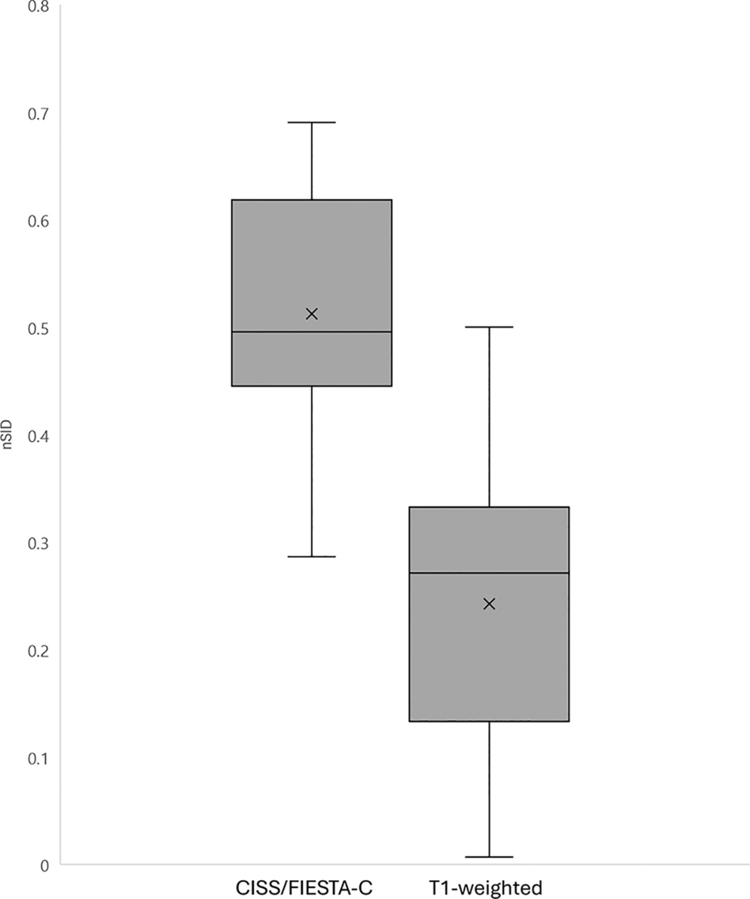
- FIG 2.
When one compares the nSID of contrast-enhanced CISS/FIESTA-C with T1-weighted imaging (P < .001), a higher nSID indicates a greater enhancement difference between the adenoma and pituitary gland.
- FIG 3.
A 44-year-old woman with Cushing disease. One year prior, the patient underwent pituitary lesion resection and radiation treatment at an outside institution; now she has persistent symptoms and laboratory values compatible with persistent Cushing disease. A, Postcontrast SPACE imaging shows hypoenhancement in the same area that was present but less conspicuous (arrow). Postcontrast coronal (B) and sagittal (C) CISS imaging show a hypoenhancing lesion in the left inferior pituitary (arrows). This lesion was resected and pathologically confirmed as a corticotroph adenoma.
- FIG 4.
An 18-year-old man with clinical symptoms and endocrine laboratory values compatible with Cushing disease. Postcontrast FIESTA-C imaging (A) demonstrates a hypoenhancing lesion in the right lateral pituitary gland (arrow) with corresponding hypoenhancement on the contrast-enhanced Cube that was less conspicuous (B, arrow). This lesion was a pathologically-confirmed corticotroph adenoma. The FIESTA-C image does show artifacts related to aeration of the sphenoid sinus (A, broken arrow). Representative circles are shown to demonstrate how the SI was measured.


Tables
Patient-specific details
Sex Age (yr) Size (mm) ST T1 Discrete nSID F 44 5 1 Y 0.376 F 54 5 1 Y 0.285 F 23 6 0.7 Y 0.354 F 69 6 1 Y 0.302 M 39 5 0.7 N 0.271 F 38 4 1 Y 0.500 F 55 6 0.6 N 0.017 F 49 4 1 Y 0.470 F 66 6 0.6 Y 0.163 F 67 2 0.8 N 0.299 F 48 5 1 Y 0.246 F 35 7 1 Y 0.187 M 18 7 0.8 Y 0.196 F 56 3 1 N 0.007 F 47 3 0.8 Y 0.311 F 63 7 0.7 N 0.035 F 38 3 9 N 0.103 Note:—ST indicates slice thickness; Y, yes; N, no; F, female; M, male.


Jump to section
Related Articles
Cited By...
- No citing articles found.