
Article Figures & Data
Figures
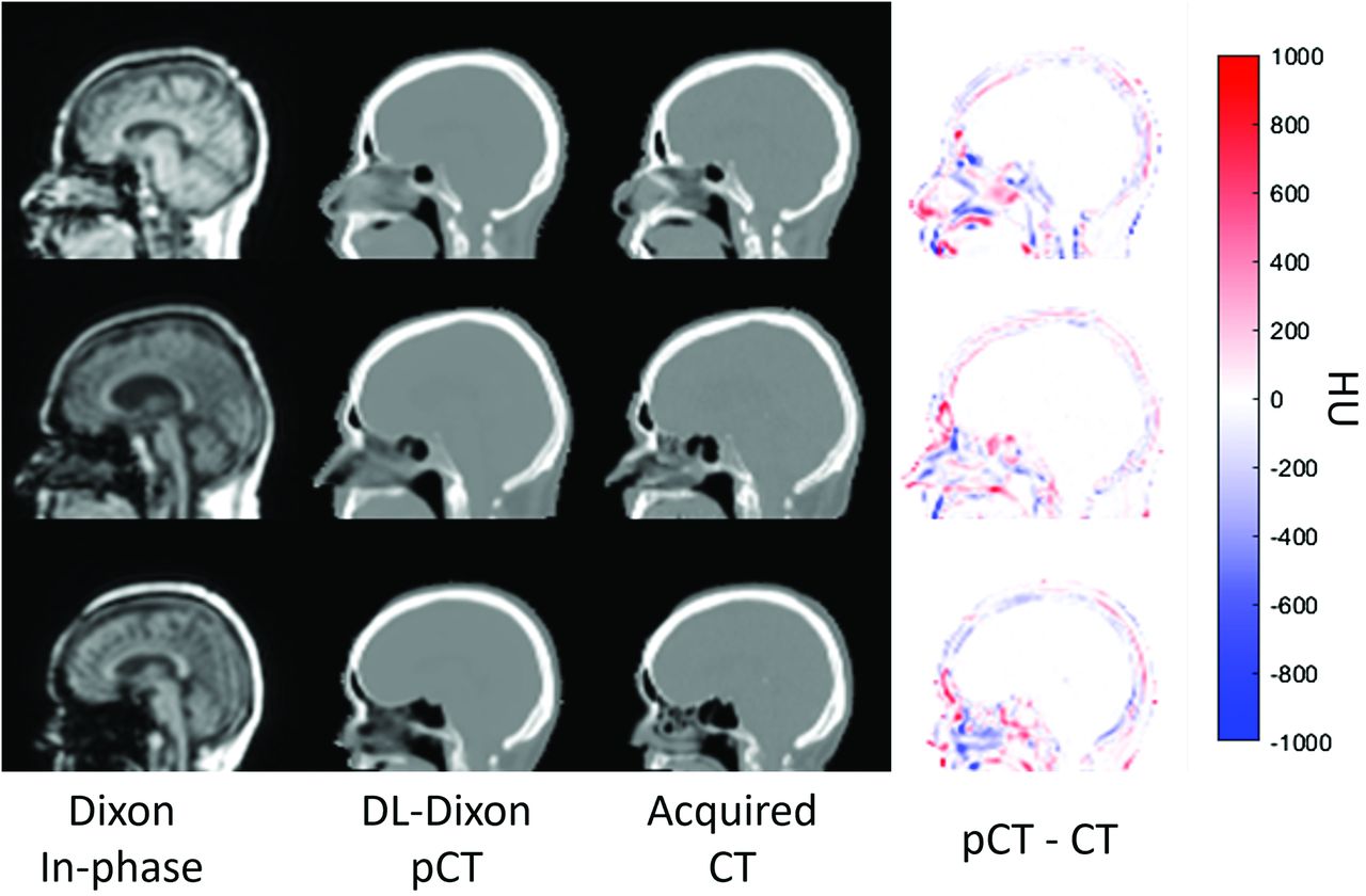
- FIG 1.
Dixon in-phase MR images (first column), DL-Dixon pCT images (second column), CT images (third column), and HU difference map between pCT and CT (fourth column) from 3 representative participants. The PET/MR scans were acquired by using the VB20P software version and a 16-channel head/neck coil (first row), the VE11P software version and a 16-channel head/neck coil (second row), and the VE11P software version and a 32-channel head coil (third row).
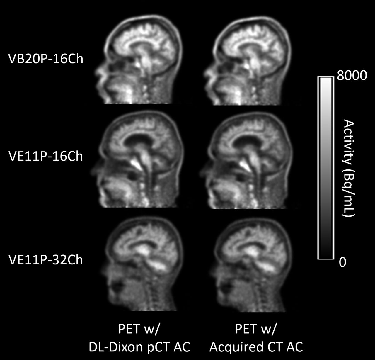
- FIG 2.
PET reconstructed with the DL-Dixon AC (first column) or the CT AC (second column) from 3 representative participants. The PET/MR scans were acquired by using the VB20P software version and a 16-channel head/neck coil (first row), the VE11P software version and a 16-channel head/neck coil (second row), and the VE11P software version and a 32-channel head coil (third row).
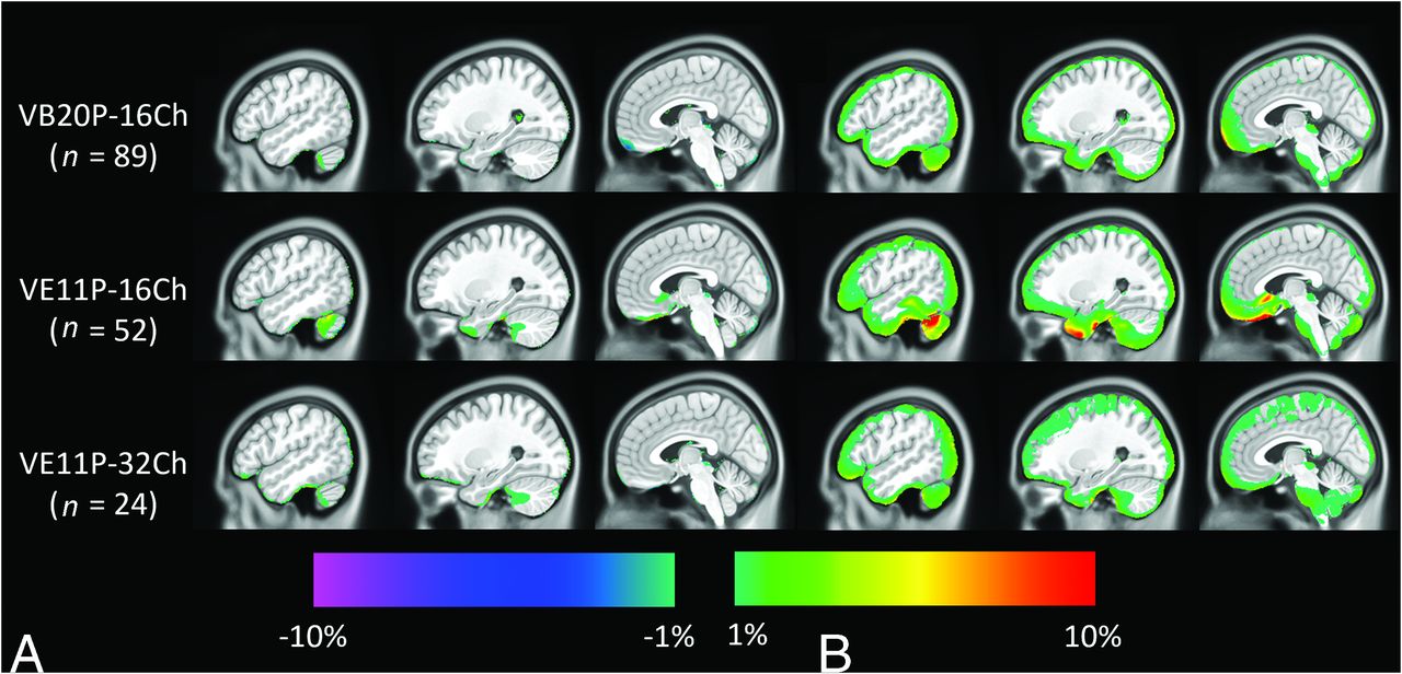
- FIG 3.
Mean (A) and SD (B) of PET relative error on the voxel basis across testing participants of the VB20P-16Ch model (n = 89), the VE11P-16Ch model (n = 52), and the VE11P-32Ch model (n = 24). The CT AC method is used as the reference.
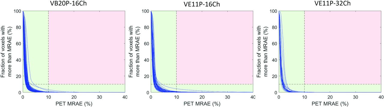
- FIG 4.
Cumulative voxelwise PET MRAE by using DL-Dixon AC. The acquired CT AC method is used as the standard reference. Each blue curve represents 1 participant. If the line stayed within the green region, the participant passed the qualification criteria.
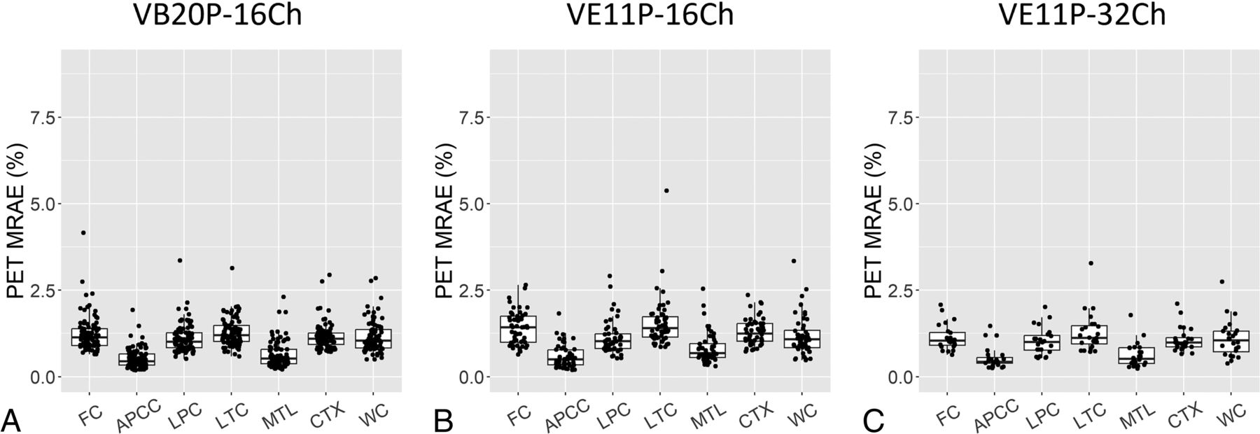
- FIG 5.
PET MRAE in 7 cerebral cortical and cerebellum ROIs. The boxplots show the 25th, 50th (median), and 75th percentiles. FC = frontal cortical region; APCC = anterior and posterior cingulate cortical region; LPC = lateral parietal cortical region; LTC = lateral temporal cortical region; MTL = medial temporal lobe; CTX = cortical summary region; WC = whole cerebellum region.
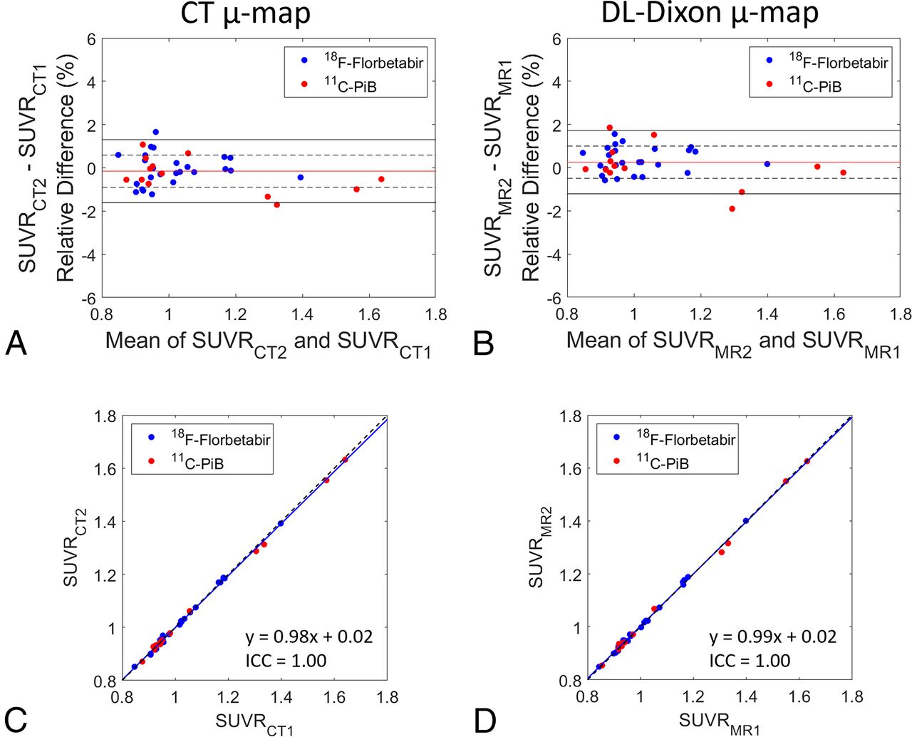
- FIG 6.
Longitudinal consistency of PET by using CT and DL-Dixon AC. The Bland-Altman plots of the PET SUVR difference between 2 CT (A) and 2 DL-Dixon (B) ACs in the cortical summary region are shown. The red horizontal line, dotted black horizontal lines, and solid black horizontal lines represent the mean, ± SD, and ±1.96 SD of the PET SUVR differences, respectively. Scatterplots of the PET SUVR between 2 CT (C) and 2 DL-Dixon (D) ACs in the cortical summary region are shown. The solid blue line and dotted black line represent the linear fitting line and line of identity, respectively. Symbol colors indicate different tracers (blue symbols: 18F-florbetapir PET, red symbols: 11C-PiB PET).
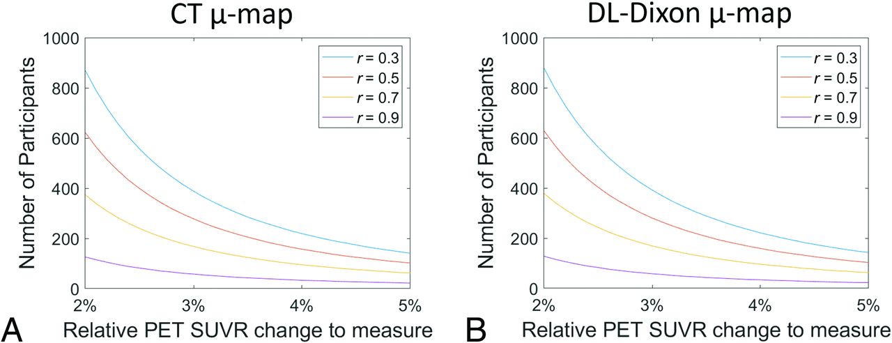
- FIG 7.
Number of participants required to detect longitudinal change in the cortical summary region with 80% power. The r is the assumed correlation between the paired measures from a participant.


Tables
n = 329 16-Channel Coil 32-Channel Coil VE20P VB20P-16Ch (n = 176) — VE11P VE11P-16Ch (n = 105) VE11P-32Ch (n = 48) Note: The total number of participants for each model is summarized.
- Table 2:
The longitudinal consistency of DL-Dixon was evaluated in 38 participants with repeated scans over approximately 3 years
n = 38 Visit 2 VB20P 16-Channel Coil VE11P 16-Channel Coil VE11P 32-Channel Coil Visit 1 VB20P 16-channel coil 10 15 4 VE11P 16-channel coil — — 9 Note: The software version and head coil used during the 2 visits are summarized.
- Table 3:
PET SUVR longitudinal consistency of CT AC and DL-Dixon AC in the cortical summary region
CT DL-Dixon SUVR difference (Mean ± SD) −0.16% ± 0.74% 0.25% ± 0.75% wCV 0.53% 0.55% ICC 1.00 1.00


Jump to section
Related Articles
Cited By...
- No citing articles found.