Advertisement

Main menu

  • Home
  • Content
    • Current Issue
    • Accepted Manuscripts
    • Article Preview
    • Past Issue Archive
    • Video Articles
    • AJNR Case Collection
    • Case of the Week Archive
    • Case of the Month Archive
    • Classic Case Archive
  • Special Collections
    • AJNR Awards
    • Low-Field MRI
    • Alzheimer Disease
    • ASNR Foundation Special Collection
    • Photon-Counting CT
    • View All
  • Multimedia
    • AJNR Podcasts
    • AJNR SCANtastic
    • Trainee Corner
    • MRI Safety Corner
    • Imaging Protocols
  • For Authors
    • Submit a Manuscript
    • Submit a Video Article
    • Submit an eLetter to the Editor/Response
    • Manuscript Submission Guidelines
    • Statistical Tips
    • Fast Publishing of Accepted Manuscripts
    • Graphical Abstract Preparation
    • Imaging Protocol Submission
    • Author Policies
  • About Us
    • About AJNR
    • Editorial Board
    • Editorial Board Alumni
  • More
    • Become a Reviewer/Academy of Reviewers
    • Subscribers
    • Permissions
    • Alerts
    • Feedback
    • Advertisers
    • ASNR Home

User menu

  • Alerts
  • Log in

Search

  • Advanced search
American Journal of Neuroradiology

American Journal of Neuroradiology

ASHNR American Society of Functional Neuroradiology ASHNR American Society of Pediatric Neuroradiology ASSR
  • Alerts
  • Log in

Advanced Search

  • Home
  • Content
    • Current Issue
    • Accepted Manuscripts
    • Article Preview
    • Past Issue Archive
    • Video Articles
    • AJNR Case Collection
    • Case of the Week Archive
    • Case of the Month Archive
    • Classic Case Archive
  • Special Collections
    • AJNR Awards
    • Low-Field MRI
    • Alzheimer Disease
    • ASNR Foundation Special Collection
    • Photon-Counting CT
    • View All
  • Multimedia
    • AJNR Podcasts
    • AJNR SCANtastic
    • Trainee Corner
    • MRI Safety Corner
    • Imaging Protocols
  • For Authors
    • Submit a Manuscript
    • Submit a Video Article
    • Submit an eLetter to the Editor/Response
    • Manuscript Submission Guidelines
    • Statistical Tips
    • Fast Publishing of Accepted Manuscripts
    • Graphical Abstract Preparation
    • Imaging Protocol Submission
    • Author Policies
  • About Us
    • About AJNR
    • Editorial Board
    • Editorial Board Alumni
  • More
    • Become a Reviewer/Academy of Reviewers
    • Subscribers
    • Permissions
    • Alerts
    • Feedback
    • Advertisers
    • ASNR Home
  • Follow AJNR on Twitter
  • Visit AJNR on Facebook
  • Follow AJNR on Instagram
  • Join AJNR on LinkedIn
  • RSS Feeds

AJNR Awards, New Junior Editors, and more. Read the latest AJNR updates

Research ArticleNeuroimaging Physics/Functional Neuroimaging/CT and MRI Technology

Whole-Brain Vascular Architecture Mapping Identifies Region-Specific Microvascular Profiles In Vivo

Anja Hohmann, Ke Zhang, Christoph M. Mooshage, Johann M.E. Jende, Lukas T. Rotkopf, Heinz-Peter Schlemmer, Martin Bendszus, Wolfgang Wick and Felix T. Kurz
American Journal of Neuroradiology September 2024, 45 (9) 1346-1354; DOI: https://doi.org/10.3174/ajnr.A8344
Anja Hohmann
aFrom the Department of Neurology (A.H., W.W.), Heidelberg University Hospital, Heidelberg, Germany
  • Find this author on Google Scholar
  • Find this author on PubMed
  • Search for this author on this site
  • ORCID record for Anja Hohmann
Ke Zhang
bDepartment of Diagnostic and Interventional Radiology (K.Z.), Heidelberg University Hospital, Heidelberg, Germany
  • Find this author on Google Scholar
  • Find this author on PubMed
  • Search for this author on this site
  • ORCID record for Ke Zhang
Christoph M. Mooshage
cDepartment of Neuroradiology (C.M.M., J.M.E.J., M.B., F.T.K.), Heidelberg University Hospital, Heidelberg, Germany
  • Find this author on Google Scholar
  • Find this author on PubMed
  • Search for this author on this site
  • ORCID record for Christoph M. Mooshage
Johann M.E. Jende
cDepartment of Neuroradiology (C.M.M., J.M.E.J., M.B., F.T.K.), Heidelberg University Hospital, Heidelberg, Germany
  • Find this author on Google Scholar
  • Find this author on PubMed
  • Search for this author on this site
  • ORCID record for Johann M.E. Jende
Lukas T. Rotkopf
dDivision of Radiology (L.T.R., H.-P.S., F.T.K.) German Cancer Research Center, Heidelberg, Germany
  • Find this author on Google Scholar
  • Find this author on PubMed
  • Search for this author on this site
Heinz-Peter Schlemmer
dDivision of Radiology (L.T.R., H.-P.S., F.T.K.) German Cancer Research Center, Heidelberg, Germany
  • Find this author on Google Scholar
  • Find this author on PubMed
  • Search for this author on this site
  • ORCID record for Heinz-Peter Schlemmer
Martin Bendszus
cDepartment of Neuroradiology (C.M.M., J.M.E.J., M.B., F.T.K.), Heidelberg University Hospital, Heidelberg, Germany
  • Find this author on Google Scholar
  • Find this author on PubMed
  • Search for this author on this site
  • ORCID record for Martin Bendszus
Wolfgang Wick
aFrom the Department of Neurology (A.H., W.W.), Heidelberg University Hospital, Heidelberg, Germany
eClinical Cooperation Unit Neurooncology (W.W.), German Cancer Research Center, Heidelberg, Germany
  • Find this author on Google Scholar
  • Find this author on PubMed
  • Search for this author on this site
  • ORCID record for Wolfgang Wick
Felix T. Kurz
cDepartment of Neuroradiology (C.M.M., J.M.E.J., M.B., F.T.K.), Heidelberg University Hospital, Heidelberg, Germany
dDivision of Radiology (L.T.R., H.-P.S., F.T.K.) German Cancer Research Center, Heidelberg, Germany
fDivision of Neuroradiology (F.T.K.), University Hospital Geneva, Geneva, Switzerland
  • Find this author on Google Scholar
  • Find this author on PubMed
  • Search for this author on this site
  • ORCID record for Felix T. Kurz
  • Article
  • Figures & Data
  • Supplemental
  • Info & Metrics
  • Responses
  • References
  • PDF
Loading

Article Figures & Data

Figures

  • Tables
  • FIG 1.
    • Download figure
    • Open in new tab
    • Download powerpoint
    FIG 1.

    Flow diagram showing the patient-selection protocol and criteria for inclusion and exclusion. The asterisk indicates that multiple scans per patient were used for further subanalyses on intrasubject test-retest reliability. VP-Shunt indicates ventriculoperitoneal shunt.
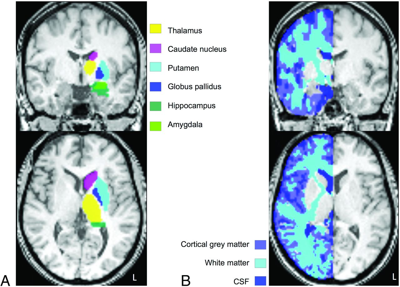
  • FIG 2.
    • Download figure
    • Open in new tab
    • Download powerpoint
    FIG 2.

    Cerebral volumes of interest. Axial representation of VOIs on spatially normalized T1-weighted images displaying 6 atlas-based anatomic VOIs (female subject, 27 years of age; right-sided lesion) (A) and individually segmented gray and white matter VOIs (female subject, 38 years of age; left-sided lesion) (B). Only the hemisphere contralateral to the lesion was analyzed for each subject, with a total of 8 VOIs per subject.

  • FIG 3.
    • Download figure
    • Open in new tab
    • Download powerpoint
    FIG 3.

    Color representations of distance map parameters I, VTI, and CBI. Parameters I, VTI, and CBI are shown as spatially normalized, averaged axial parametric maps. In MNI standard space, slice coordinates are as follows for all maps (from left to right, Z-axis): -40, -14, 0, 8, 24, 44. Maps of parameters I and VTI are closely correlated and depict differences between brain regions with predominantly venous outflow, eg, close to the transverse and rectus sinus, and predominantly arterial inflow, eg, in the insular cortex. Parameter CBI contrasts brain regions with increased capillary vascularization, such as the GP and hippocampus.

  • FIG 4.
    • Download figure
    • Open in new tab
    • Download powerpoint
    FIG 4.

    Parametric maps for CBV fraction, VIPS, and rCBV. Parameters BVF, VIPS, and rCBV are shown as spatially normalized, averaged axial parametric maps in MNI standard space as above. Because BVF identifies regions with increased blood volume, it is similar to rCBV with moderate correlations between both parameter values in the cGM and WM, respectively (Online Supplemental Data). The parameter VIPS is highly increased in the GP and thalamus (Table 2), as well as in parts of the WM along the pyramidal tracts.
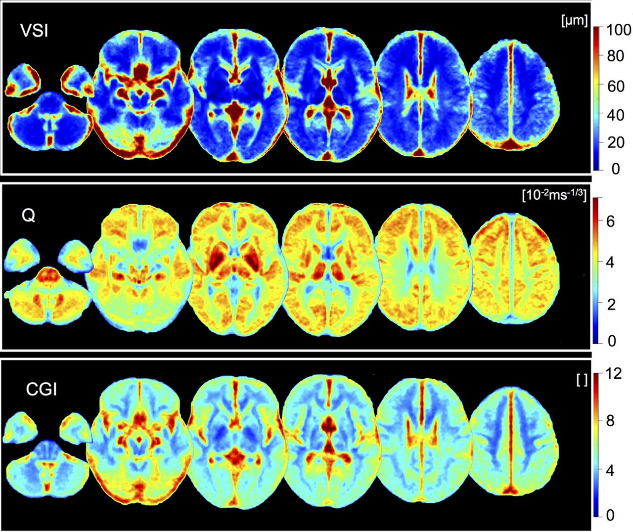
  • FIG 5.
    • Download figure
    • Open in new tab
    • Download powerpoint
    FIG 5.

    Color maps of VSI, microvessel density Q, and CGI. Parameters VSI, Q, and CGI are shown as spatially normalized, averaged axial parametric maps in MNI standard space as above. Parameters VSI and CGI indicate larger vessel calibers in the cGM compared with WM (Table 2). Microvessel density Q is increased in the lenticular nucleus (GP and putamen) and thalamus, compared with WM.

Tables

  • Figures
    • View popup
    Table 1:

    Demographics and distribution of individual vascular risk factors within the cohort (n = 106)a

    Cohort
    Variable
     Age (mean) (range) (yr) 39.2 (SD, 12.5) (20–70)
     Sex (% female) 52.8
     BMI (mean) (range) (kg/m2)25.7 (SD, 4.2) (18.4–37.7)
    Vascular risk factors (No.) (% of 106)
     Obesity (BMI >30) 19 (17.9%)
     Arterial hypertension 16 (15.1%)
     Diabetes mellitus type 24 (3.8%)
     Smoking25 (23.6%)
    KPS (mean) (range)95.2 (SD, 6.8) (80–100)
    • ↵a Data are displayed as mean values (SD) and (range) except where otherwise indicated.

    • View popup
    Table 2:

    VAM parameter value ranges within cerebral VOIsa

    Mean [95% CI]Cortical GMCNThalamusPutamenGPHippocampusAmygdalaWM
    Average mask size (voxel)38,3548331107103128793923436,322
    I−2.27−2.42−1.06−1.89−0.95−2.39−1.79−1.33
    [s−1][−2.45 to −2.08][−2.79 to −2.04][−1.37 to −0.75][−2.11 to −1.67][−1.23 to −0.67][−2.69 to −2.09][−2.31 to −1.26][−1.44 to −1.22]
    VTI−5.41−5.66−2.47−3.64−2.81−6.60−5.13−2.60
    [s−2][−5.96 to −4.86][−6.58 to −4.75][−.25 to −1.7][−4.24 to −3.04][−3.81 to −1.82][−7.5 to −5.7][−7.23 to −3.02][−2.84 to −2.35]
    CBI1.441.341.731.632.461.652.171.16
    [s−1][1.38–1.49][1.28–1.4][1.67–1.79][1.57–1.7][2.36–2.56][1.6–1.71][2.08–2.27][1.12–1.19]
    BVF20.4016.3918.2815.5215.6022.0523.9211.05
    [s−1][19.53–21.26][15.63–17.15][17.46–19.1][14.7–16.33][14.88–16.32][21.1–22.99][22.67–25.16][10.58–11.52]
    VIPS−3.97−2.820.02−2.236.68−5.39−7.17−3.19
    [10−1 s][−4.34 to −3.60][-4.25 to −1.38][−0.75–0.79][−2.84 to –1.61][4.91–8.44][−6.29 to –4.49][−8.89 to –5.46][−3.64 to −2.74]
    rCBV3.032.442.791.971.804.204.161.61
    [10−2][3.01–3.05][2.37–2.51][2.69–2.88][1.93–2.02][1.74–1.85][4.07–4.34][3.95–4.36][1.58–1.63]
    VSI37.3243.2029.1112.9310.9345.2045.2519.29
    [µm][36.43–38.22][40.2–46.2][27.31–30.91][12.53–13.32][10.13–11.73][43.3–47.1][42.37–48.13][18.85–19.74]
    Q4.253.364.504.915.903.813.904.23
    [10−2·ms−1/3][4.20–4.30][3.24–3.48][4.42–4.57][4.85–4.97][5.75–6.05][3.74–3.88][3.8–4][4.18–4.27]
    CGI5.806.195.104.143.146.105.684.25
    [ ][5.68–5.91][5.96–6.42][4.91–5.28][4.01–4.28][2.99–3.29][5.93–6.26][5.47–5.89][4.13–4.36]
    • Note:—[ ] indicates 95% confidence intervals.

    • a Data are presented as mean values with 95% confidence intervals for mean values.

    • View popup
    Table 3:

    ICCs between 2 scans (n = 27)a

    ParameterICC95% CIFP
    I0.4895[0.3814–0.5844]2.9241<.0001
    VTI0.3623[0.2406–0.4727]2.1337<.0001
    CBI0.7297[0.6591–0.7873]6.5456<.0001
    BVF0.7771[0.7182–0.8250]7.9463<.0001
    VIPS0.5689[0.4713–0.6527]3.6274<.0001
    rCBV0.9274[0.9061–0.9440]26.4302<.0001
    VSI0.9163[0.892–0.9353]22.969<.0001
    Q0.853[0.8046–0.8890]13.3324<.0001
    CGI0.8109[0.7598–0.8520]9.5721<.0001
    • ↵a ICC estimates with 95% confidence intervals for each parameter and the F test for the hypothesis that ICC = 0. Mean values for 8 VOIs were compared between n = 27 individual subjects at 2 different time points (n = 216 values per time point).

PreviousNext
Back to top

In this issue

American Journal of Neuroradiology: 45 (9)
American Journal of Neuroradiology
Vol. 45, Issue 9
1 Sep 2024
  • Table of Contents
  • Index by author
  • Complete Issue (PDF)
Advertisement
Print
Download PDF
Email Article

Thank you for your interest in spreading the word on American Journal of Neuroradiology.

NOTE: We only request your email address so that the person you are recommending the page to knows that you wanted them to see it, and that it is not junk mail. We do not capture any email address.

Enter multiple addresses on separate lines or separate them with commas.
Whole-Brain Vascular Architecture Mapping Identifies Region-Specific Microvascular Profiles In Vivo
(Your Name) has sent you a message from American Journal of Neuroradiology
(Your Name) thought you would like to see the American Journal of Neuroradiology web site.
CAPTCHA
This question is for testing whether or not you are a human visitor and to prevent automated spam submissions.
Cite this article
Anja Hohmann, Ke Zhang, Christoph M. Mooshage, Johann M.E. Jende, Lukas T. Rotkopf, Heinz-Peter Schlemmer, Martin Bendszus, Wolfgang Wick, Felix T. Kurz
Whole-Brain Vascular Architecture Mapping Identifies Region-Specific Microvascular Profiles In Vivo
American Journal of Neuroradiology Sep 2024, 45 (9) 1346-1354; DOI: 10.3174/ajnr.A8344

Citation Manager Formats

  • BibTeX
  • Bookends
  • EasyBib
  • EndNote (tagged)
  • EndNote 8 (xml)
  • Medlars
  • Mendeley
  • Papers
  • RefWorks Tagged
  • Ref Manager
  • RIS
  • Zotero
0 Responses
Respond to this article
Share
Bookmark this article
Whole-Brain Vascular Mapping
Anja Hohmann, Ke Zhang, Christoph M. Mooshage, Johann M.E. Jende, Lukas T. Rotkopf, Heinz-Peter Schlemmer, Martin Bendszus, Wolfgang Wick, Felix T. Kurz
American Journal of Neuroradiology Sep 2024, 45 (9) 1346-1354; DOI: 10.3174/ajnr.A8344
del.icio.us logo Twitter logo Facebook logo Mendeley logo
  • Tweet Widget
  • Facebook Like
  • Google Plus One
Purchase

Jump to section

  • Article
    • Abstract
    • ABBREVIATIONS:
    • MATERIALS AND METHODS
    • RESULTS
    • DISCUSSION
    • CONCLUSIONS
    • Footnotes
    • References
  • Figures & Data
  • Supplemental
  • Info & Metrics
  • Responses
  • References
  • PDF

Related Articles

  • PubMed
  • Google Scholar

Cited By...

  • No citing articles found.
  • Crossref
  • Google Scholar

This article has not yet been cited by articles in journals that are participating in Crossref Cited-by Linking.

More in this TOC Section

  • A Comprehensive Review of IA Imaging Modalities
  • Synthetic MRI based on 3D-QALAS MR Quantification
  • Delta wave MRI
Show more Neuroimaging Physics/Functional Neuroimaging/CT and MRI Technology

Similar Articles

Advertisement

Indexed Content

  • Current Issue
  • Accepted Manuscripts
  • Article Preview
  • Past Issues
  • Editorials
  • Editor's Choice
  • Fellows' Journal Club
  • Letters to the Editor
  • Video Articles

Cases

  • Case Collection
  • Archive - Case of the Week
  • Archive - Case of the Month
  • Archive - Classic Case

More from AJNR

  • Trainee Corner
  • Imaging Protocols
  • MRI Safety Corner
  • Book Reviews

Multimedia

  • AJNR Podcasts
  • AJNR Scantastics

Resources

  • Turnaround Time
  • Submit a Manuscript
  • Submit a Video Article
  • Submit an eLetter to the Editor/Response
  • Manuscript Submission Guidelines
  • Statistical Tips
  • Fast Publishing of Accepted Manuscripts
  • Graphical Abstract Preparation
  • Imaging Protocol Submission
  • Evidence-Based Medicine Level Guide
  • Publishing Checklists
  • Author Policies
  • Become a Reviewer/Academy of Reviewers
  • News and Updates

About Us

  • About AJNR
  • Editorial Board
  • Editorial Board Alumni
  • Alerts
  • Permissions
  • Not an AJNR Subscriber? Join Now
  • Advertise with Us
  • Librarian Resources
  • Feedback
  • Terms and Conditions
  • AJNR Editorial Board Alumni

American Society of Neuroradiology

  • Not an ASNR Member? Join Now

© 2025 by the American Society of Neuroradiology All rights, including for text and data mining, AI training, and similar technologies, are reserved.
Print ISSN: 0195-6108 Online ISSN: 1936-959X

Powered by HighWire