Advertisement

Main menu

  • Home
  • Content
    • Current Issue
    • Accepted Manuscripts
    • Article Preview
    • Past Issue Archive
    • Video Articles
    • AJNR Case Collection
    • Case of the Week Archive
    • Case of the Month Archive
    • Classic Case Archive
  • Special Collections
    • AJNR Awards
    • Low-Field MRI
    • Alzheimer Disease
    • ASNR Foundation Special Collection
    • Photon-Counting CT
    • View All
  • Multimedia
    • AJNR Podcasts
    • AJNR SCANtastic
    • Trainee Corner
    • MRI Safety Corner
    • Imaging Protocols
  • For Authors
    • Submit a Manuscript
    • Submit a Video Article
    • Submit an eLetter to the Editor/Response
    • Manuscript Submission Guidelines
    • Statistical Tips
    • Fast Publishing of Accepted Manuscripts
    • Graphical Abstract Preparation
    • Imaging Protocol Submission
    • Author Policies
  • About Us
    • About AJNR
    • Editorial Board
    • Editorial Board Alumni
  • More
    • Become a Reviewer/Academy of Reviewers
    • Subscribers
    • Permissions
    • Alerts
    • Feedback
    • Advertisers
    • ASNR Home

User menu

  • Alerts
  • Log in

Search

  • Advanced search
American Journal of Neuroradiology

American Journal of Neuroradiology

ASHNR American Society of Functional Neuroradiology ASHNR American Society of Pediatric Neuroradiology ASSR
  • Alerts
  • Log in

Advanced Search

  • Home
  • Content
    • Current Issue
    • Accepted Manuscripts
    • Article Preview
    • Past Issue Archive
    • Video Articles
    • AJNR Case Collection
    • Case of the Week Archive
    • Case of the Month Archive
    • Classic Case Archive
  • Special Collections
    • AJNR Awards
    • Low-Field MRI
    • Alzheimer Disease
    • ASNR Foundation Special Collection
    • Photon-Counting CT
    • View All
  • Multimedia
    • AJNR Podcasts
    • AJNR SCANtastic
    • Trainee Corner
    • MRI Safety Corner
    • Imaging Protocols
  • For Authors
    • Submit a Manuscript
    • Submit a Video Article
    • Submit an eLetter to the Editor/Response
    • Manuscript Submission Guidelines
    • Statistical Tips
    • Fast Publishing of Accepted Manuscripts
    • Graphical Abstract Preparation
    • Imaging Protocol Submission
    • Author Policies
  • About Us
    • About AJNR
    • Editorial Board
    • Editorial Board Alumni
  • More
    • Become a Reviewer/Academy of Reviewers
    • Subscribers
    • Permissions
    • Alerts
    • Feedback
    • Advertisers
    • ASNR Home
  • Follow AJNR on Twitter
  • Visit AJNR on Facebook
  • Follow AJNR on Instagram
  • Join AJNR on LinkedIn
  • RSS Feeds

AJNR Awards, New Junior Editors, and more. Read the latest AJNR updates

Research ArticleNeurodegenerative Disorder Imaging

Quantifying Brain Iron in Hereditary Hemochromatosis Using R2* and Susceptibility Mapping

S.K. Sethi, S. Sharma, S. Gharabaghi, D. Reese, Y. Chen, P. Adams, M.S. Jog and E.M. Haacke
American Journal of Neuroradiology July 2022, 43 (7) 991-997; DOI: https://doi.org/10.3174/ajnr.A7560
S.K. Sethi
aFrom the Department of Radiology (S.K.S., E.M.H.), Wayne State University, Detroit, Michigan
bSpinTech MRI Inc (S.K.S., S.G., E.M.H.), Bingham Farms, Michigan
  • Find this author on Google Scholar
  • Find this author on PubMed
  • Search for this author on this site
  • ORCID record for S.K. Sethi
S. Sharma
cDepartment of Clinical Neurological Sciences (S.S., M.S.J.), London Health Sciences Centre
  • Find this author on Google Scholar
  • Find this author on PubMed
  • Search for this author on this site
  • ORCID record for S. Sharma
S. Gharabaghi
bSpinTech MRI Inc (S.K.S., S.G., E.M.H.), Bingham Farms, Michigan
  • Find this author on Google Scholar
  • Find this author on PubMed
  • Search for this author on this site
  • ORCID record for S. Gharabaghi
D. Reese
eImaging Research Laboratories (D.R.), Robarts Research Institute, London, Ontario, Canada
  • Find this author on Google Scholar
  • Find this author on PubMed
  • Search for this author on this site
  • ORCID record for D. Reese
Y. Chen
fDepartment of Neurology (Y.C.), Wayne State University School of Medicine, Detroit, Michigan
  • Find this author on Google Scholar
  • Find this author on PubMed
  • Search for this author on this site
  • ORCID record for Y. Chen
P. Adams
dDivision of Gastroenterology (P.A.), Department of Medicine, Western University, London, Ontario, Canada
  • Find this author on Google Scholar
  • Find this author on PubMed
  • Search for this author on this site
  • ORCID record for P. Adams
M.S. Jog
cDepartment of Clinical Neurological Sciences (S.S., M.S.J.), London Health Sciences Centre
  • Find this author on Google Scholar
  • Find this author on PubMed
  • Search for this author on this site
  • ORCID record for M.S. Jog
E.M. Haacke
aFrom the Department of Radiology (S.K.S., E.M.H.), Wayne State University, Detroit, Michigan
bSpinTech MRI Inc (S.K.S., S.G., E.M.H.), Bingham Farms, Michigan
  • Find this author on Google Scholar
  • Find this author on PubMed
  • Search for this author on this site
  • ORCID record for E.M. Haacke
  • Article
  • Figures & Data
  • Info & Metrics
  • Responses
  • References
  • PDF
Loading

Article Figures & Data

Figures

  • Tables
  • FIG 1.
    • Download figure
    • Open in new tab
    • Download powerpoint
    FIG 1.

    Upper row: QSM showing 2 HC (A and B) and 2 subjects with HH (C and D). Lower row: Corresponding R2* maps. Elevated striatal and PT iron is depicted in both subjects with HH (C and D, F and G). For 1 subject (C and F), high iron can be seen in the cuneus and striatal cortex.

  • FIG 2.
    • Download figure
    • Open in new tab
    • Download powerpoint
    FIG 2.

    Comparison of susceptibility (parts per billion) between patients with HH and HC. Asterisk indicates P < .05; double asterisks, P < .01; triple asterisks, P < .001.
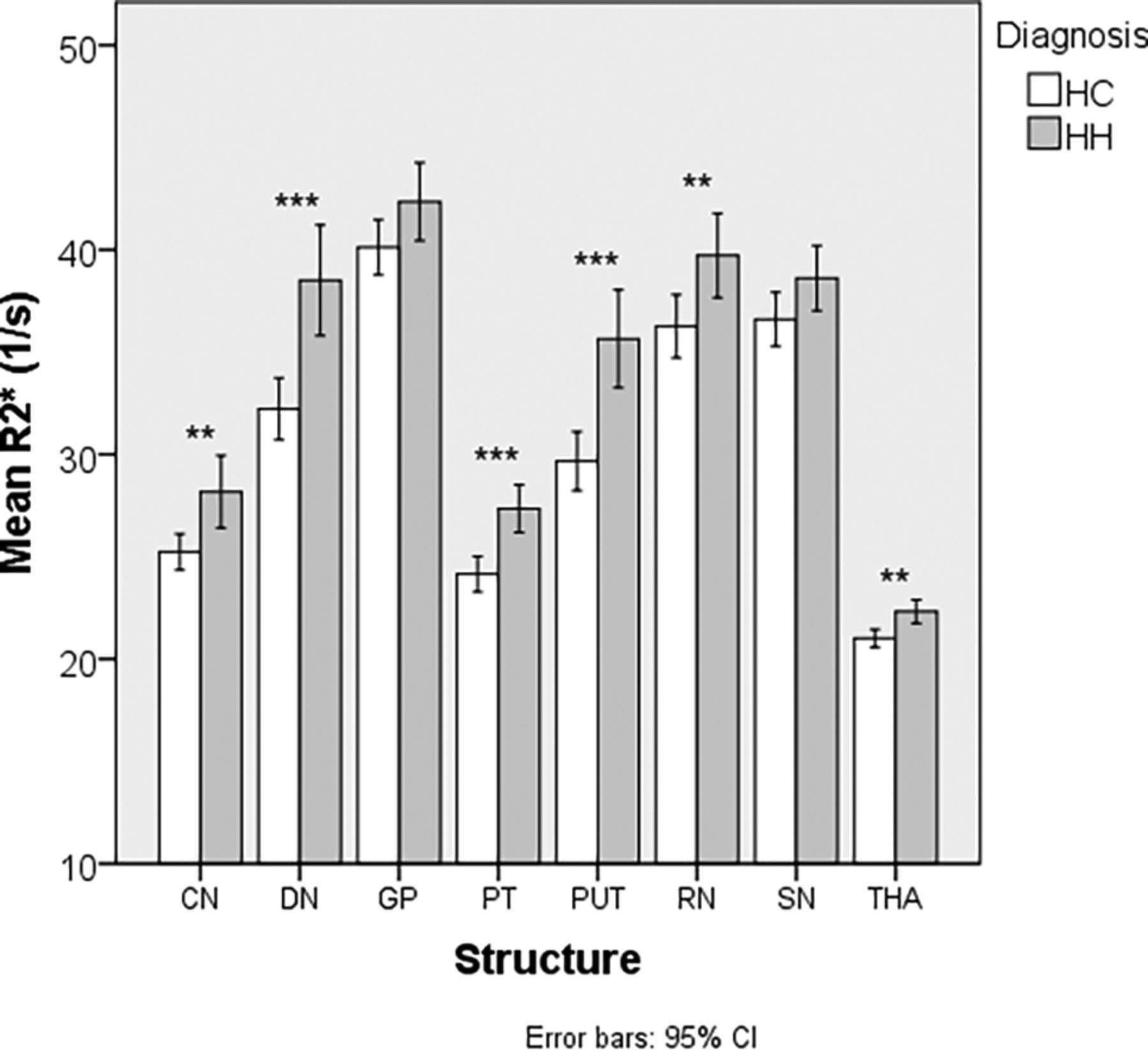
  • FIG 3.
    • Download figure
    • Open in new tab
    • Download powerpoint
    FIG 3.

    Comparison of R2* (s−1) between patients with HH and HC. Double asterisks indicate P < .01; triple asterisks, P < .001.

Tables

  • Figures
    • View popup
    Table 1:

    Mean age of subjects with HH and healthy controls

    Subjects with HH
    (n = 52)
    No.Mean Age (yr)95% CIP Value
    Men
     HH3058.23 (SD, 12.29)–12.29–2.52.19
     HC1953.32 (SD, 12.82)
    Women
     HH2258.00 (SD, 14.91)–11.59–4.52.38
     HC2854.46 (SD, 13.38)
    • View popup
    Table 2:

    Demographics of patients with HH who underwent MR imaging

    Demographics of Patients with HH
    Age at HH diagnosis (mean) (yr)51.09 (SD, 12.92) (n = 52)
    HandednessRight: 50, left: 2
    Family history of HHYes: 30, no: 21, unknown: 1
    Average duration from time of diagnosis to MR imaging (yr)7.29 (SD, 5.22) (n = 51)
    Genetic diagnosis (n = 60)C282Y homozygous: 41
    H63D homozygous: 2
    C282Y heterozygous: 2
    H63D heterozygous: 1
    Compound heterozygous: 3
    Unknown status: 3
    • View popup
    Table 3:

    Comparative analysis of mean susceptibility (parts per billion) in the DGM nuclei between patients with HH and HCa

    Group StatisticsT Test for Equality of Means
    StructureDiagnosisNo.MeanSDStandard Error of the MeantdfSignificant (2-Tailed)Mean DifferenceStandard Error Difference95% CI of the Difference
    LowerUpper
    CNHC4724.438.71.3−2.0897.040a−6.863.30−13.41−0.31
     HH5231.2921.02.9
    GPHC4785.4820.12.91.0997.2784.574.19−3.7412.89
     HH5280.9121.43.0
    PUTHC4733.0614.42.1−3.3797.001b−19.635.83−31.20−8.07
     HH5252.7037.55.2
    THAHC473.453.60.5−1.7897.079−1.580.89−3.360.19
     HH525.045.10.7
    PTHC4736.4013.52.0−2.6697.009b−9.793.68−17.09−2.48
     HH5246.1921.73.0
    RNHC4794.9124.13.5−4.3297<.001c−23.405.41−34.15−12.66
     HH52118.3229.24.1
    SNHC47129.6922.83.3−2.2697.026a−11.665.15−21.88−1.43
     HH52141.3527.93.9
    DNHC4792.5324.23.5−5.1097<.001c−32.096.3044.59−19.59
     HH52124.6236.55.1
    • ↵a P < .05.

    • ↵b P < .01.

    • ↵c P < .001.

    • View popup
    Table 4:

    Comparative analysis of R2* (s−1) in the deep gray matter nuclei between subjects with HH and HC

    Group StatisticsT Test for Equality of Means
    StructureDiagnosisNo.MeanSDStandard Error of the MeantdfSignificant (2-Tailed)Mean DifferenceStandard Error Difference95% Confidence Interval of the Difference
    LowerUpper
    CNHC4725.233.00.42.8997.005a−2.941.02−4.95−0.92
     HH5228.176.30.9
    GPHC4740.124.60.71.8897.064−2.221.18−4.570.13
     HH5242.356.80.9
    PUTHC4729.674.90.74.1997<.001b−5.981.43−8.81−3.15
     HH5235.658.61.2
    THAHC4721.011.50.2−3.5997.001a−1.310.37−2.04−0.59
     HH5222.322.10.3
    PTHC4724.153.00.4−4.3697<.001b−3.200.73−4.66−1.74
     HH5227.354.20.6
    RNHC4736.265.30.8−2.6597.009a−3.461.31−6.05−0.87
     HH5239.727.41.0
    SNHC4736.604.50.7−1.9497.056−2.021.04-4.080.05
     HH5238.625.70.8
    DNHC4732.235.20.8−3.9597<.001b−6.281.59−9.43−3.13
     HH5238.509.71.3
    • ↵a P <.01.

    • ↵b P <.001.

PreviousNext
Back to top

In this issue

American Journal of Neuroradiology: 43 (7)
American Journal of Neuroradiology
Vol. 43, Issue 7
1 Jul 2022
  • Table of Contents
  • Index by author
  • Complete Issue (PDF)
Advertisement
Print
Download PDF
Email Article

Thank you for your interest in spreading the word on American Journal of Neuroradiology.

NOTE: We only request your email address so that the person you are recommending the page to knows that you wanted them to see it, and that it is not junk mail. We do not capture any email address.

Enter multiple addresses on separate lines or separate them with commas.
Quantifying Brain Iron in Hereditary Hemochromatosis Using R2* and Susceptibility Mapping
(Your Name) has sent you a message from American Journal of Neuroradiology
(Your Name) thought you would like to see the American Journal of Neuroradiology web site.
CAPTCHA
This question is for testing whether or not you are a human visitor and to prevent automated spam submissions.
Cite this article
S.K. Sethi, S. Sharma, S. Gharabaghi, D. Reese, Y. Chen, P. Adams, M.S. Jog, E.M. Haacke
Quantifying Brain Iron in Hereditary Hemochromatosis Using R2* and Susceptibility Mapping
American Journal of Neuroradiology Jul 2022, 43 (7) 991-997; DOI: 10.3174/ajnr.A7560

Citation Manager Formats

  • BibTeX
  • Bookends
  • EasyBib
  • EndNote (tagged)
  • EndNote 8 (xml)
  • Medlars
  • Mendeley
  • Papers
  • RefWorks Tagged
  • Ref Manager
  • RIS
  • Zotero
0 Responses
Respond to this article
Share
Bookmark this article
Quantifying Brain Iron in Hemochromatosis
S.K. Sethi, S. Sharma, S. Gharabaghi, D. Reese, Y. Chen, P. Adams, M.S. Jog, E.M. Haacke
American Journal of Neuroradiology Jul 2022, 43 (7) 991-997; DOI: 10.3174/ajnr.A7560
del.icio.us logo Twitter logo Facebook logo Mendeley logo
  • Tweet Widget
  • Facebook Like
  • Google Plus One
Purchase

Jump to section

  • Article
    • Abstract
    • ABBREVIATIONS:
    • MATERIALS AND METHODS
    • RESULTS
    • DISCUSSION
    • CONCLUSIONS
    • ACKNOWLEDGMENTS
    • Footnotes
    • References
  • Figures & Data
  • Info & Metrics
  • Responses
  • References
  • PDF

Related Articles

  • No related articles found.
  • PubMed
  • Google Scholar

Cited By...

  • The Role of Labile Iron on Brain Proteostasis; Could it be an Early Event of Neurodegenerative Disease?
  • Crossref
  • Google Scholar

This article has not yet been cited by articles in journals that are participating in Crossref Cited-by Linking.

More in this TOC Section

  • ASL Perfusion Metric Correlates with SVD Severity
  • Anti-Amyloid Therapy & CBF as a Response Biomarker
  • ICP Sign: New Imaging Marker for MSA-C vs SCA
Show more NEURODEGENERATIVE DISORDER IMAGING

Similar Articles

Advertisement

Indexed Content

  • Current Issue
  • Accepted Manuscripts
  • Article Preview
  • Past Issues
  • Editorials
  • Editor's Choice
  • Fellows' Journal Club
  • Letters to the Editor
  • Video Articles

Cases

  • Case Collection
  • Archive - Case of the Week
  • Archive - Case of the Month
  • Archive - Classic Case

More from AJNR

  • Trainee Corner
  • Imaging Protocols
  • MRI Safety Corner
  • Book Reviews

Multimedia

  • AJNR Podcasts
  • AJNR Scantastics

Resources

  • Turnaround Time
  • Submit a Manuscript
  • Submit a Video Article
  • Submit an eLetter to the Editor/Response
  • Manuscript Submission Guidelines
  • Statistical Tips
  • Fast Publishing of Accepted Manuscripts
  • Graphical Abstract Preparation
  • Imaging Protocol Submission
  • Evidence-Based Medicine Level Guide
  • Publishing Checklists
  • Author Policies
  • Become a Reviewer/Academy of Reviewers
  • News and Updates

About Us

  • About AJNR
  • Editorial Board
  • Editorial Board Alumni
  • Alerts
  • Permissions
  • Not an AJNR Subscriber? Join Now
  • Advertise with Us
  • Librarian Resources
  • Feedback
  • Terms and Conditions
  • AJNR Editorial Board Alumni

American Society of Neuroradiology

  • Not an ASNR Member? Join Now

© 2025 by the American Society of Neuroradiology All rights, including for text and data mining, AI training, and similar technologies, are reserved.
Print ISSN: 0195-6108 Online ISSN: 1936-959X

Powered by HighWire