Advertisement

Main menu

  • Home
  • Content
    • Current Issue
    • Accepted Manuscripts
    • Article Preview
    • Past Issue Archive
    • Video Articles
    • AJNR Case Collection
    • Case of the Week Archive
    • Case of the Month Archive
    • Classic Case Archive
  • Special Collections
    • AJNR Awards
    • Low-Field MRI
    • Alzheimer Disease
    • ASNR Foundation Special Collection
    • Photon-Counting CT
    • View All
  • Multimedia
    • AJNR Podcasts
    • AJNR SCANtastic
    • Trainee Corner
    • MRI Safety Corner
    • Imaging Protocols
  • For Authors
    • Submit a Manuscript
    • Submit a Video Article
    • Submit an eLetter to the Editor/Response
    • Manuscript Submission Guidelines
    • Statistical Tips
    • Fast Publishing of Accepted Manuscripts
    • Graphical Abstract Preparation
    • Imaging Protocol Submission
    • Author Policies
  • About Us
    • About AJNR
    • Editorial Board
    • Editorial Board Alumni
  • More
    • Become a Reviewer/Academy of Reviewers
    • Subscribers
    • Permissions
    • Alerts
    • Feedback
    • Advertisers
    • ASNR Home

User menu

  • Alerts
  • Log in

Search

  • Advanced search
American Journal of Neuroradiology

American Journal of Neuroradiology

ASHNR American Society of Functional Neuroradiology ASHNR American Society of Pediatric Neuroradiology ASSR
  • Alerts
  • Log in

Advanced Search

  • Home
  • Content
    • Current Issue
    • Accepted Manuscripts
    • Article Preview
    • Past Issue Archive
    • Video Articles
    • AJNR Case Collection
    • Case of the Week Archive
    • Case of the Month Archive
    • Classic Case Archive
  • Special Collections
    • AJNR Awards
    • Low-Field MRI
    • Alzheimer Disease
    • ASNR Foundation Special Collection
    • Photon-Counting CT
    • View All
  • Multimedia
    • AJNR Podcasts
    • AJNR SCANtastic
    • Trainee Corner
    • MRI Safety Corner
    • Imaging Protocols
  • For Authors
    • Submit a Manuscript
    • Submit a Video Article
    • Submit an eLetter to the Editor/Response
    • Manuscript Submission Guidelines
    • Statistical Tips
    • Fast Publishing of Accepted Manuscripts
    • Graphical Abstract Preparation
    • Imaging Protocol Submission
    • Author Policies
  • About Us
    • About AJNR
    • Editorial Board
    • Editorial Board Alumni
  • More
    • Become a Reviewer/Academy of Reviewers
    • Subscribers
    • Permissions
    • Alerts
    • Feedback
    • Advertisers
    • ASNR Home
  • Follow AJNR on Twitter
  • Visit AJNR on Facebook
  • Follow AJNR on Instagram
  • Join AJNR on LinkedIn
  • RSS Feeds

AJNR Awards, New Junior Editors, and more. Read the latest AJNR updates

Research ArticleHead and Neck Imaging
Open Access

Correlation between Histopathology and Signal Loss on Spin-Echo T2-Weighted MR Images of the Inner Ear: Distinguishing Artifacts from Anatomy

B.K. Ward, A. Mair, N. Nagururu, M. Bauer and B. Büki
American Journal of Neuroradiology October 2022, 43 (10) 1464-1469; DOI: https://doi.org/10.3174/ajnr.A7625
B.K. Ward
aFrom the Department of Otolaryngology-Head and Neck Surgery (B.K.W., N.N.), The Johns Hopkins University School of Medicine, Baltimore, Maryland
  • Find this author on Google Scholar
  • Find this author on PubMed
  • Search for this author on this site
  • ORCID record for B.K. Ward
A. Mair
bDepartment of Otolaryngology (A.M., B.B.)
  • Find this author on Google Scholar
  • Find this author on PubMed
  • Search for this author on this site
  • ORCID record for A. Mair
N. Nagururu
aFrom the Department of Otolaryngology-Head and Neck Surgery (B.K.W., N.N.), The Johns Hopkins University School of Medicine, Baltimore, Maryland
  • Find this author on Google Scholar
  • Find this author on PubMed
  • Search for this author on this site
  • ORCID record for N. Nagururu
M. Bauer
cRadiology (M.B.), Karl Landsteiner University Hospital Krems, Krems an der Donau, Austria
  • Find this author on Google Scholar
  • Find this author on PubMed
  • Search for this author on this site
  • ORCID record for M. Bauer
B. Büki
bDepartment of Otolaryngology (A.M., B.B.)
  • Find this author on Google Scholar
  • Find this author on PubMed
  • Search for this author on this site
  • ORCID record for B. Büki
  • Article
  • Figures & Data
  • Info & Metrics
  • Responses
  • References
  • PDF
Loading

Article Figures & Data

Figures

  • Tables
  • FIG 1.
    • Download figure
    • Open in new tab
    • Download powerpoint
    FIG 1.

    Demonstration of a morphologic fitting procedure, which was applied to search for correlations between MR images and anatomy observed in histologic sections. A, Inner ear fluid-filled spaces (right ear) as seen in the SPACE images were reconstructed in 3D (the 3D reconstruction has been mirrored to fit the anatomy of the left ear). Red: Note 2 landmarks in the MR images: an indentation in the region of the lateral canal crista (green arrow) and a diamond-shaped area of low signal (blue arrow). B, 3D reconstruction of the histologic sections (left ear) showing the lateral canal crista (LC), utricular macula (UM), and saccular macula (S). C, The two 3D reconstructions (MR imaging and histology) are fitted together according to their outer (perilymphatic) fluid boundary. D, Results of the fitting (green: histologic endolymphatic space of the utricle and semicircular canals). E, The composite fitted MR imaging and histologic 3D compartments are re-sectioned in the plane of the MR imaging sections to create a virtual MR image based on histology in which the regions with low signal intensity are embedded. In this image, the indentation in the region of the lateral canal crista on MR imaging (red) is colocalized with the same structure from histology (blue) and the diamond-shaped area shown in red projected more cranially to the utricular macula (blue).

  • FIG 2.
    • Download figure
    • Open in new tab
    • Download powerpoint
    FIG 2.

    Landmarks identified in the MR images (upper row) and in the histologic images (middle row; red: foci of low intensity from the MR imaging projected into the histologic section after the 3D fitting procedure). Red: MR imaging hypointensities reconstructed in 3D. Dark blue: utricular macula. Transparent green: endolymphatic space. Light blue, saccule (for an explanation also see D). Lower row: Schematic drawing depicting the 5 areas of interest. A, Lateral canal crista. B, Utricular root region. C, Utricular “diamond.” D, Focus of low intensity in the saccular region. E, Focus of low intensity in the posterior vestibulum. Red: ROIs, statistically evaluated.
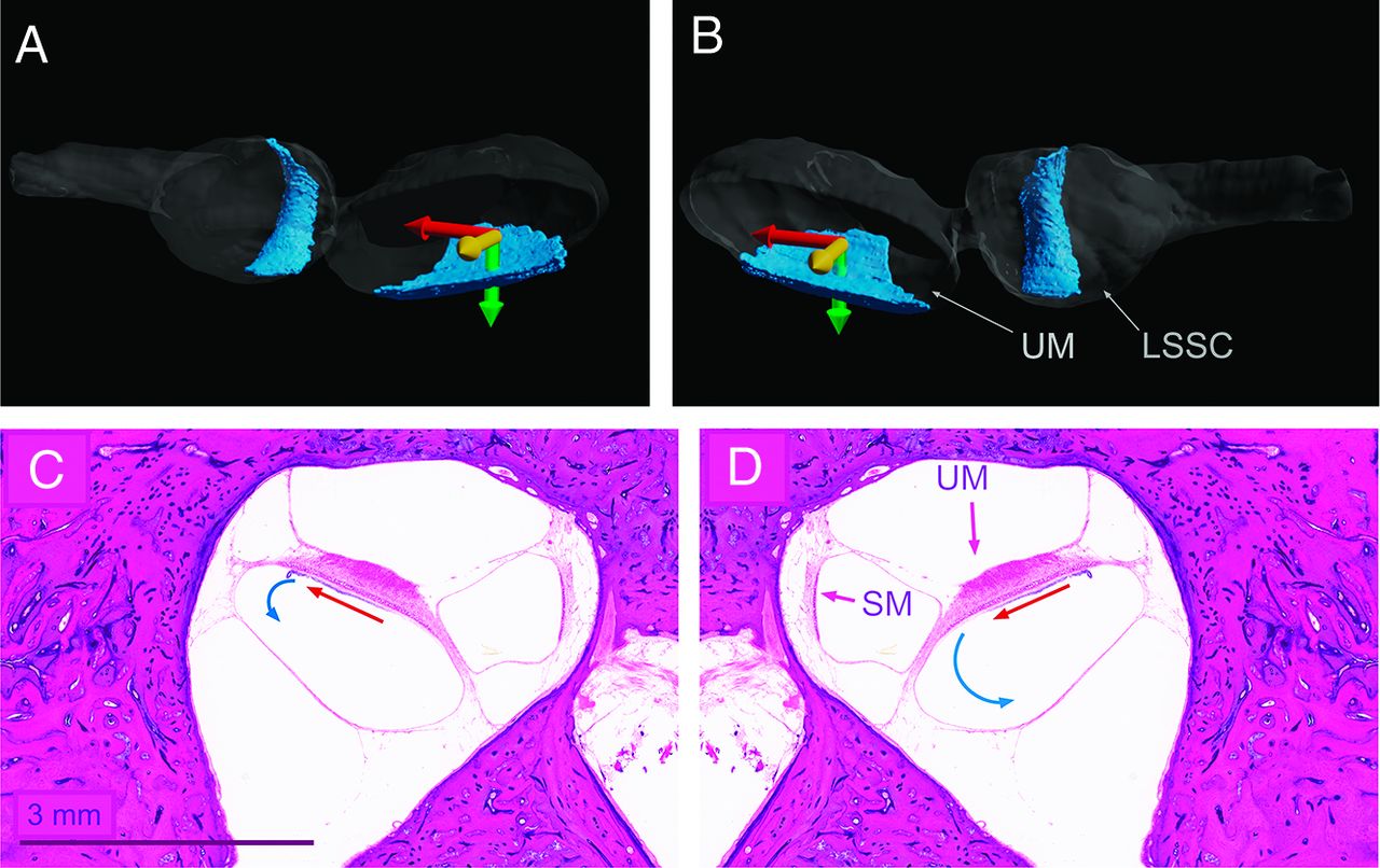
  • FIG 3.
    • Download figure
    • Open in new tab
    • Download powerpoint
    FIG 3.

    Cartoon presentation of the hypothetical mechanism of asymmetric turbulent fluid movements in the left and right ears caused by Lorentz forces. The Lorentz force (red arrow) is evoked, the direction determined by the right-hand rule, by the combination of the magnetic field (yellow arrow) and the utricle current (green arrow) as described in Roberts et al.16 The anterior part of the utricle and lateral semicircular canal viewed from posterior on the left (A) and right (B) side. 3D reconstruction of the endolymphatic space from the histologic specimen. UM indicates utricular macula; LSSC, lateral semicircular canal crista in the ampulla. C and D, Original axial histologic section of the left inner ear across the utricular macula. Image C was mirrored horizontally to create the impression of the right inner ear. UM indicates utricular macula; SM, saccular macula; red arrows, magneto-hydrodynamic Lorentz force; blue arrows, hypothetical turbulent endolymph movements.

Tables

  • Figures
  • Frequency of foci of low signal intensity among vestibular ROIs

    ROIs (No.)
    Lateral Semicircular Canal CristaUtricular Root RegionUtricular “Diamond”Saccular RegionPosterior Vestibular Region
    Right side25/27 (93%)17/27 (63%)23/27 (85%)0/27 (0%)25/27 (93%)
    Left side26/27 (96%)17/27 (63%)13/27a (48%)0/27 (0%)20/27 (74%)
    • ↵a Significant difference between left and right sides (Fisher exact test, 2-tailed P value = .0084).

PreviousNext
Back to top

In this issue

American Journal of Neuroradiology: 43 (10)
American Journal of Neuroradiology
Vol. 43, Issue 10
1 Oct 2022
  • Table of Contents
  • Index by author
  • Complete Issue (PDF)
Advertisement
Print
Download PDF
Email Article

Thank you for your interest in spreading the word on American Journal of Neuroradiology.

NOTE: We only request your email address so that the person you are recommending the page to knows that you wanted them to see it, and that it is not junk mail. We do not capture any email address.

Enter multiple addresses on separate lines or separate them with commas.
Correlation between Histopathology and Signal Loss on Spin-Echo T2-Weighted MR Images of the Inner Ear: Distinguishing Artifacts from Anatomy
(Your Name) has sent you a message from American Journal of Neuroradiology
(Your Name) thought you would like to see the American Journal of Neuroradiology web site.
CAPTCHA
This question is for testing whether or not you are a human visitor and to prevent automated spam submissions.
Cite this article
B.K. Ward, A. Mair, N. Nagururu, M. Bauer, B. Büki
Correlation between Histopathology and Signal Loss on Spin-Echo T2-Weighted MR Images of the Inner Ear: Distinguishing Artifacts from Anatomy
American Journal of Neuroradiology Oct 2022, 43 (10) 1464-1469; DOI: 10.3174/ajnr.A7625

Citation Manager Formats

  • BibTeX
  • Bookends
  • EasyBib
  • EndNote (tagged)
  • EndNote 8 (xml)
  • Medlars
  • Mendeley
  • Papers
  • RefWorks Tagged
  • Ref Manager
  • RIS
  • Zotero
0 Responses
Respond to this article
Share
Bookmark this article
Histopathology vs. MRI Signal Loss in Inner Ear
B.K. Ward, A. Mair, N. Nagururu, M. Bauer, B. Büki
American Journal of Neuroradiology Oct 2022, 43 (10) 1464-1469; DOI: 10.3174/ajnr.A7625
del.icio.us logo Twitter logo Facebook logo Mendeley logo
  • Tweet Widget
  • Facebook Like
  • Google Plus One
Purchase

Jump to section

  • Article
    • Abstract
    • MATERIALS AND METHODS
    • RESULTS
    • DISCUSSION
    • CONCLUSIONS
    • Acknowledgments
    • Footnotes
    • References
  • Figures & Data
  • Info & Metrics
  • Responses
  • References
  • PDF

Related Articles

  • No related articles found.
  • PubMed
  • Google Scholar

Cited By...

  • No citing articles found.
  • Crossref
  • Google Scholar

This article has not yet been cited by articles in journals that are participating in Crossref Cited-by Linking.

More in this TOC Section

  • MRI of the Parasellar Ligaments
  • ASL Sensitivity for Head and Neck Paraganglioma
  • Post SRS Peritumoral Hyperintense Signal of VSs
Show more Head and Neck Imaging

Similar Articles

Advertisement

Indexed Content

  • Current Issue
  • Accepted Manuscripts
  • Article Preview
  • Past Issues
  • Editorials
  • Editor's Choice
  • Fellows' Journal Club
  • Letters to the Editor
  • Video Articles

Cases

  • Case Collection
  • Archive - Case of the Week
  • Archive - Case of the Month
  • Archive - Classic Case

More from AJNR

  • Trainee Corner
  • Imaging Protocols
  • MRI Safety Corner
  • Book Reviews

Multimedia

  • AJNR Podcasts
  • AJNR Scantastics

Resources

  • Turnaround Time
  • Submit a Manuscript
  • Submit a Video Article
  • Submit an eLetter to the Editor/Response
  • Manuscript Submission Guidelines
  • Statistical Tips
  • Fast Publishing of Accepted Manuscripts
  • Graphical Abstract Preparation
  • Imaging Protocol Submission
  • Evidence-Based Medicine Level Guide
  • Publishing Checklists
  • Author Policies
  • Become a Reviewer/Academy of Reviewers
  • News and Updates

About Us

  • About AJNR
  • Editorial Board
  • Editorial Board Alumni
  • Alerts
  • Permissions
  • Not an AJNR Subscriber? Join Now
  • Advertise with Us
  • Librarian Resources
  • Feedback
  • Terms and Conditions
  • AJNR Editorial Board Alumni

American Society of Neuroradiology

  • Not an ASNR Member? Join Now

© 2025 by the American Society of Neuroradiology All rights, including for text and data mining, AI training, and similar technologies, are reserved.
Print ISSN: 0195-6108 Online ISSN: 1936-959X

Powered by HighWire