
Article Figures & Data
Figures
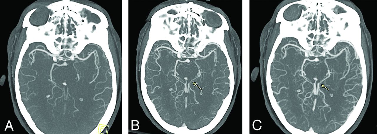
- FIG 1.
Delayed vessel sign. Peak arterial (A), early venous (B), and late venous (C) phase axial MIP mCTA images show occlusion of a proximal M2 segment (arrow in A) of the left MCA. The delayed vessel sign is demonstrated in the early venous and late venous phases (arrows in B and C), assisting in detection of occlusion.
- FIG 2.
Axial arterial phase mCTA MIP image of a left PCA occlusion (A), which is rendered more conspicuous by delayed vessel opacification in the second and third phases (arrows in B and C).
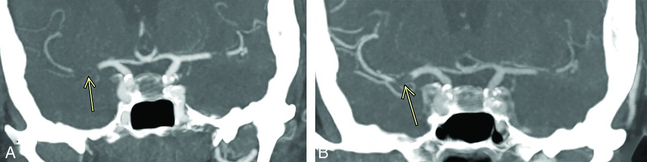
- FIG 3.
Coronal mCTA MIP images from the arterial phase (A) and late venous phase (B) show a right MCA M1 segment occlusion. The M1 segment thrombus length is estimated more accurately during the venous phase (7 mm in B, arrow) than in the arterial phase (11 mm in A, arrow).
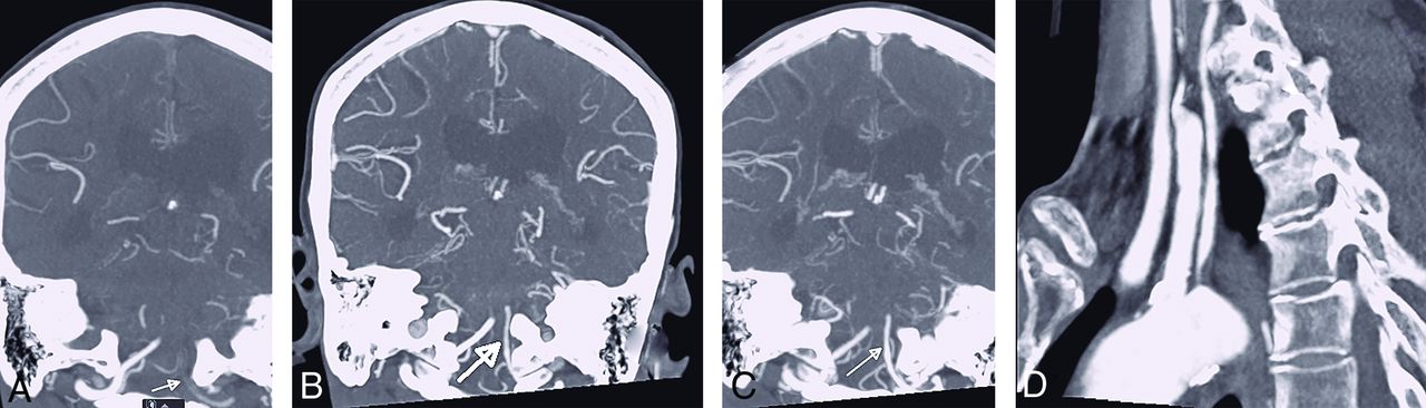
- FIG 4.
Pseudoocclusion of the left vertebral artery. The left vertebral artery appears occluded at its intracranial V4 segment during the arterial phase of mCTA (arrow in A). This is related to severe stenosis at its origin (D). The previously unopacified intracranial V4 segment is shown to enhance in the delayed phases of mCTA (arrows in B and C). If only single-phase CTA had been used (analogous to the arterial phase of the mCTA, A), a V4 segment occlusion would have been diagnosed.
- FIG 5.
Coronal arterial phase mCTA MIP image demonstrates right M1 segment occlusion (A). Axial arterial phase mCTA MIP image (B) shows filling of at most 50% of distal branches. Axial delayed phase mCTA MIP image (C) shows filling of most distal branches. The patient underwent mechanical thrombectomy with restoration of TICI 2c flow. Follow-up diffusion-weighted MR image shows a small infarct core. If only sCTA had been used in this case (analogous to the arterial phase of the mCTA, B), the collateral status would have been classified as “poor to moderate distal collaterals” instead of “good collaterals.”
- FIG 6.
Right M1 segment occlusion. mCTA axial MIP images from the arterial phase (A), early venous phase (B), and late venous phase (C) show progressive collateral filling of distal MCA branches. The patient underwent endovascular recanalization with restoration of TICI 3 flow. Follow-up diffusion-weighted MR image shows a small infarct core.
- FIG 7.
Multiphase CTA arterial phase axial MIP image shows occlusion of the right M1 segment (A, arrow) with no right MCA distal branches. The arterial phase of mCTA is analogous to sCTA and would have been classified as “poor collaterals” in this patient, if considered without the benefit of delayed-phase CTA images. mCTA delayed phase axial MIP in the same patient (B) shows filling of about 50% of right MCA distal branches, resulting in a more accurate assessment of collateral status.
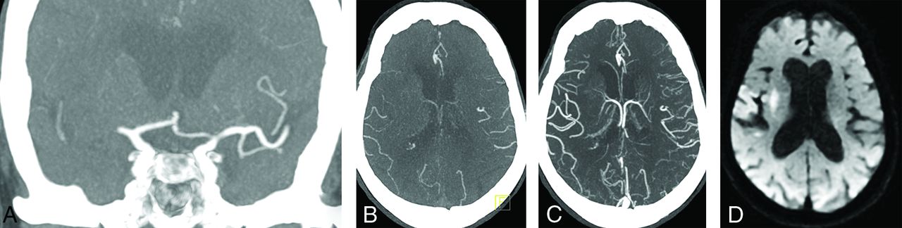
- FIG 8.
Correlation between mCTA and CT perfusion imaging in a patient with left MCA M2 segment occlusion. A region of hypoattenuation in an arterial phase mCTA source image (A, circled area) closely correlates with a region of delayed Tmax in a CT perfusion image (B), suggestive of tissue at risk. Absence of hypoattenuation in a delayed venous phase mCTA source image (C) is matched by preservation of cerebral blood volume in a CT perfusion image (D), suggestive of no infarct core. The patient underwent endovascular intervention with restoration of TICI 2b antegrade flow. Subsequent diffusion-weighted MR imaging showed only small foci of acute ischemia in the frontoparietal region (not shown).


Jump to section
Related Articles
Cited By...
- The Cortical Vein Opacification Score Is Independently Associated with Good and Excellent Functional Outcomes at 90 Days in Patients with Minor Stroke with Anterior Circulation Large-Vessel Occlusion: A Multicenter Study
- Role of Hypoperfusion Intensity Ratio in Vessel Occlusions: A Review on Safety and Clinical Outcomes
- The Cortical Vein Opacification Score (COVES) Is Independently Associated with DSA ASITN Collateral Score
- Thrombus imaging characteristics within acute ischemic stroke: similarities and interdependence
- Stroke imaging prior to thrombectomy in the late window: results from a pooled multicentre analysis