Advertisement

Main menu

  • Home
  • Content
    • Current Issue
    • Accepted Manuscripts
    • Article Preview
    • Past Issue Archive
    • Video Articles
    • AJNR Case Collection
    • Case of the Week Archive
    • Case of the Month Archive
    • Classic Case Archive
  • Special Collections
    • AJNR Awards
    • Low-Field MRI
    • Alzheimer Disease
    • ASNR Foundation Special Collection
    • Photon-Counting CT
    • View All
  • Multimedia
    • AJNR Podcasts
    • AJNR SCANtastic
    • Trainee Corner
    • MRI Safety Corner
    • Imaging Protocols
  • For Authors
    • Submit a Manuscript
    • Submit a Video Article
    • Submit an eLetter to the Editor/Response
    • Manuscript Submission Guidelines
    • Statistical Tips
    • Fast Publishing of Accepted Manuscripts
    • Graphical Abstract Preparation
    • Imaging Protocol Submission
    • Author Policies
  • About Us
    • About AJNR
    • Editorial Board
    • Editorial Board Alumni
  • More
    • Become a Reviewer/Academy of Reviewers
    • Subscribers
    • Permissions
    • Alerts
    • Feedback
    • Advertisers
    • ASNR Home

User menu

  • Alerts
  • Log in

Search

  • Advanced search
American Journal of Neuroradiology

American Journal of Neuroradiology

ASHNR American Society of Functional Neuroradiology ASHNR American Society of Pediatric Neuroradiology ASSR
  • Alerts
  • Log in

Advanced Search

  • Home
  • Content
    • Current Issue
    • Accepted Manuscripts
    • Article Preview
    • Past Issue Archive
    • Video Articles
    • AJNR Case Collection
    • Case of the Week Archive
    • Case of the Month Archive
    • Classic Case Archive
  • Special Collections
    • AJNR Awards
    • Low-Field MRI
    • Alzheimer Disease
    • ASNR Foundation Special Collection
    • Photon-Counting CT
    • View All
  • Multimedia
    • AJNR Podcasts
    • AJNR SCANtastic
    • Trainee Corner
    • MRI Safety Corner
    • Imaging Protocols
  • For Authors
    • Submit a Manuscript
    • Submit a Video Article
    • Submit an eLetter to the Editor/Response
    • Manuscript Submission Guidelines
    • Statistical Tips
    • Fast Publishing of Accepted Manuscripts
    • Graphical Abstract Preparation
    • Imaging Protocol Submission
    • Author Policies
  • About Us
    • About AJNR
    • Editorial Board
    • Editorial Board Alumni
  • More
    • Become a Reviewer/Academy of Reviewers
    • Subscribers
    • Permissions
    • Alerts
    • Feedback
    • Advertisers
    • ASNR Home
  • Follow AJNR on Twitter
  • Visit AJNR on Facebook
  • Follow AJNR on Instagram
  • Join AJNR on LinkedIn
  • RSS Feeds

AJNR Awards, New Junior Editors, and more. Read the latest AJNR updates

Research ArticleAdult Brain
Open Access

Identification of BRAF p. V600E-Mutant and Wild-Type by MR Imaging in Pleomorphic Xanthoastrocytoma and Anaplastic Pleomorphic Xanthoastrocytoma

W. Huang, J. Cai, N. Lin, Y. Xu, H. Wang, Z. Wu and D. Kang
American Journal of Neuroradiology December 2021, 42 (12) 2152-2159; DOI: https://doi.org/10.3174/ajnr.A7324
W. Huang
aFrom the Department of Neurosurgery (W.H., J.C., Y.X., H.W., Z.W., D.K.), The First Affiliated Hospital of Fujian Medical University, Fuzhou, Fujian, China
bThe First Clinical Medical College of Fujian Medical University (W.H., J.C., Y.X., H.W., Z.W., D.K.), Fuzhou, Fujian, China
  • Find this author on Google Scholar
  • Find this author on PubMed
  • Search for this author on this site
  • ORCID record for W. Huang
J. Cai
aFrom the Department of Neurosurgery (W.H., J.C., Y.X., H.W., Z.W., D.K.), The First Affiliated Hospital of Fujian Medical University, Fuzhou, Fujian, China
bThe First Clinical Medical College of Fujian Medical University (W.H., J.C., Y.X., H.W., Z.W., D.K.), Fuzhou, Fujian, China
  • Find this author on Google Scholar
  • Find this author on PubMed
  • Search for this author on this site
  • ORCID record for J. Cai
N. Lin
cThe School of Medical Technology and Engineering (N.L.), Fujian Medical University, Fuzhou, Fujian, China
  • Find this author on Google Scholar
  • Find this author on PubMed
  • Search for this author on this site
  • ORCID record for N. Lin
Y. Xu
aFrom the Department of Neurosurgery (W.H., J.C., Y.X., H.W., Z.W., D.K.), The First Affiliated Hospital of Fujian Medical University, Fuzhou, Fujian, China
bThe First Clinical Medical College of Fujian Medical University (W.H., J.C., Y.X., H.W., Z.W., D.K.), Fuzhou, Fujian, China
  • Find this author on Google Scholar
  • Find this author on PubMed
  • Search for this author on this site
  • ORCID record for Y. Xu
H. Wang
aFrom the Department of Neurosurgery (W.H., J.C., Y.X., H.W., Z.W., D.K.), The First Affiliated Hospital of Fujian Medical University, Fuzhou, Fujian, China
bThe First Clinical Medical College of Fujian Medical University (W.H., J.C., Y.X., H.W., Z.W., D.K.), Fuzhou, Fujian, China
  • Find this author on Google Scholar
  • Find this author on PubMed
  • Search for this author on this site
  • ORCID record for H. Wang
Z. Wu
aFrom the Department of Neurosurgery (W.H., J.C., Y.X., H.W., Z.W., D.K.), The First Affiliated Hospital of Fujian Medical University, Fuzhou, Fujian, China
bThe First Clinical Medical College of Fujian Medical University (W.H., J.C., Y.X., H.W., Z.W., D.K.), Fuzhou, Fujian, China
  • Find this author on Google Scholar
  • Find this author on PubMed
  • Search for this author on this site
  • ORCID record for Z. Wu
D. Kang
aFrom the Department of Neurosurgery (W.H., J.C., Y.X., H.W., Z.W., D.K.), The First Affiliated Hospital of Fujian Medical University, Fuzhou, Fujian, China
bThe First Clinical Medical College of Fujian Medical University (W.H., J.C., Y.X., H.W., Z.W., D.K.), Fuzhou, Fujian, China
  • Find this author on Google Scholar
  • Find this author on PubMed
  • Search for this author on this site
  • ORCID record for D. Kang
  • Article
  • Figures & Data
  • Info & Metrics
  • Responses
  • References
  • PDF
Loading

Article Figures & Data

Figures

  • Tables
  • FIG 1.
    • Download figure
    • Open in new tab
    • Download powerpoint
    FIG 1.

    Boxplots representing tumor size and the presence and degree of peritumoral edema of PXA and APXA. A significant difference (P < . 05) between PXA and APXA is demonstrated with both tumor size and the presence and degree of peritumoral edema.
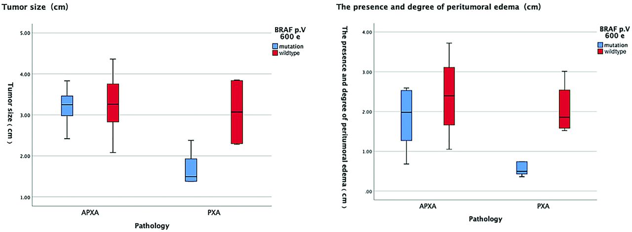
  • FIG 2.
    • Download figure
    • Open in new tab
    • Download powerpoint
    FIG 2.

    A significant difference (P < .05) between the BRAF p. V600E-mutant type PXA group and the BRAF p. V600E wild-type PXA group is demonstrated with both tumor size and the presence and degree of peritumoral edema. However, there was no significant difference in both tumor size and the maximum diameter of peritumoral edema between the BRAF p. V600E-mutant type APXA group and BRAF p. V600E wild-type APXA group.
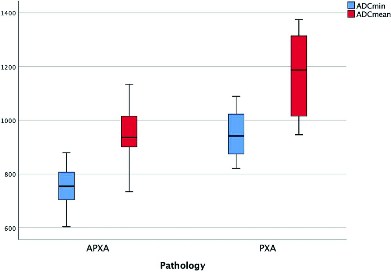
  • FIG 3.
    • Download figure
    • Open in new tab
    • Download powerpoint
    FIG 3.

    Boxplots representing ADCmin and ADCmean of PXA and APXA. A significant difference (P < .05) between PXA and APXA is demonstrated with both ADCmin and ADCmean.

  • FIG 4.
    • Download figure
    • Open in new tab
    • Download powerpoint
    FIG 4.

    A significant difference (P < .05) between the BRAF p. V600E-mutant type PXA group and the BRAF p. V600E wild-type PXA group is demonstrated with both ADCmin and ADCmean. However, there was no significant difference in both ADCmin and ADCmean between the BRAF p. V600E-mutant type APXA group and BRAF p. V600E wild-type APXA group.

  • FIG 5.
    • Download figure
    • Open in new tab
    • Download powerpoint
    FIG 5.

    Conventional and advanced MR images of BRAF p. V600E-mutant and wild-type PXA. Upper row: A 27-year-old female patient with a BRAF p. V600E-mutant pleomorphic astrocytoma. T2WI (A) and contrast-enhanced T1WI (B) show a homogeneous contrast-enhancing solid tumor with mild perilesional edema located in the temporal lobe. C, A correlative ADC map shows the tumor with an elevated ADC value (ADCmin = 1089). Second row: A 53-year-old male patient with a BRAF p. V600E wild-type pleomorphic astrocytoma. T2WI (A) and contrast-enhanced T1WI (B) show a heterogeneous contrast-enhancing tumor with cystic degeneration and marked perilesional edema located in the insula. C, A correlative ADC map shows the lesion with a decreased ADC value (ADCmin = 875). Third row: A 46-year-old female patient with a BRAF p. V600E wild-type anaplastic pleomorphic astrocytoma. T2WI (A) and contrast-enhanced T1WI (B) show a heterogeneous contrast-enhancing tumor with cystic degeneration and marked perilesional edema located in the insula. C, A correlative ADC map shows the lesion with diffusion restriction (ADCmin = 704). CE indicates contrast-enhanced.

Tables

  • Figures
    • View popup
    Table 1:

    Demographic data and conventional MR imaging characteristics of PXA and APXA

    PXAAPXAP Value
    Clinical data
     Male sex (No.) (%)3 (30%)6 (46.1%).363
     Mean age (yr)34 (SD, 10)41 (SD, 10).232
    Presenting symptoms (No.).899
     Seizures69
     Headache or increased ICP22
     Neurologic deficit22
    BRAF p. V600E-mutant6 (60%)5 (38.5%).273
    Location (No.).526
     Frontal lobe03
     Temporal lobe55
     Occipital lobe11
     Parietal lobe23
     Insula21
     Superficial location (No.) (%)8 (80%)11 (84.6%).596
    Conventional MR imaging
     Mean size (cm)2.2 (SD, 0.9)3.2 (SD, 0.6)<.01
     Presence of cystic degeneration (No.) (%)6 (60%)9 (69.2%).490
     Peritumoral edema (mean) (cm)1.2 (SD, 0.8)2.1 (SD, 0.9).021
     Heterogeneous enhancement (No.) (%)4 (40%)11 (84.6%)<.001
     Leptomeningeal contact (No.) (%)8 (80%)11 (84.6%).596
    • Note:—ICP indicates intracranial pressure.

    • View popup
    Table 2:

    Demographic data and conventional MR imaging characteristics of BRAF p. V600E-mutant and BRAF p. V600E wild-type PXA

    BRAF p. V600E- Mutant PXABRAF p. V600E Wild-Type PXAP Value
    Clinical data
     Male sex (No.) (%)2 (33.3%)1 (25%).667
     Mean age (yr)32 (SD, 8.6)37(SD, 13).762
    Presenting symptoms (No.).870
     Seizures42
     Headache or increased ICP11
     Neurologic deficit11
    Location (No.).405
     Frontal lobe00
     Temporal lobe32
     Occipital lobe01
     Parietal lobe20
     Insula11
     Superficial location (No.) (%)4 (66.6%)4 (100%).333
    Conventional MR imaging
     Mean size (cm)1.7 (SD, 0.4)3.1 (SD, 0.9).038
     Presence of cystic degeneration (No.) (%)2 (33.3%)4 (100%).071
     Peritumoral edema (mean) (cm)0.6 ± 0.22.1 ± 0.7<.01
     Heterogeneous enhancement (No.) (%)0 (0%)4 (100%)<.001
     Leptomeningeal contact (No.) (%)6 (100%)2 (50%).03
    • Note:— ICP indicates intracranial pressure.

    • View popup
    Table 3:

    Demographic data and conventional MR imaging characteristics of BRAF p. V600E-mutant and BRAF p. V600E wild-type APXA

    BRAF p. V600E-Mutant APXABRAF p. V600E Wild-Type APXAP Value
    Clinical data
     Male sex (No.) (%)3 (60%)3 (37.5%).413
     Mean age (yr)33 (SD, 13)43 (SD, 6).284
    Presenting symptoms (No.).850
     Seizures36
     Headache or increased ICP11
     Neurologic deficit11
    Location (No.).276
     Frontal lobe03
     Temporal lobe23
     Occipital lobe10
     Parietal lobe21
     Insula01
     Superficial location (No.) (%)4 (80.0%)7 (87.5%).641
    Conventional MR imaging
     Mean size (cm)3.2 (SD, 0.5)3.3 (SD, 0.7).943
     Presence of cystic degeneration (No.) (%)3 (60%)6 (75%).343
     Peritumoral edema (mean) (cm)1.8 (SD, 0.4)2.4 (SD, 0.9).354
     Heterogeneous enhancement (No.) (%)5 (100%)6 (75%).359
     Leptomeningeal contact (No.) (%)4 (80%)7 (87.5%).641
    • Note:— ICP indicates intracranial pressure.

    • View popup
    Table 4:

    Advanced MR imaging characteristics of the PXA and APXA groups

    PXAAPXAP Value
    ADCmin (×10−6 mm2/s)951 (SD, 88)755 (SD, 77)<.001
    ADCmean (×10−6 mm2/s)1173 (SD, 159)956 (SD, 106)<.001
    • View popup
    Table 5:

    Advanced MR imaging characteristics of the BRAF p. V600E-mutant PXA and BRAF p. V600E wild-type PXA groups

    BRAF p. V600E-Mutant PXABRAF p. V600E Wild-Type PXAP Value
    ADCmin (×10−6 mm2/s)988 (SD, 73)896 (SD, 86).047
    ADCmean (×10−6 mm2/s)1248 (SD, 116)1060 (SD, 159).049
    • View popup
    Table 6:

    Advanced MR imaging characteristics of the BRAF p. V600E-mutant APXA and BRAF p. V600E wild-type APXA groups

    BRAF p. V600E-Mutant APXABRAF p. V600E Wild-Type APXAP Value
    ADCmin (×10−6 mm2/s)762 (SD, 92)750 (SD, 73)1
    ADCmean (×10−6 mm2/s)925 (SD, 131)974 (SD, 92).724
PreviousNext
Back to top

In this issue

American Journal of Neuroradiology: 42 (12)
American Journal of Neuroradiology
Vol. 42, Issue 12
1 Dec 2021
  • Table of Contents
  • Index by author
  • Complete Issue (PDF)
Advertisement
Print
Download PDF
Email Article

Thank you for your interest in spreading the word on American Journal of Neuroradiology.

NOTE: We only request your email address so that the person you are recommending the page to knows that you wanted them to see it, and that it is not junk mail. We do not capture any email address.

Enter multiple addresses on separate lines or separate them with commas.
Identification of BRAF p. V600E-Mutant and Wild-Type by MR Imaging in Pleomorphic Xanthoastrocytoma and Anaplastic Pleomorphic Xanthoastrocytoma
(Your Name) has sent you a message from American Journal of Neuroradiology
(Your Name) thought you would like to see the American Journal of Neuroradiology web site.
CAPTCHA
This question is for testing whether or not you are a human visitor and to prevent automated spam submissions.
Cite this article
W. Huang, J. Cai, N. Lin, Y. Xu, H. Wang, Z. Wu, D. Kang
Identification of BRAF p. V600E-Mutant and Wild-Type by MR Imaging in Pleomorphic Xanthoastrocytoma and Anaplastic Pleomorphic Xanthoastrocytoma
American Journal of Neuroradiology Dec 2021, 42 (12) 2152-2159; DOI: 10.3174/ajnr.A7324

Citation Manager Formats

  • BibTeX
  • Bookends
  • EasyBib
  • EndNote (tagged)
  • EndNote 8 (xml)
  • Medlars
  • Mendeley
  • Papers
  • RefWorks Tagged
  • Ref Manager
  • RIS
  • Zotero
0 Responses
Respond to this article
Share
Bookmark this article
Identification of BRAF p. V600E-Mutant and Wild-Type by MR Imaging in Pleomorphic Xanthoastrocytoma and Anaplastic Pleomorphic Xanthoastrocytoma
W. Huang, J. Cai, N. Lin, Y. Xu, H. Wang, Z. Wu, D. Kang
American Journal of Neuroradiology Dec 2021, 42 (12) 2152-2159; DOI: 10.3174/ajnr.A7324
del.icio.us logo Twitter logo Facebook logo Mendeley logo
  • Tweet Widget
  • Facebook Like
  • Google Plus One
Purchase

Jump to section

  • Article
    • Abstract
    • ABBREVIATIONS:
    • MATERIALS AND METHODS
    • RESULTS
    • DISCUSSION
    • CONCLUSIONS
    • Footnotes
    • References
  • Figures & Data
  • Info & Metrics
  • Responses
  • References
  • PDF

Related Articles

  • No related articles found.
  • PubMed
  • Google Scholar

Cited By...

  • No citing articles found.
  • Crossref
  • Google Scholar

This article has not yet been cited by articles in journals that are participating in Crossref Cited-by Linking.

More in this TOC Section

  • Diagnostic Neuroradiology of Monoclonal Antibodies
  • Cerebral ADC Changes in Fabry Disease
  • ML for Glioma Molecular Subtype Prediction
Show more Adult Brain

Similar Articles

Advertisement

Indexed Content

  • Current Issue
  • Accepted Manuscripts
  • Article Preview
  • Past Issues
  • Editorials
  • Editor's Choice
  • Fellows' Journal Club
  • Letters to the Editor
  • Video Articles

Cases

  • Case Collection
  • Archive - Case of the Week
  • Archive - Case of the Month
  • Archive - Classic Case

More from AJNR

  • Trainee Corner
  • Imaging Protocols
  • MRI Safety Corner
  • Book Reviews

Multimedia

  • AJNR Podcasts
  • AJNR Scantastics

Resources

  • Turnaround Time
  • Submit a Manuscript
  • Submit a Video Article
  • Submit an eLetter to the Editor/Response
  • Manuscript Submission Guidelines
  • Statistical Tips
  • Fast Publishing of Accepted Manuscripts
  • Graphical Abstract Preparation
  • Imaging Protocol Submission
  • Evidence-Based Medicine Level Guide
  • Publishing Checklists
  • Author Policies
  • Become a Reviewer/Academy of Reviewers
  • News and Updates

About Us

  • About AJNR
  • Editorial Board
  • Editorial Board Alumni
  • Alerts
  • Permissions
  • Not an AJNR Subscriber? Join Now
  • Advertise with Us
  • Librarian Resources
  • Feedback
  • Terms and Conditions
  • AJNR Editorial Board Alumni

American Society of Neuroradiology

  • Not an ASNR Member? Join Now

© 2025 by the American Society of Neuroradiology All rights, including for text and data mining, AI training, and similar technologies, are reserved.
Print ISSN: 0195-6108 Online ISSN: 1936-959X

Powered by HighWire