
Article Figures & Data
Figures
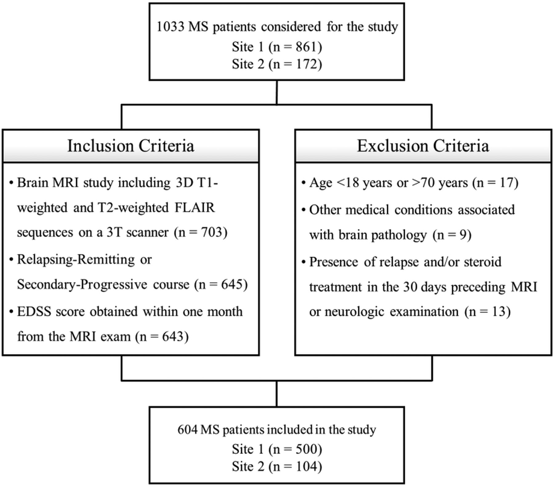
- FIG 1.
Flow diagram showing inclusion and exclusion criteria.
- FIG 2.
Workflow summarizing the main MR imaging data-processing and data-mining steps. Image illustrates the data set composition as well as the major steps performed for feature extraction, feature selection, and regression modeling. LL indicates lesion load; NAWM, normal-appearing white matter; ChaCo, change in connectivity; T1w, T1-weighted imaging.


Tables
- Table 1:
Clinicodemographic characteristics of the studied population, along with MR imaging–derived global brain volumesa
Site 1 (n = 500) Site 2 (n = 104) P Value
(Site 1 vs Site 2)Age (mean) (yr) 37.5 (SD, 10.9) 38.3 (SD, 9.8) .49 Female sex (No.) (%) 349 (69.8) 80 (76.9) .16 Secondary-progressive course (No.) (%) 72 (14.4) 20 (19.2) .23 DD (mean) (yr) 9.3 (SD, 8.1) 9.2 (SD, 8.5) .83 EDSS (median) (IQR) 2.5 (2.0–4.0) 2.0 (1.5–4.0) .03 TLV (mean) (mL) 10.6 (SD, 13.4) 7.2 (SD, 8.6) .05 WBV (mean) (mL) 1026.1 (SD, 116.3) 1042.6 (SD, 117.4) .48 Note:—TLV indicates total lesion volume.
↵a Between-group differences regarding MR imaging measures are adjusted for age, sex, and estimated total intracranial volume.
Anatomic Label Feature Class Class Characteristics Feature Feature Characteristics Right frontal superior orbital cortex First order Describes the distribution of voxel intensities Median The median gray level intensity Left amygdala Gray level co-occurrence matrix Quantifies how often pairs of pixels with specific values occur in a specified spatial range Correlation Measures the linear dependency of gray level values to their respective voxels in the matrix Left caudate nucleus Gray level co-occurrence matrix Quantifies how often pairs of pixels with specific values occur in a specified spatial range Informational measure of correlation 1 Quantifies the complexity of the texture Right thalamus First order Describes the distribution of voxel intensities Energy Measures the magnitude of voxel values Left cerebellar lobule VIII Gray level dependence matrix Quantifies gray level dependencies (ie, the number of connected voxels within a set distance that are dependent on the center voxel) Small dependence low gray level emphasis Measures the joint distribution of small dependence with higher gray-level values Cerebellar vermis (lobules IV–V) Gray level size-zone matrix Quantifies gray level zones (ie, the number of connected voxels sharing the same intensity value) Size-zone non-uniformity Measures the variability of size-zone volumes Left cerebellar crus First order Describes the distribution of voxel intensities Median Median gray level intensity ↵a Characteristics of each selected feature and relative class according to PyRadiomics official documentation (https://pyradiomics.readthedocs.io/en/latest/features.html) are presented, along with the anatomic location (according to Tzourio-Mazoyer et al20) of the corresponding ROI.
Cohort Ridge Regression Gaussian Process Support-Vector Machine Random Forest P Value r R2 MAE r R2 MAE r R2 MAE r R2 MAE Training 0.797 0.636 0.651 0.795 0.632 0.814 0.797 0.635 0.710 0.790 0.624 0.656 – Internal test 0.741 0.549 0.725 0.733 0.537 0.874 0.734 0.538 0.754 0.734 0.539 0.740 .001a External test 0.755 0.570 1.155 0.799 0.638 1.247 0.794 0.631 1.112 0.775 0.600 1.162 .009b


