Advertisement

Main menu

  • Home
  • Content
    • Current Issue
    • Accepted Manuscripts
    • Article Preview
    • Past Issue Archive
    • Video Articles
    • AJNR Case Collection
    • Case of the Week Archive
    • Case of the Month Archive
    • Classic Case Archive
  • Special Collections
    • AJNR Awards
    • Low-Field MRI
    • Alzheimer Disease
    • ASNR Foundation Special Collection
    • Photon-Counting CT
    • View All
  • Multimedia
    • AJNR Podcasts
    • AJNR SCANtastic
    • Trainee Corner
    • MRI Safety Corner
    • Imaging Protocols
  • For Authors
    • Submit a Manuscript
    • Submit a Video Article
    • Submit an eLetter to the Editor/Response
    • Manuscript Submission Guidelines
    • Statistical Tips
    • Fast Publishing of Accepted Manuscripts
    • Graphical Abstract Preparation
    • Imaging Protocol Submission
    • Author Policies
  • About Us
    • About AJNR
    • Editorial Board
    • Editorial Board Alumni
  • More
    • Become a Reviewer/Academy of Reviewers
    • Subscribers
    • Permissions
    • Alerts
    • Feedback
    • Advertisers
    • ASNR Home

User menu

  • Alerts
  • Log in

Search

  • Advanced search
American Journal of Neuroradiology

American Journal of Neuroradiology

ASHNR American Society of Functional Neuroradiology ASHNR American Society of Pediatric Neuroradiology ASSR
  • Alerts
  • Log in

Advanced Search

  • Home
  • Content
    • Current Issue
    • Accepted Manuscripts
    • Article Preview
    • Past Issue Archive
    • Video Articles
    • AJNR Case Collection
    • Case of the Week Archive
    • Case of the Month Archive
    • Classic Case Archive
  • Special Collections
    • AJNR Awards
    • Low-Field MRI
    • Alzheimer Disease
    • ASNR Foundation Special Collection
    • Photon-Counting CT
    • View All
  • Multimedia
    • AJNR Podcasts
    • AJNR SCANtastic
    • Trainee Corner
    • MRI Safety Corner
    • Imaging Protocols
  • For Authors
    • Submit a Manuscript
    • Submit a Video Article
    • Submit an eLetter to the Editor/Response
    • Manuscript Submission Guidelines
    • Statistical Tips
    • Fast Publishing of Accepted Manuscripts
    • Graphical Abstract Preparation
    • Imaging Protocol Submission
    • Author Policies
  • About Us
    • About AJNR
    • Editorial Board
    • Editorial Board Alumni
  • More
    • Become a Reviewer/Academy of Reviewers
    • Subscribers
    • Permissions
    • Alerts
    • Feedback
    • Advertisers
    • ASNR Home
  • Follow AJNR on Twitter
  • Visit AJNR on Facebook
  • Follow AJNR on Instagram
  • Join AJNR on LinkedIn
  • RSS Feeds

AJNR Awards, New Junior Editors, and more. Read the latest AJNR updates

Research ArticleSpine Imaging and Spine Image-Guided Interventions

Spine MRI in Spontaneous Intracranial Hypotension for CSF Leak Detection: Nonsuperiority of Intrathecal Gadolinium to Heavily T2-Weighted Fat-Saturated Sequences

T. Dobrocky, A. Winklehner, P.S. Breiding, L. Grunder, G. Peschi, L. Häni, P.J. Mosimann, M. Branca, J. Kaesmacher, P. Mordasini, A. Raabe, C.T. Ulrich, J. Beck, J. Gralla and E.I. Piechowiak
American Journal of Neuroradiology July 2020, 41 (7) 1309-1315; DOI: https://doi.org/10.3174/ajnr.A6592
T. Dobrocky
aFrom the University Institute of Diagnostic and Interventional Neuroradiology (T.D., A.W., P.S.B., L.G., G.P., P.J.M., J.K., P.M., J.G., E.I.P.)
  • Find this author on Google Scholar
  • Find this author on PubMed
  • Search for this author on this site
  • ORCID record for T. Dobrocky
A. Winklehner
aFrom the University Institute of Diagnostic and Interventional Neuroradiology (T.D., A.W., P.S.B., L.G., G.P., P.J.M., J.K., P.M., J.G., E.I.P.)
  • Find this author on Google Scholar
  • Find this author on PubMed
  • Search for this author on this site
  • ORCID record for A. Winklehner
P.S. Breiding
aFrom the University Institute of Diagnostic and Interventional Neuroradiology (T.D., A.W., P.S.B., L.G., G.P., P.J.M., J.K., P.M., J.G., E.I.P.)
  • Find this author on Google Scholar
  • Find this author on PubMed
  • Search for this author on this site
  • ORCID record for P.S. Breiding
L. Grunder
aFrom the University Institute of Diagnostic and Interventional Neuroradiology (T.D., A.W., P.S.B., L.G., G.P., P.J.M., J.K., P.M., J.G., E.I.P.)
  • Find this author on Google Scholar
  • Find this author on PubMed
  • Search for this author on this site
  • ORCID record for L. Grunder
G. Peschi
aFrom the University Institute of Diagnostic and Interventional Neuroradiology (T.D., A.W., P.S.B., L.G., G.P., P.J.M., J.K., P.M., J.G., E.I.P.)
bDepartment of Interventional, Pediatric, and Diagnostic Radiology (G.P., J.K.)
  • Find this author on Google Scholar
  • Find this author on PubMed
  • Search for this author on this site
  • ORCID record for G. Peschi
L. Häni
cNeurosurgery (L.H., A.R., C.T.U., J.B.), University of Bern, Inselspital, Bern, Switzerland
  • Find this author on Google Scholar
  • Find this author on PubMed
  • Search for this author on this site
  • ORCID record for L. Häni
P.J. Mosimann
aFrom the University Institute of Diagnostic and Interventional Neuroradiology (T.D., A.W., P.S.B., L.G., G.P., P.J.M., J.K., P.M., J.G., E.I.P.)
  • Find this author on Google Scholar
  • Find this author on PubMed
  • Search for this author on this site
  • ORCID record for P.J. Mosimann
M. Branca
dClinical Trials Unit (M.B.), University of Bern, Bern, Switzerland
  • Find this author on Google Scholar
  • Find this author on PubMed
  • Search for this author on this site
  • ORCID record for M. Branca
J. Kaesmacher
aFrom the University Institute of Diagnostic and Interventional Neuroradiology (T.D., A.W., P.S.B., L.G., G.P., P.J.M., J.K., P.M., J.G., E.I.P.)
bDepartment of Interventional, Pediatric, and Diagnostic Radiology (G.P., J.K.)
  • Find this author on Google Scholar
  • Find this author on PubMed
  • Search for this author on this site
  • ORCID record for J. Kaesmacher
P. Mordasini
aFrom the University Institute of Diagnostic and Interventional Neuroradiology (T.D., A.W., P.S.B., L.G., G.P., P.J.M., J.K., P.M., J.G., E.I.P.)
  • Find this author on Google Scholar
  • Find this author on PubMed
  • Search for this author on this site
  • ORCID record for P. Mordasini
A. Raabe
cNeurosurgery (L.H., A.R., C.T.U., J.B.), University of Bern, Inselspital, Bern, Switzerland
  • Find this author on Google Scholar
  • Find this author on PubMed
  • Search for this author on this site
  • ORCID record for A. Raabe
C.T. Ulrich
cNeurosurgery (L.H., A.R., C.T.U., J.B.), University of Bern, Inselspital, Bern, Switzerland
  • Find this author on Google Scholar
  • Find this author on PubMed
  • Search for this author on this site
  • ORCID record for C.T. Ulrich
J. Beck
cNeurosurgery (L.H., A.R., C.T.U., J.B.), University of Bern, Inselspital, Bern, Switzerland
eDepartment of Neurosurgery (J.B.), Medical Center, University of Freiburg, Freiburg, Germany.
  • Find this author on Google Scholar
  • Find this author on PubMed
  • Search for this author on this site
  • ORCID record for J. Beck
J. Gralla
aFrom the University Institute of Diagnostic and Interventional Neuroradiology (T.D., A.W., P.S.B., L.G., G.P., P.J.M., J.K., P.M., J.G., E.I.P.)
  • Find this author on Google Scholar
  • Find this author on PubMed
  • Search for this author on this site
  • ORCID record for J. Gralla
E.I. Piechowiak
aFrom the University Institute of Diagnostic and Interventional Neuroradiology (T.D., A.W., P.S.B., L.G., G.P., P.J.M., J.K., P.M., J.G., E.I.P.)
  • Find this author on Google Scholar
  • Find this author on PubMed
  • Search for this author on this site
  • ORCID record for E.I. Piechowiak
  • Article
  • Figures & Data
  • Info & Metrics
  • Responses
  • References
  • PDF
Loading

Article Figures & Data

Figures

  • Tables
  • FIG 1.
    • Download figure
    • Open in new tab
    • Download powerpoint
    FIG 1.

    Transversal, isotropic, unenhanced, 3D T2WI FS (A) versus a transversal (3 mm), T1-weighted sequence with fat saturation after intrathecal gadolinium (B). The extrathecal fluid and contrast media are visible posterior to the dura mater (arrows).

  • FIG 2.
    • Download figure
    • Open in new tab
    • Download powerpoint
    FIG 2.

    Multiplanar reconstruction of 3D T2WI FS sequence showing a large nerve root diverticulum, which demonstrated a leakage on conventional dynamic myelography (not shown).

  • FIG 3.
    • Download figure
    • Open in new tab
    • Download powerpoint
    FIG 3.

    A 57-year-old woman with orthostatic headache and tinnitus. A, The sagittal, T2-weighted sequence demonstrates extensive extrathecal CSF and multiple suspicious disc protrusions (black arrowheads). B–D, Conventional dynamic myelography demonstrates a CSF leak at the T10/11 level with progressive contrast media distribution (arrowheads). Even in retrospect, the MR imaging did not demonstrate any suspicious lesion at the corresponding level (A, white arrowhead). The leak was confirmed intraoperatively and surgically closed. The follow-up spine MR imaging did not show any residual epidural CSF collection (not shown).
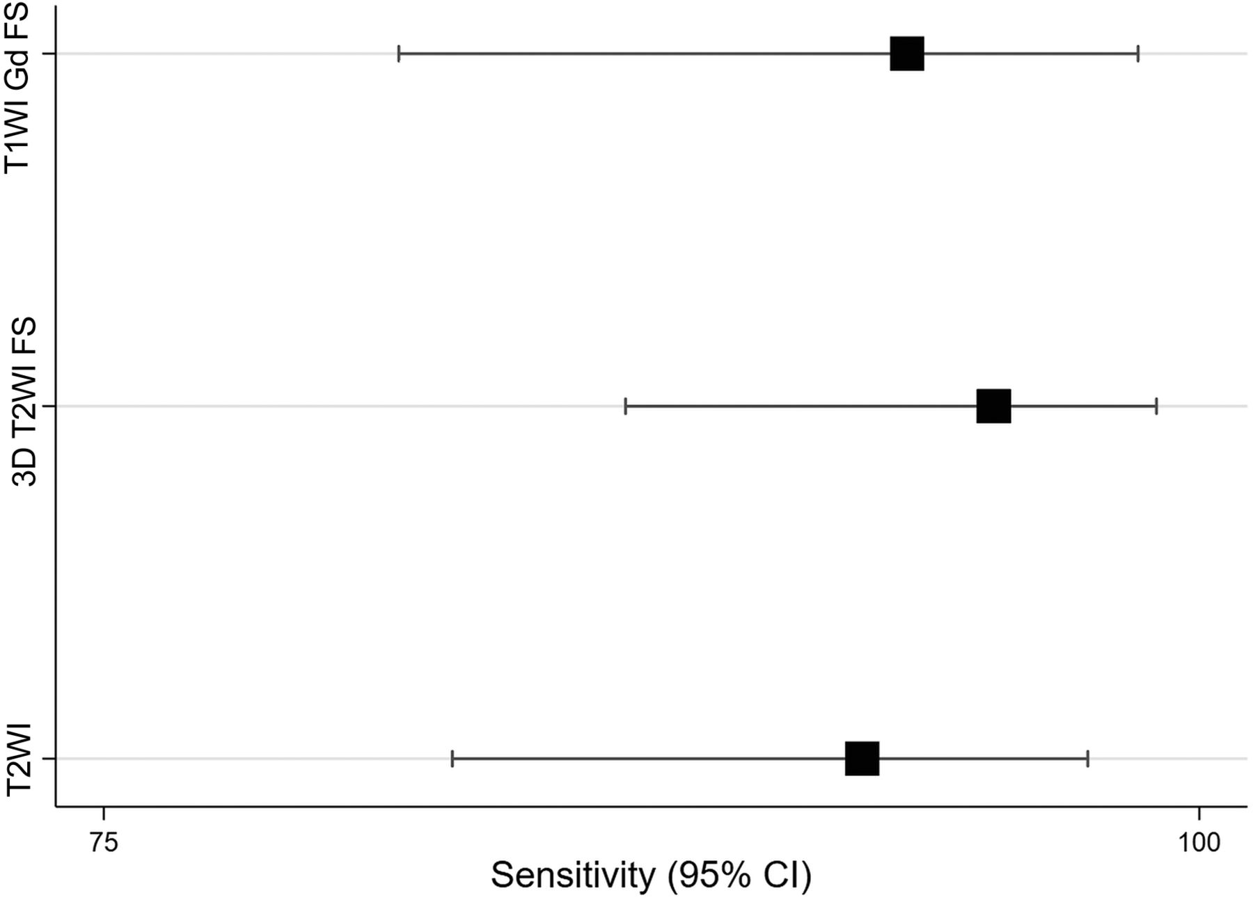
  • FIG 4.
    • Download figure
    • Open in new tab
    • Download powerpoint
    FIG 4.

    Sensitivity and specificity for the 3 different sequences with corresponding confidence intervals. T2WI indicates T2-weighted; 3D T2WI FS, 3D, isotropic, T2-weighted sequence with fat saturation; and T1WI Gd FS, isotropic, T1-weighted sequence with fat saturation after intrathecal application of gadolinium.

  • FIG 5.
    • Download figure
    • Open in new tab
    • Download powerpoint
    FIG 5.

    Scatterplots showing the accuracy of each sequence compared with the true location. T2-weighted (A); 3D T2WI FS, 3D T2-weighted sequence with fat saturation (B); and T1WI GD (C). On the x-axis is the location as reported by the readers. On the y-axis is the true location as found intraoperatively, −57 of 70 (81%); or as determined with multimodal imaging in conservatively managed patients −13 of 70 (19%). Patients in whom the leak has been correctly localized based on the corresponding MR image lie on the reference line.
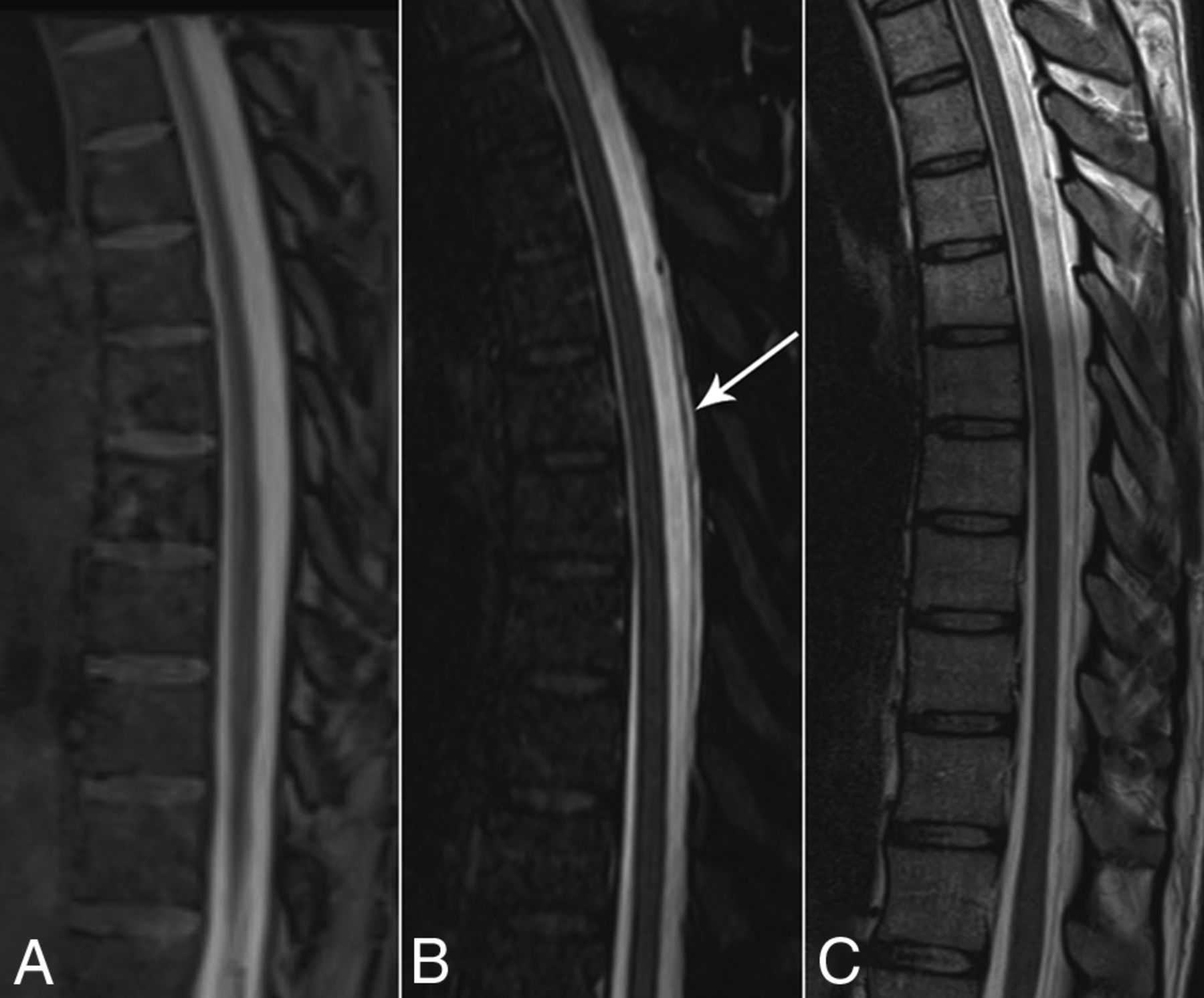
  • FIG 6.
    • Download figure
    • Open in new tab
    • Download powerpoint
    FIG 6.

    A 38-year-old woman with orthostatic headache and intracranial signs of hypotension (not shown). A, Sagittal, T1-weighted, isotropic sequence after intrathecal gadolinium injection, without proof of epidural contrast media distribution. B, Unenhanced, sagittal, heavily T2-weighted isotropic sequence with fat saturation shows a CSF collection in the posterior epidural space and the prominent dural membrane (arrow). C, Due to lack of fat saturation, the epidural CSF collection could not be discerned on the sagittal T2-weighted sequence.

Tables

  • Figures
    • View popup
    Table 1:

    Spine MR imaging findings for three different sequences in the patient cohort

    T2WI3D T2WI FST1WI Gd FS
    Sequence available in all patients96/103 (93%)96/103 (93%)75/103 (73%)
    Sequence available in patients with leaks65/70 (93%)64/70 (91%)45/70 (64%)
    Craniocaudal extent of the CSF leak (mean No. of vertebrae)7.7 ± 6.28.5 ± 6.97.5 ± 6.7
    Interrater agreement for the presence of a CSF leak0.840.910.82
    Interrater agreement for the location of the CSF leak0.290.670.62
    Mean number of suspicious lesions potentially causing CSF leakage4.3 ± 3.05.6 ± 3.95.1 ± 4.2
    • Note:—T2WI indicates T2-weighted; 3D T2WI FS, three-dimensional, isotropic, T2-weighted turbo SE sequence with fat saturation; T1WI Gd FS, isotropic T1-weighted blocks with fat saturation after intrathecal application of gadolinium.

    • View popup
    Table 2:

    Sensitivity and specificity or three different MR imaging sequences for detection of epidural CSFa

    SensitivitySpecificityYouden Index
    T2WI92.3% (83.0%–97.5%)93.5% (78.6%–99.2%)0.858
    3D T2WI FS95.3% (86.9%–99.0%)96.9% (83.8%–99.9%)0.922
    T1WI Gd FS93.3% (81.7%–98.6%)90.0% (73.5%–97.9%)0.833
    • ↵a The numbers in parentheses are confidence intervals.

PreviousNext
Back to top

In this issue

American Journal of Neuroradiology: 41 (7)
American Journal of Neuroradiology
Vol. 41, Issue 7
1 Jul 2020
  • Table of Contents
  • Index by author
  • Complete Issue (PDF)
Advertisement
Print
Download PDF
Email Article

Thank you for your interest in spreading the word on American Journal of Neuroradiology.

NOTE: We only request your email address so that the person you are recommending the page to knows that you wanted them to see it, and that it is not junk mail. We do not capture any email address.

Enter multiple addresses on separate lines or separate them with commas.
Spine MRI in Spontaneous Intracranial Hypotension for CSF Leak Detection: Nonsuperiority of Intrathecal Gadolinium to Heavily T2-Weighted Fat-Saturated Sequences
(Your Name) has sent you a message from American Journal of Neuroradiology
(Your Name) thought you would like to see the American Journal of Neuroradiology web site.
CAPTCHA
This question is for testing whether or not you are a human visitor and to prevent automated spam submissions.
Cite this article
T. Dobrocky, A. Winklehner, P.S. Breiding, L. Grunder, G. Peschi, L. Häni, P.J. Mosimann, M. Branca, J. Kaesmacher, P. Mordasini, A. Raabe, C.T. Ulrich, J. Beck, J. Gralla, E.I. Piechowiak
Spine MRI in Spontaneous Intracranial Hypotension for CSF Leak Detection: Nonsuperiority of Intrathecal Gadolinium to Heavily T2-Weighted Fat-Saturated Sequences
American Journal of Neuroradiology Jul 2020, 41 (7) 1309-1315; DOI: 10.3174/ajnr.A6592

Citation Manager Formats

  • BibTeX
  • Bookends
  • EasyBib
  • EndNote (tagged)
  • EndNote 8 (xml)
  • Medlars
  • Mendeley
  • Papers
  • RefWorks Tagged
  • Ref Manager
  • RIS
  • Zotero
0 Responses
Respond to this article
Share
Bookmark this article
Spine MRI in Spontaneous Intracranial Hypotension for CSF Leak Detection: Nonsuperiority of Intrathecal Gadolinium to Heavily T2-Weighted Fat-Saturated Sequences
T. Dobrocky, A. Winklehner, P.S. Breiding, L. Grunder, G. Peschi, L. Häni, P.J. Mosimann, M. Branca, J. Kaesmacher, P. Mordasini, A. Raabe, C.T. Ulrich, J. Beck, J. Gralla, E.I. Piechowiak
American Journal of Neuroradiology Jul 2020, 41 (7) 1309-1315; DOI: 10.3174/ajnr.A6592
del.icio.us logo Twitter logo Facebook logo Mendeley logo
  • Tweet Widget
  • Facebook Like
  • Google Plus One
Purchase

Jump to section

  • Article
    • Abstract
    • ABBREVIATIONS:
    • MATERIALS AND METHODS
    • RESULTS
    • DISCUSSION
    • CONCLUSIONS
    • Footnotes
    • References
  • Figures & Data
  • Info & Metrics
  • Responses
  • References
  • PDF

Related Articles

  • No related articles found.
  • PubMed
  • Google Scholar

Cited By...

  • Mesoscale CISS Imaging for the Detection of Dural Defects in Spinal CSF Leaks
  • Spinal CSF Leaks: The Neuroradiologist Transforming Care
  • Modified Dynamic CT Myelography for Type 1 and 2 CSF Leaks: A Procedural Approach
  • Phase Contrast Spine MRI for the Evaluation of CSF Leak, and Why It Matters
  • Multiple Spinal CSF Leaks in Spontaneous Intracranial Hypotension: Do They Exist?
  • Crossref
  • Google Scholar

This article has not yet been cited by articles in journals that are participating in Crossref Cited-by Linking.

More in this TOC Section

  • Optimization of Photon Counting CT Myelography
  • Characteristics of SIH Type I Culprit Lesions
  • Management Outcomes For VO Spine Biopsy
Show more Spine Imaging and Spine Image-Guided Interventions

Similar Articles

Advertisement

Indexed Content

  • Current Issue
  • Accepted Manuscripts
  • Article Preview
  • Past Issues
  • Editorials
  • Editor's Choice
  • Fellows' Journal Club
  • Letters to the Editor
  • Video Articles

Cases

  • Case Collection
  • Archive - Case of the Week
  • Archive - Case of the Month
  • Archive - Classic Case

More from AJNR

  • Trainee Corner
  • Imaging Protocols
  • MRI Safety Corner
  • Book Reviews

Multimedia

  • AJNR Podcasts
  • AJNR Scantastics

Resources

  • Turnaround Time
  • Submit a Manuscript
  • Submit a Video Article
  • Submit an eLetter to the Editor/Response
  • Manuscript Submission Guidelines
  • Statistical Tips
  • Fast Publishing of Accepted Manuscripts
  • Graphical Abstract Preparation
  • Imaging Protocol Submission
  • Evidence-Based Medicine Level Guide
  • Publishing Checklists
  • Author Policies
  • Become a Reviewer/Academy of Reviewers
  • News and Updates

About Us

  • About AJNR
  • Editorial Board
  • Editorial Board Alumni
  • Alerts
  • Permissions
  • Not an AJNR Subscriber? Join Now
  • Advertise with Us
  • Librarian Resources
  • Feedback
  • Terms and Conditions
  • AJNR Editorial Board Alumni

American Society of Neuroradiology

  • Not an ASNR Member? Join Now

© 2025 by the American Society of Neuroradiology All rights, including for text and data mining, AI training, and similar technologies, are reserved.
Print ISSN: 0195-6108 Online ISSN: 1936-959X

Powered by HighWire