Advertisement

Main menu

  • Home
  • Content
    • Current Issue
    • Accepted Manuscripts
    • Article Preview
    • Past Issue Archive
    • Video Articles
    • AJNR Case Collection
    • Case of the Week Archive
    • Case of the Month Archive
    • Classic Case Archive
  • Special Collections
    • AJNR Awards
    • Low-Field MRI
    • Alzheimer Disease
    • ASNR Foundation Special Collection
    • Photon-Counting CT
    • View All
  • Multimedia
    • AJNR Podcasts
    • AJNR SCANtastic
    • Trainee Corner
    • MRI Safety Corner
    • Imaging Protocols
  • For Authors
    • Submit a Manuscript
    • Submit a Video Article
    • Submit an eLetter to the Editor/Response
    • Manuscript Submission Guidelines
    • Statistical Tips
    • Fast Publishing of Accepted Manuscripts
    • Graphical Abstract Preparation
    • Imaging Protocol Submission
    • Author Policies
  • About Us
    • About AJNR
    • Editorial Board
    • Editorial Board Alumni
  • More
    • Become a Reviewer/Academy of Reviewers
    • Subscribers
    • Permissions
    • Alerts
    • Feedback
    • Advertisers
    • ASNR Home

User menu

  • Alerts
  • Log in

Search

  • Advanced search
American Journal of Neuroradiology

American Journal of Neuroradiology

ASHNR American Society of Functional Neuroradiology ASHNR American Society of Pediatric Neuroradiology ASSR
  • Alerts
  • Log in

Advanced Search

  • Home
  • Content
    • Current Issue
    • Accepted Manuscripts
    • Article Preview
    • Past Issue Archive
    • Video Articles
    • AJNR Case Collection
    • Case of the Week Archive
    • Case of the Month Archive
    • Classic Case Archive
  • Special Collections
    • AJNR Awards
    • Low-Field MRI
    • Alzheimer Disease
    • ASNR Foundation Special Collection
    • Photon-Counting CT
    • View All
  • Multimedia
    • AJNR Podcasts
    • AJNR SCANtastic
    • Trainee Corner
    • MRI Safety Corner
    • Imaging Protocols
  • For Authors
    • Submit a Manuscript
    • Submit a Video Article
    • Submit an eLetter to the Editor/Response
    • Manuscript Submission Guidelines
    • Statistical Tips
    • Fast Publishing of Accepted Manuscripts
    • Graphical Abstract Preparation
    • Imaging Protocol Submission
    • Author Policies
  • About Us
    • About AJNR
    • Editorial Board
    • Editorial Board Alumni
  • More
    • Become a Reviewer/Academy of Reviewers
    • Subscribers
    • Permissions
    • Alerts
    • Feedback
    • Advertisers
    • ASNR Home
  • Follow AJNR on Twitter
  • Visit AJNR on Facebook
  • Follow AJNR on Instagram
  • Join AJNR on LinkedIn
  • RSS Feeds

AJNR Awards, New Junior Editors, and more. Read the latest AJNR updates

Research ArticlePediatric Neuroimaging
Open Access

Neonatal Developmental Venous Anomalies: Clinicoradiologic Characterization and Follow-Up

A.F. Geraldo, S.S. Messina, D. Tortora, A. Parodi, M. Malova, G. Morana, C. Gandolfo, A. D’Amico, E. Herkert, P. Govaert, L.A. Ramenghi, A. Rossi and M. Severino
American Journal of Neuroradiology December 2020, 41 (12) 2370-2376; DOI: https://doi.org/10.3174/ajnr.A6829
A.F. Geraldo
aFrom the Neuroradiology Unit (A.F.G.), Centro Hospitalar de Vila Nova de Gaia/Espinho, Vila Nova de Gaia, Portugal
bNeuroradiology Unit (A.F.G., D.T., G.M., A.R., M.S.)
  • Find this author on Google Scholar
  • Find this author on PubMed
  • Search for this author on this site
  • ORCID record for A.F. Geraldo
S.S. Messina
eRadiology Unit (S.S.M.), Casa di Cura Regina Pacis, Palermo, Italy
  • Find this author on Google Scholar
  • Find this author on PubMed
  • Search for this author on this site
  • ORCID record for S.S. Messina
D. Tortora
bNeuroradiology Unit (A.F.G., D.T., G.M., A.R., M.S.)
  • Find this author on Google Scholar
  • Find this author on PubMed
  • Search for this author on this site
  • ORCID record for D. Tortora
A. Parodi
cNeonatal Intensive Care Unit (A.P., M.M., L.A.R.)
  • Find this author on Google Scholar
  • Find this author on PubMed
  • Search for this author on this site
  • ORCID record for A. Parodi
M. Malova
cNeonatal Intensive Care Unit (A.P., M.M., L.A.R.)
  • Find this author on Google Scholar
  • Find this author on PubMed
  • Search for this author on this site
  • ORCID record for M. Malova
G. Morana
bNeuroradiology Unit (A.F.G., D.T., G.M., A.R., M.S.)
  • Find this author on Google Scholar
  • Find this author on PubMed
  • Search for this author on this site
  • ORCID record for G. Morana
C. Gandolfo
dInterventional Unit (C.G.), IRCCS Istituto Giannina Gaslini, Genova, Italy
  • Find this author on Google Scholar
  • Find this author on PubMed
  • Search for this author on this site
  • ORCID record for C. Gandolfo
A. D’Amico
fDipartimento di Scienze Biomediche Avanzate (A.D.), Universita’ Federico II, Napoli, Italy
  • Find this author on Google Scholar
  • Find this author on PubMed
  • Search for this author on this site
  • ORCID record for A. D’Amico
E. Herkert
gDivision of Neonatology (E.H., P.G.), Department of Paediatrics, Erasmus University Medical Centre, Rotterdam, the Netherlands.
  • Find this author on Google Scholar
  • Find this author on PubMed
  • Search for this author on this site
  • ORCID record for E. Herkert
P. Govaert
gDivision of Neonatology (E.H., P.G.), Department of Paediatrics, Erasmus University Medical Centre, Rotterdam, the Netherlands.
  • Find this author on Google Scholar
  • Find this author on PubMed
  • Search for this author on this site
  • ORCID record for P. Govaert
L.A. Ramenghi
cNeonatal Intensive Care Unit (A.P., M.M., L.A.R.)
  • Find this author on Google Scholar
  • Find this author on PubMed
  • Search for this author on this site
  • ORCID record for L.A. Ramenghi
A. Rossi
bNeuroradiology Unit (A.F.G., D.T., G.M., A.R., M.S.)
  • Find this author on Google Scholar
  • Find this author on PubMed
  • Search for this author on this site
  • ORCID record for A. Rossi
M. Severino
bNeuroradiology Unit (A.F.G., D.T., G.M., A.R., M.S.)
  • Find this author on Google Scholar
  • Find this author on PubMed
  • Search for this author on this site
  • ORCID record for M. Severino
  • Article
  • Figures & Data
  • Supplemental
  • Info & Metrics
  • Responses
  • References
  • PDF
Loading

Article Figures & Data

Figures

  • Tables
  • FIG 1.
    • Download figure
    • Open in new tab
    • Download powerpoint
    FIG 1.

    Neonatal developmental venous anomaly complicated by focal areas of venous ischemia. A, Axial SWI shows a left parietal developmental venous anomaly with superficial drainage. B, Axial T2WI reveals small linear hypointense lesions in the surrounding WM (arrow), with corresponding hyperintensity on b = 1000 image (C, arrow) and low ADC values on the ADC map (D, arrow).

  • FIG 2.
    • Download figure
    • Open in new tab
    • Download powerpoint
    FIG 2.

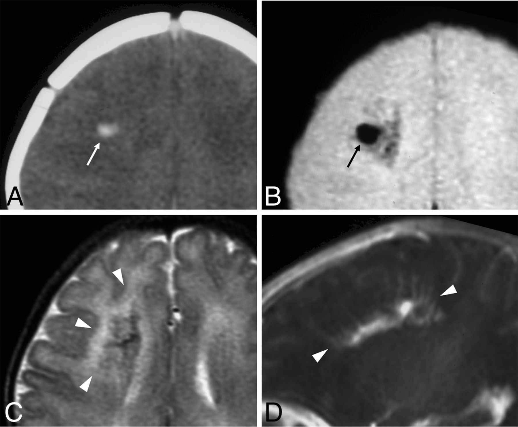
    Neonatal developmental venous anomaly complicated by focal hemorrhage and diffuse WM signal abnormalities likely related to venous congestion. A, Unenhanced head CT scan demonstrates a focal area of spontaneous hyperdensity (white arrow) in the right frontal region, suggestive of recent hemorrhage. Corresponding axial gradient-echo T2*-weighted image (B) and T2WI (C) show a blooming artifact (black arrow) in the region corresponding to the hemorrhage, which subsequently regressed (not shown), and diffuse hyperintensity of the surrounding WM (arrowheads), in keeping with venous congestion. D, Sagittal contrast-enhanced T1WI reveals a large developmental venous anomaly characterized by several radially-oriented dilated veins with a caput medusae morphology and deep venous drainage (arrowheads).

  • FIG 3.
    • Download figure
    • Open in new tab
    • Download powerpoint
    FIG 3.

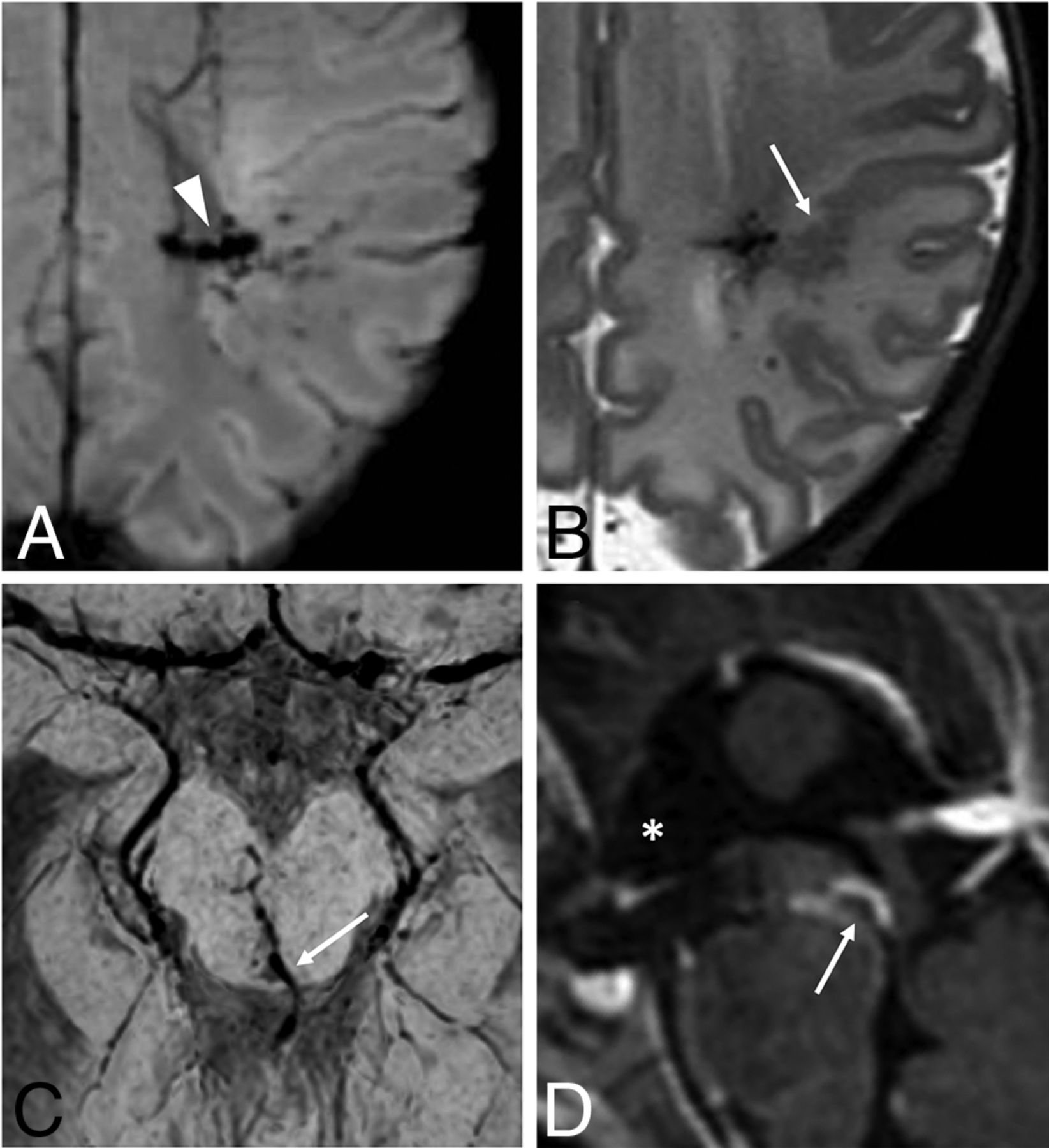
    Neonatal developmental venous anomalies associated with focal polymicrogyria (A and B) and supratentorial hydrocephalus (C and D) in 2 different patients. Axial SWI (A) and T2WI (B) depict a developmental venous anomaly with deep venous drainage (arrowhead) and an adjacent area of cortical abnormality consistent with focal polymicrogyria (arrow). Axial SWI (C) and sagittal postgadolinium T1WI (D) demonstrate a mesencephalic developmental venous anomaly with the venous collector (arrows) causing focal compression of the inferior third of the cerebral aqueduct and consequent dilation of the anterior recesses of the third ventricle (asterisk), in keeping with supratentorial obstructive hydrocephalus (see also On-line Fig 8).

Tables

  • Figures
    • View popup
    Table 1:

    Location and angioarchitecture characteristics of developmental venous anomalies

    Total(n = 58)Complicated DVA(n = 21) (36.2%)Uncomplicated DVA (n = 37) (63.2%)P Valuea
    Location (%).44
     Frontal24 (41.4)9 (42.9)15 (40.5)
     Parieto-occipital16 (27.7)6 (28.6)10 (27)
     Temporal8 (13.8)3 (14.3)5 (13.5)
     Basal ganglia/thalami5 (8.6)0 (0)5 (13.5)
     Brain stem2 (3.4)1 (4.8)1 (2.7)
     Cerebellum3 (5.2)2 (9.5)1 (2.7)
    Infratentorial (%)5 (8.6)3 (14.3)2 (5.4).34
    Right side (%)33 (56.9)13 (61.9)20 (54.1).59
    Multiple collectors (%)9 (15.5)7 (33.3)2 (5.4).008b
    Main collector caliber (median) (IQR) (mm)1.6 (1.18–2.10)2.1 (1.95–2.30)1.2 (1–1.6)<.001b
    Drainage (%).70
     Deep31 (53.4)11 (52.4)20 (54.1)
     Superficial19 (32.8)6 (28.6)13 (35.1)
     Both8 (13.8)4 (19)4 (10.8)
    • Note:—IQR indicates interquartile range.

    • ↵a P values for group comparisons were determined by χ2 or Fisher exact tests for categoric variables or by Mann-Whitney U tests for continuous variables, as appropriate.

    • ↵b Value statistically significant (statistical significance was set at P < .0083 after Bonferroni correction for multiple comparisons).

    • View popup
    Table 2:

    Neuroimaging abnormalities associated with developmental venous anomalies

    MRI AbnormalitiesNeonatal Perioda (n = 15)Last Follow-Upa (n = 10)
    WM T2 signal abnormalities7Reduced 2/5
    Stable 1/5
    Complete regression, 2/5
    Restricted diffusion foci4Total regression, 2/2
    Hemorrhagic foci8Gliosis with or without hemosiderin deposits, 3/3
    Multiple CCM2bStable, 1/2
    Growth, 1/2
    PMG2Stable, 2/2
    Calcifications2Stable, 2/2
    Triventricular hydrocephalus1Resolution, 1/1c
    Draining venous varix thrombosis1Recanalization, 1/1
    • Note:—PMG indicates polymicrogyria.

    • ↵a Some patients presented with ≥1 DVA-related complication.

    • ↵b Includes 1 neonate with cerebrofacial venous metameric syndrome.

    • c Postendoscopic third ventriculostomy.

PreviousNext
Back to top

In this issue

American Journal of Neuroradiology: 41 (12)
American Journal of Neuroradiology
Vol. 41, Issue 12
1 Dec 2020
  • Table of Contents
  • Index by author
  • Complete Issue (PDF)
Advertisement
Print
Download PDF
Email Article

Thank you for your interest in spreading the word on American Journal of Neuroradiology.

NOTE: We only request your email address so that the person you are recommending the page to knows that you wanted them to see it, and that it is not junk mail. We do not capture any email address.

Enter multiple addresses on separate lines or separate them with commas.
Neonatal Developmental Venous Anomalies: Clinicoradiologic Characterization and Follow-Up
(Your Name) has sent you a message from American Journal of Neuroradiology
(Your Name) thought you would like to see the American Journal of Neuroradiology web site.
CAPTCHA
This question is for testing whether or not you are a human visitor and to prevent automated spam submissions.
Cite this article
A.F. Geraldo, S.S. Messina, D. Tortora, A. Parodi, M. Malova, G. Morana, C. Gandolfo, A. D’Amico, E. Herkert, P. Govaert, L.A. Ramenghi, A. Rossi, M. Severino
Neonatal Developmental Venous Anomalies: Clinicoradiologic Characterization and Follow-Up
American Journal of Neuroradiology Dec 2020, 41 (12) 2370-2376; DOI: 10.3174/ajnr.A6829

Citation Manager Formats

  • BibTeX
  • Bookends
  • EasyBib
  • EndNote (tagged)
  • EndNote 8 (xml)
  • Medlars
  • Mendeley
  • Papers
  • RefWorks Tagged
  • Ref Manager
  • RIS
  • Zotero
0 Responses
Respond to this article
Share
Bookmark this article
Neonatal Developmental Venous Anomalies: Clinicoradiologic Characterization and Follow-Up
A.F. Geraldo, S.S. Messina, D. Tortora, A. Parodi, M. Malova, G. Morana, C. Gandolfo, A. D’Amico, E. Herkert, P. Govaert, L.A. Ramenghi, A. Rossi, M. Severino
American Journal of Neuroradiology Dec 2020, 41 (12) 2370-2376; DOI: 10.3174/ajnr.A6829
del.icio.us logo Twitter logo Facebook logo Mendeley logo
  • Tweet Widget
  • Facebook Like
  • Google Plus One
Purchase

Jump to section

  • Article
    • Abstract
    • ABBREVIATIONS:
    • MATERIALS AND METHODS
    • RESULTS
    • DISCUSSION
    • CONCLUSIONS
    • Footnotes
    • References
  • Figures & Data
  • Supplemental
  • Info & Metrics
  • Responses
  • References
  • PDF

Related Articles

  • PubMed
  • Google Scholar

Cited By...

  • No citing articles found.
  • Crossref
  • Google Scholar

This article has not yet been cited by articles in journals that are participating in Crossref Cited-by Linking.

More in this TOC Section

  • Frontal Paraventricular Cysts
  • Sodium MRI in Pediatric Brain Tumors
  • FRACTURE MR in Congenital Vertebral Anomalies
Show more Pediatric Neuroimaging

Similar Articles

Advertisement

Indexed Content

  • Current Issue
  • Accepted Manuscripts
  • Article Preview
  • Past Issues
  • Editorials
  • Editor's Choice
  • Fellows' Journal Club
  • Letters to the Editor
  • Video Articles

Cases

  • Case Collection
  • Archive - Case of the Week
  • Archive - Case of the Month
  • Archive - Classic Case

More from AJNR

  • Trainee Corner
  • Imaging Protocols
  • MRI Safety Corner
  • Book Reviews

Multimedia

  • AJNR Podcasts
  • AJNR Scantastics

Resources

  • Turnaround Time
  • Submit a Manuscript
  • Submit a Video Article
  • Submit an eLetter to the Editor/Response
  • Manuscript Submission Guidelines
  • Statistical Tips
  • Fast Publishing of Accepted Manuscripts
  • Graphical Abstract Preparation
  • Imaging Protocol Submission
  • Evidence-Based Medicine Level Guide
  • Publishing Checklists
  • Author Policies
  • Become a Reviewer/Academy of Reviewers
  • News and Updates

About Us

  • About AJNR
  • Editorial Board
  • Editorial Board Alumni
  • Alerts
  • Permissions
  • Not an AJNR Subscriber? Join Now
  • Advertise with Us
  • Librarian Resources
  • Feedback
  • Terms and Conditions
  • AJNR Editorial Board Alumni

American Society of Neuroradiology

  • Not an ASNR Member? Join Now

© 2025 by the American Society of Neuroradiology All rights, including for text and data mining, AI training, and similar technologies, are reserved.
Print ISSN: 0195-6108 Online ISSN: 1936-959X

Powered by HighWire