
Article Figures & Data
Figures
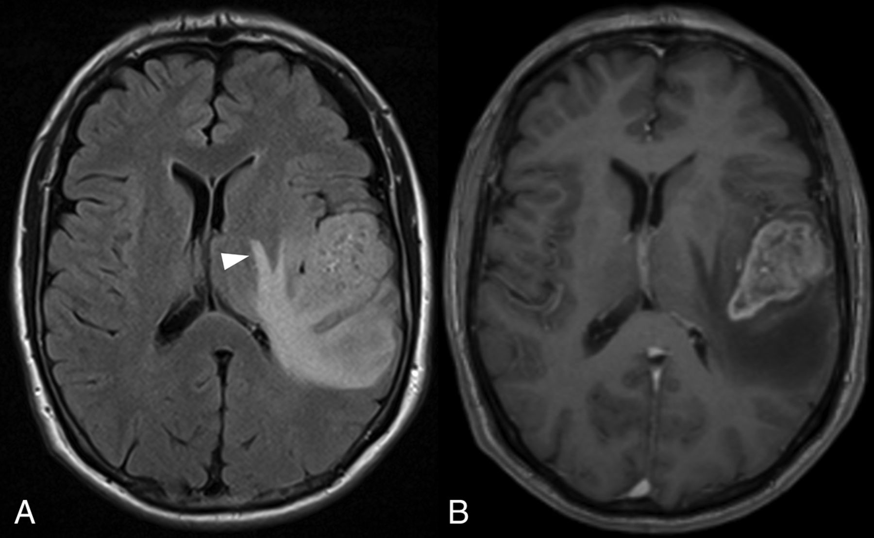
- Fig 1.
FLAIR imaging (A) showing edema around the CET component (B) sparing the cortex (arrowhead), producing the characteristic fingerlike appearance.
- Fig 2.
FLAIR imaging (A) showing edema extending around the left lentiform nucleus. The CET component is shown in B.
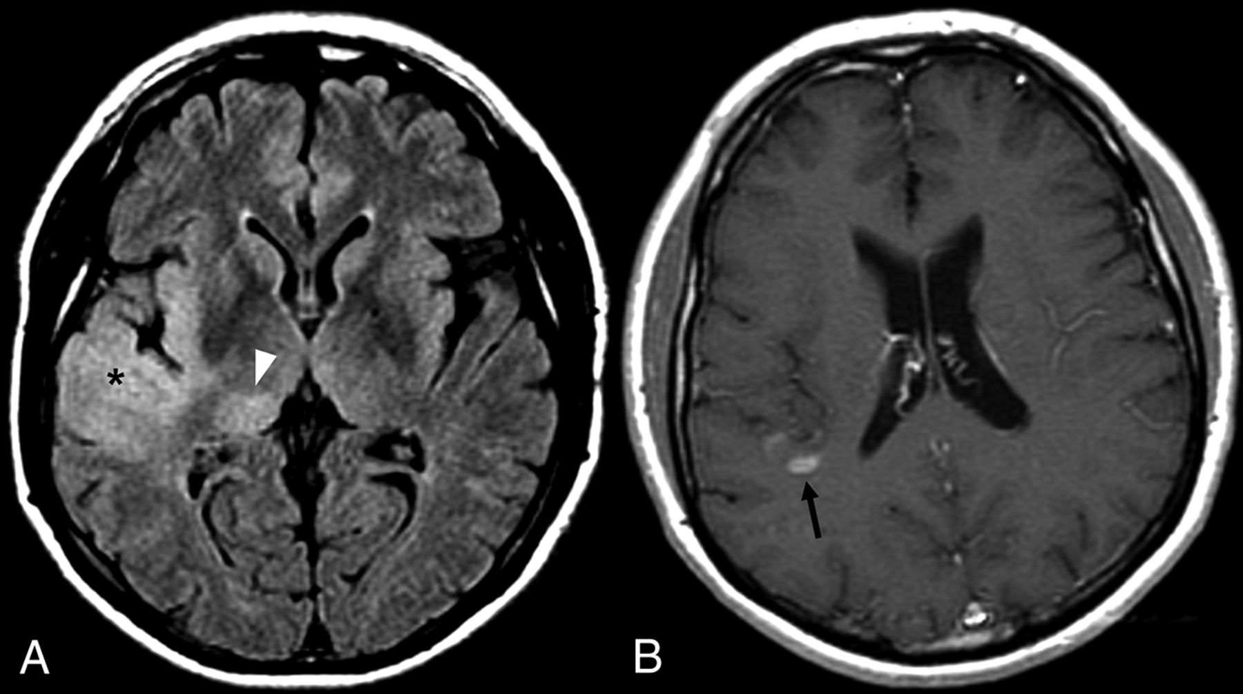
- Fig 3.
FLAIR imaging (A) demonstrating nCET involving the anterior right temporal cortex (asterisk), insula, and thalamus (arrowhead). A narrow window width has been used to improve conspicuity. The small CET component lies further superiorly (B, arrow).
- Fig 4.
FLAIR imaging (A) showing eccentric extension of nCET across the corpus callosum, with associated expansion (arrowhead). Rounded hyperintensity in the left thalamus (arrow) is also consistent with nCET. Note the paucity of edema in the white matter immediately adjacent to the CET component (B).


Tables
Conventional and advanced MRI features useful for differentiating between nCET and edema
nCET Edema Conventional MRI features Gray matter involvement Spares the gray matter Eccentric Relatively concentric around enhancing lesions Relatively mild FLAIR and T2 hyperintensity More marked FLAIR and T2 hyperintensity Focal parenchymal expansion More diffuse mass effect if marked edema Advanced MRI sequences Relative diffusion restriction Facilitated diffusion Choline elevation, NAA depletion Normal MRS findings Elevated rCBV around CET rCBV elevation confined to CET Note:—rCBV indicates relative cerebral blood volume.


Jump to section
Related Articles
Cited By...
- Revealing the Infiltration: Prognostic Value of Automated Segmentation of Non-Contrast-Enhancing Tumor in Glioblastoma
- Anatomy-guided, modality-agnostic segmentation of neuroimaging abnormalities
- Cluster Analysis of DSC MRI, Dynamic Contrast-Enhanced MRI, and DWI Parameters Associated with Prognosis in Patients with Glioblastoma after Removal of the Contrast-Enhancing Component: A Preliminary Study
- Radio-Pathomic Maps of Cell Density Identify Brain Tumor Invasion beyond Traditional MRI-Defined Margins
- LUMOS - Low and Intermediate Grade Glioma Umbrella Study of Molecular Guided TherapieS at relapse: Protocol for a pilot study
- Localized blood-brain barrier opening in infiltrating gliomas with MRI-guided acoustic emissions-controlled focused ultrasound
- MGMT Promoter Methylation Status in Initial and Recurrent Glioblastoma: Correlation Study with DWI and DSC PWI Features
- Radio-pathomic maps of cell density identify glioma invasion beyond traditional MR imaging defined margins
- Neuro-Oncology and Radiogenomics: Time to Integrate?
- Response Assessment in Neuro-Oncology Criteria for Gliomas: Practical Approach Using Conventional and Advanced Techniques