
Article Figures & Data
Figures
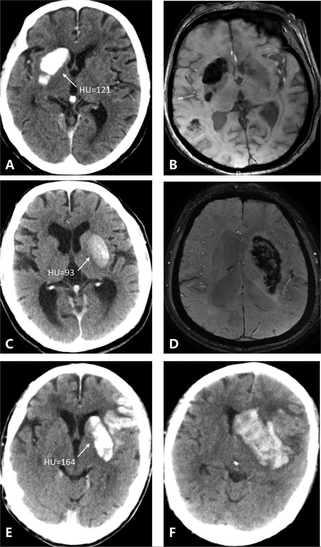
- Fig 1.
Three patients with 3 kinds of metallic hyperdensity signs. A, C, and E, NCCT images obtained immediately after mechanical thrombectomy. B and D, SWI at 24 hours after mechanical thrombectomy. F, NCCT image at 24 hours after mechanical thrombectomy. A 77-year-old woman with a hyperdense lesion and a maximum CT density of 121 HU in the right caudate nucleus on NCCT images immediately after mechanical thrombectomy (A) has parenchymal hemorrhage on SWI at 24 hours (B). A 71-year-old man with a hyperdense lesion and a maximum CT density of 93 HU in the left lenticular nucleus on NCCT images immediately after mechanical thrombectomy (C) has parenchymal hemorrhage on SWI at 24 hours (D). An 81-year-old man with a hyperdense lesion and a maximum CT density of 164 HU in the left lenticular nucleus on NCCT images immediately after mechanical thrombectomy (E) has parenchymal hemorrhage on the NCCT image at 24 hours (F).
- Fig 2.
Two patients who show the typical cortical hyperdense lesion on NCCT immediately after MT (A and C) have no HT on follow-up SWI (B and D).
- Fig 3.
Two patients who show a petechial hyperdense lesion in the basal ganglia on NCCT immediately after MT (A and C) have no HT on follow-up SWI (B) and the remaining low densities on follow-up NCCT (D).
- Fig 4.
A patient who shows a nonpetechial hyperdense lesion on NCCT immediately after MT (A) has no HT on follow-up NCCT and the remaining low densities (B).
- Fig 5.
A patient who shows the subcortical hyperdense lesion involving both the basal ganglia and subcortical white matter on NCCT immediately after MT (A) has no HT on NCCT immediately after MT and the remaining low densities on follow-up NCCT (B).
- Fig 6.
A patient who shows the subcortical hyperdense lesion on NCCT immediately after MT (A) but has no HT on follow-up NCCT and the remaining low densities (B).


Tables
- Table 1:
Predictive value of characteristics of metallic hyperdensity for parenchymal hemorrhage
AUC 95% CI P Value Sensitivity Specificity Positive Predictive Value Negative Predictive Value Presence of metallic hyperdensity sign 0.894 0.835–0.952 <.001 0.882 0.905 0.763 0.957 Note:—AUC indicates area under the curve.
- Table 2:
Comparison of characteristics between patients with and without the metallic hyperdensity sign
Characteristics Presence of Metallic Hyperdensity Sign (n = 59) Absence of Metallic Hyperdensity Sign (n = 139) P Value Age (mean) (yr) 70.5 ± 10.5 69.4 ± 12.2 .568 Female (No.) (%) 26 (44.1) 56 (40.3) .639 Comorbid conditions Hypertension (No.) (%) 37 (62.7) 84 (60.4) .874 Diabetes mellitus (No.) (%) 14 (23.7) 24 (17.3) .326 Atrial fibrillation (No.) (%) 35 (59.3) 50 (35.9) .003 Clinical variables Baseline NIHSS (median) (IQR) 15 (12–18) 14 (10–17) .116 Onset-to-puncture time (median) (IQR) (min) 334 (237–425) 306 (213–415) .289 Retrieval attempts (median) (IQR) 2 (1–3) 1 (1–3) .039 Radiologic data Baseline infarct volume (median) (IQR) (mL) 59.9 (34.7–80.9) 46.3 (27.5–80.0) .096 Baseline hypoperfusion volume (median) (IQR) (mL) 108.9 (79.2–153.6) 107.1 (68.0–160.0) .639 Recanalization (No.) (%) 48 (81.4) 123 (88.4) .181 24-Hour hemorrhagic transformation (No.) (%) 58 (98.3) 54 (38.8) <.001 24-Hour parenchymal hemorrhage (No.) (%) 45 (76.3) 6 (4.3) <.001 Note:—IQR indicates interquartile range.


Jump to section
Related Articles
Cited By...
- Combinations of Clinical Factors, CT Signs, and Radiomics for Differentiating High-Density Areas after Mechanical Thrombectomy in Patients with Acute Ischemic Stroke
- Incidence, Risk Factors, and Clinical Implications of Subarachnoid Hyperdensities on Flat-Panel Detector CT following Mechanical Thrombectomy in Patients with Anterior Circulation Acute Ischemic Stroke
- Contrast extravasation and outcome of endovascular therapy in acute ischaemic stroke: a systematic review and meta-analysis
- Association of maximal systolic blood pressure with poor outcome in patients with hyperattenuated lesions on immediate NCCT after mechanical thrombectomy
- Prediction of Hemorrhage after Successful Recanalization in Patients with Acute Ischemic Stroke: Improved Risk Stratification Using Dual-Energy CT Parenchymal Iodine Concentration Ratio Relative to the Superior Sagittal Sinus