
Article Figures & Data
Figures
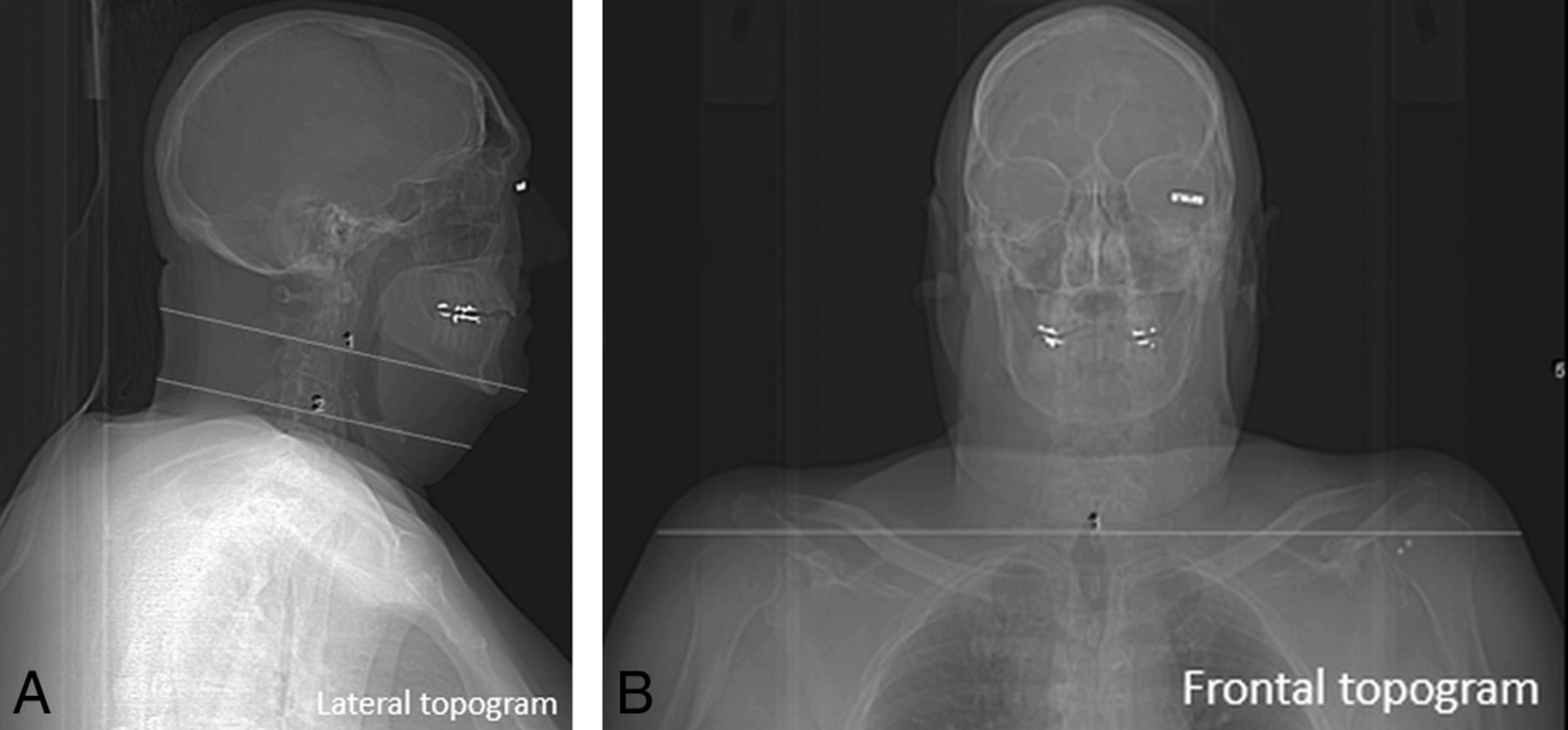
- Fig 1.
Lateral (A) and frontal (B) projections of CT topogram images with measurements of anteroposterior diameter at the level of C2–3 and C4–5 and transverse diameters at the shoulder.
- Fig 2.
Gadolinium-enhanced T1-weighted MR images with SPIR (A) and mDixon (B) techniques for fat suppression. ROIs are placed on the spinal cord and fat to obtain a signal intensity ratio.
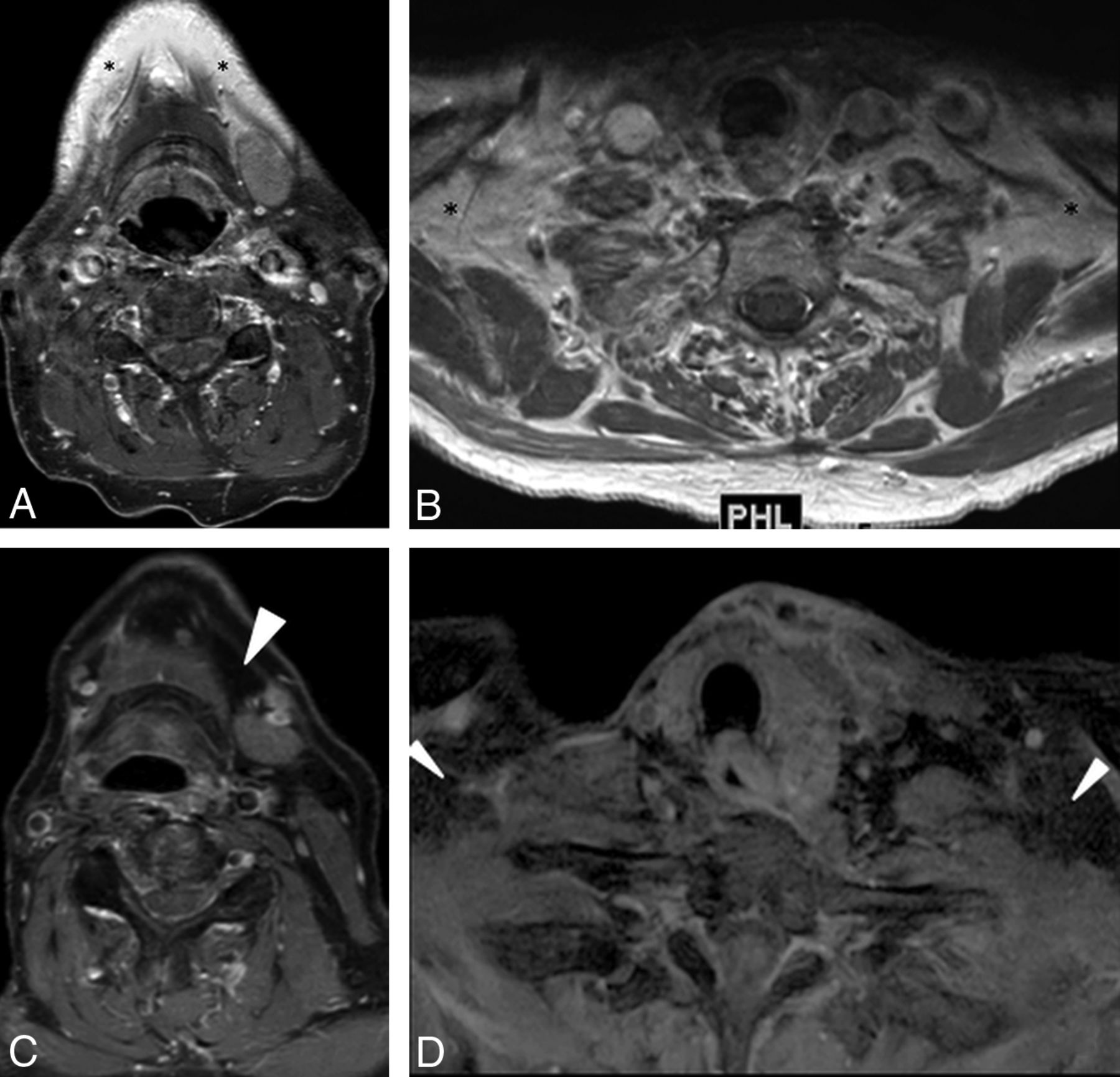
- Fig 3.
Axial STIR (A and B) and mDixon T2-weighted (C and D) MR images. Note incomplete fat suppression (asterisks) in the maxillary and supraclavicular regions on the STIR technique and complete uniform fat suppression (arrowheads) in the submandibular and supraclavicular regions on the mDixon technique.
- Fig 4.
Gadolinium-enhanced axial T1-weighted MR images with SPIR (A and B) and mDixon (C and D) techniques for fat suppression. Note incomplete fat suppression (asterisks) in the submandibular and supraclavicular regions on the SPIR technique and complete uniform fat suppression (arrowheads) in similar regions on the mDixon technique.
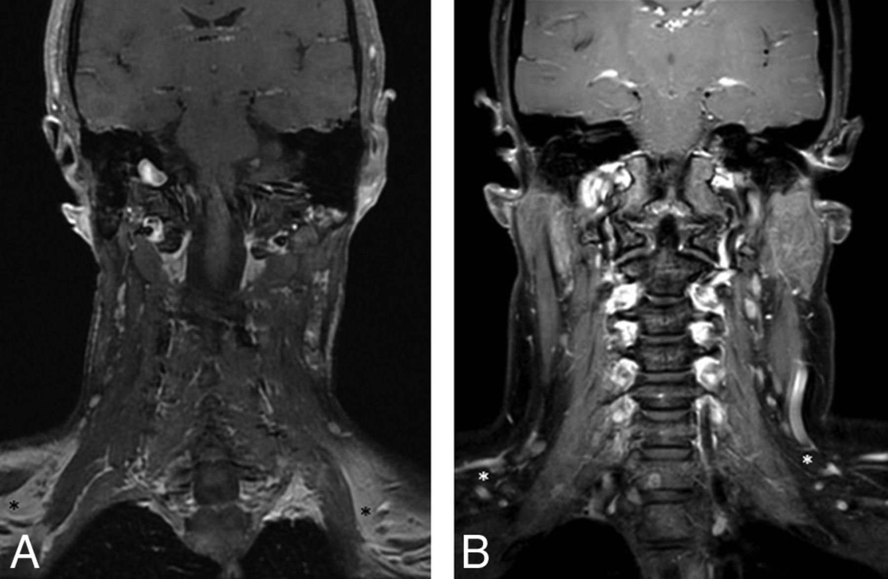
- Fig 5.
Gadolinium-enhanced coronal T1-weighted MR images with SPIR (A) and mDixon (B) techniques for fat suppression. Note incomplete fat suppression (dark asterisk) in the supraclavicular regions on the SPIR technique and complete uniform fat suppression (white asterisk) in similar regions on the mDixon technique.


Tables
Axial STIR Axial T2WI Axial Gad-T1WI Axial Gad-T1WI TSE mDixon TSE SPIR TSE mDixon TSE Coil 16 Channel 16 Channel 16 Channel 16 Channel SENSE NV SENSE NV SENSE NV SENSE NV TR/TE 3000/15 ms 3000/80 ms 600/9.2 ms 500/10 ms Section thickness/intersection gap 3/1 mm 3/1 mm 3/1 mm 3/1 mm No. of axial images 40 40 40 40 FS technique Inversion recovery (TI = 200 ms) mDixon SPIR mDixon Acquisition matrix 200 × 141 232 × 232 288 × 196 204 × 199 NEX 2 1 1 1 Acquisition time 4 min, 56 sec 2 min, 2 sec 3 min, 2 sec 2 min, 8 sec Parallel imaging Yes Yes Yes Yes Gadolinium contrast N/A N/A 0.1 mmol/kg gadodiamide (Gd-DTPA) (ProHance) 0.1 mmol/kg gadodiamide (Gd-DTPA) (ProHance) Note:—SENSE indicates sensitivity encoding; N/A, not applicable; NV, NeuroVascular.
↵a Achieva; Philips Healthcare.
Variable Sequence Group P Valueb Group B (n = 31) Group A (n = 33) Sex Male 20 (64.5) 23 (69.7) .79 Female 11 (35.5) 10 (30.3) Age (yr) 61 ± 15 55 ± 17 .15 Body habitus AP neck diameter at the level of mandible (C2–3) (mm) 185 ± 21 188 ± 21 .28 AP diameter of midneck (C4–5) (mm) 127 ± 22 124 ± 19 .90 Shoulder width (mm) 394 ± 35 394 ± 45 .84 AP neck diameter at the level of mandible-to-shoulder width ratio 1.48 ± 0.15 1.53 ± 0.16 .35 Shoulder width-to midneck AP diameter ratio 3.16 ± 0.40 3.21 ± 0.36 .88 Variable Sequence Group P Valueb Group B (n = 31) Group A (n = 33) T2WI spinal cord–to-fat ratio Submandibular level 5.7 ± 1.6 3.5 ± 3.4 <.001 Supraclavicular level 7.4 ± 2.4 3.3 ± 3.4 <.001 Post-Gad-T1WI spinal cord–to-fat ratio Submandibular level 3.7 ± 1.4 0.9 ± 0.7 <.001 Supraclavicular level 4.3 ± 2.0 0.5 ± 0.3 <.001 - Table 4:
Subjective assessment of image quality, fat suppression, and susceptibility artifactsa
Variable Sequence Group P Valueb Group B (n = 31) Group A (n = 33) T2WI/STIR images Overall image-quality grade 3.9 ± 0.5 3.6 ± 0.7 .022 Fat-saturation grade Maxillary region 4.6 ± 0.4 4.3 ± 0.5 .013 Mandibular region 4.4 ± 0.5 4.0 ± 0.6 .007 Lower neck region 4.7 ± 0.4 4.3 ± 0.4 .001 Dental amalgam susceptibility artifacts (%) 38.7% 22.7% .056 Post-Gad-T1WIs Overall image-quality grade 4.0 ± 0.4 2.6 ± 0.6 <.001 Fat-saturation grade Maxillary region 4.8 ± 0.3 3.8 ± 0.7 <.001 Mandibular region 4.7 ± 0.3 2.8 ± 0.5 <.001 Lower neck region 4.8 ± 0.3 1.4 ± 0.7 <.001 Dental amalgam susceptibility artifacts (%) 37.1% 31.8% .50


Jump to section
Related Articles
Cited By...
- Development and implementation of optimized endogenous contrast sequences for delineation in adaptive radiotherapy on a 1.5T MR-Linear-accelerator (MR-Linac): A prospective R-IDEAL Stage 0-2a quantitative/qualitative evaluation of in vivo site-specific quality-assurance using a 3D T2 fat-suppressed platform for head and neck cancer
- 4D-Dynamic Contrast-Enhanced MRI for Preoperative Localization in Patients with Primary Hyperparathyroidism