Advertisement

Main menu

  • Home
  • Content
    • Current Issue
    • Accepted Manuscripts
    • Article Preview
    • Past Issue Archive
    • Video Articles
    • AJNR Case Collection
    • Case of the Week Archive
    • Case of the Month Archive
    • Classic Case Archive
  • Special Collections
    • AJNR Awards
    • Low-Field MRI
    • Alzheimer Disease
    • ASNR Foundation Special Collection
    • Photon-Counting CT
    • View All
  • Multimedia
    • AJNR Podcasts
    • AJNR SCANtastic
    • Trainee Corner
    • MRI Safety Corner
    • Imaging Protocols
  • For Authors
    • Submit a Manuscript
    • Submit a Video Article
    • Submit an eLetter to the Editor/Response
    • Manuscript Submission Guidelines
    • Statistical Tips
    • Fast Publishing of Accepted Manuscripts
    • Graphical Abstract Preparation
    • Imaging Protocol Submission
    • Author Policies
  • About Us
    • About AJNR
    • Editorial Board
    • Editorial Board Alumni
  • More
    • Become a Reviewer/Academy of Reviewers
    • Subscribers
    • Permissions
    • Alerts
    • Feedback
    • Advertisers
    • ASNR Home

User menu

  • Alerts
  • Log in

Search

  • Advanced search
American Journal of Neuroradiology

American Journal of Neuroradiology

ASHNR American Society of Functional Neuroradiology ASHNR American Society of Pediatric Neuroradiology ASSR
  • Alerts
  • Log in

Advanced Search

  • Home
  • Content
    • Current Issue
    • Accepted Manuscripts
    • Article Preview
    • Past Issue Archive
    • Video Articles
    • AJNR Case Collection
    • Case of the Week Archive
    • Case of the Month Archive
    • Classic Case Archive
  • Special Collections
    • AJNR Awards
    • Low-Field MRI
    • Alzheimer Disease
    • ASNR Foundation Special Collection
    • Photon-Counting CT
    • View All
  • Multimedia
    • AJNR Podcasts
    • AJNR SCANtastic
    • Trainee Corner
    • MRI Safety Corner
    • Imaging Protocols
  • For Authors
    • Submit a Manuscript
    • Submit a Video Article
    • Submit an eLetter to the Editor/Response
    • Manuscript Submission Guidelines
    • Statistical Tips
    • Fast Publishing of Accepted Manuscripts
    • Graphical Abstract Preparation
    • Imaging Protocol Submission
    • Author Policies
  • About Us
    • About AJNR
    • Editorial Board
    • Editorial Board Alumni
  • More
    • Become a Reviewer/Academy of Reviewers
    • Subscribers
    • Permissions
    • Alerts
    • Feedback
    • Advertisers
    • ASNR Home
  • Follow AJNR on Twitter
  • Visit AJNR on Facebook
  • Follow AJNR on Instagram
  • Join AJNR on LinkedIn
  • RSS Feeds

AJNR Awards, New Junior Editors, and more. Read the latest AJNR updates

Research ArticlePeripheral Nervous System

Gadolinium DTPA Enhancement Characteristics of the Rat Sciatic Nerve after Crush Injury at 4.7T

B.J. Hill, K.R. Padgett, V. Kalra, A. Marcillo, B. Bowen, P. Pattany, D. Dietrich and R. Quencer
American Journal of Neuroradiology January 2018, 39 (1) 177-183; DOI: https://doi.org/10.3174/ajnr.A5437
B.J. Hill
aFrom the Departments of Radiology (B.J.H., K.R.P., B.B., P.P., R.Q.)
  • Find this author on Google Scholar
  • Find this author on PubMed
  • Search for this author on this site
  • ORCID record for B.J. Hill
K.R. Padgett
aFrom the Departments of Radiology (B.J.H., K.R.P., B.B., P.P., R.Q.)
bRadiation Oncology (K.R.P.)
cBiomedical Engineering (K.R.P.)
  • Find this author on Google Scholar
  • Find this author on PubMed
  • Search for this author on this site
  • ORCID record for K.R. Padgett
V. Kalra
eMiller School of Medicine (V.K.), University of Miami, Miami, Florida.
  • Find this author on Google Scholar
  • Find this author on PubMed
  • Search for this author on this site
  • ORCID record for V. Kalra
A. Marcillo
dMiami Project to Cure Paralysis (A.M., D.D.)
  • Find this author on Google Scholar
  • Find this author on PubMed
  • Search for this author on this site
  • ORCID record for A. Marcillo
B. Bowen
aFrom the Departments of Radiology (B.J.H., K.R.P., B.B., P.P., R.Q.)
  • Find this author on Google Scholar
  • Find this author on PubMed
  • Search for this author on this site
P. Pattany
aFrom the Departments of Radiology (B.J.H., K.R.P., B.B., P.P., R.Q.)
  • Find this author on Google Scholar
  • Find this author on PubMed
  • Search for this author on this site
  • ORCID record for P. Pattany
D. Dietrich
dMiami Project to Cure Paralysis (A.M., D.D.)
  • Find this author on Google Scholar
  • Find this author on PubMed
  • Search for this author on this site
  • ORCID record for D. Dietrich
R. Quencer
aFrom the Departments of Radiology (B.J.H., K.R.P., B.B., P.P., R.Q.)
  • Find this author on Google Scholar
  • Find this author on PubMed
  • Search for this author on this site
  • ORCID record for R. Quencer
  • Article
  • Figures & Data
  • Supplemental
  • Info & Metrics
  • Responses
  • References
  • PDF
Loading

Article Figures & Data

Figures

  • Tables
  • Fig 1.
    • Download figure
    • Open in new tab
    • Download powerpoint
    Fig 1.

    Axial T1WI with fat saturation after Gd-DTPA. Areas outlined in color represent manually defined ROIs corresponding to injured nerve (red), contralateral nerve (yellow), muscle (green), and background/air (blue). Precontrast and nonenhancing nerves were localized by correlating T1WI and T2WI findings.
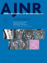
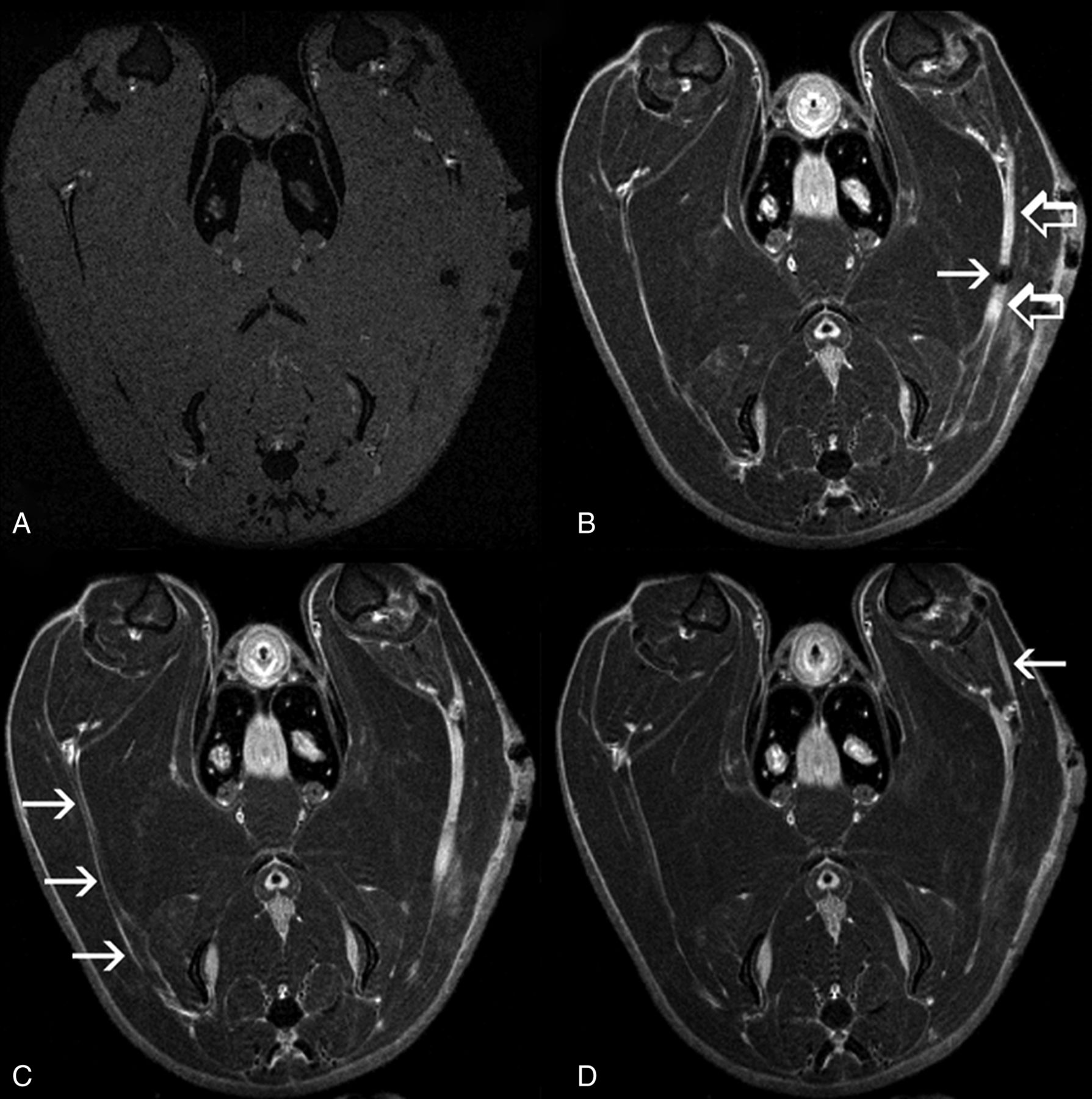
  • Fig 2.
    • Download figure
    • Open in new tab
    • Download powerpoint
    Fig 2.

    Axial T2WI with fat saturation (#5 Jeweler forceps injury, day 13). A, Injured sciatic nerve (arrows) shows increased caliber and signal compared with the nonoperative contralateral nerve. A small focus of susceptibility artifact (open arrow) is seen at the site of injury, presumably representing blood products. B, The adjacent section shows T2 hyperintensity in a distal branch (arrow) of the injured sciatic nerve.
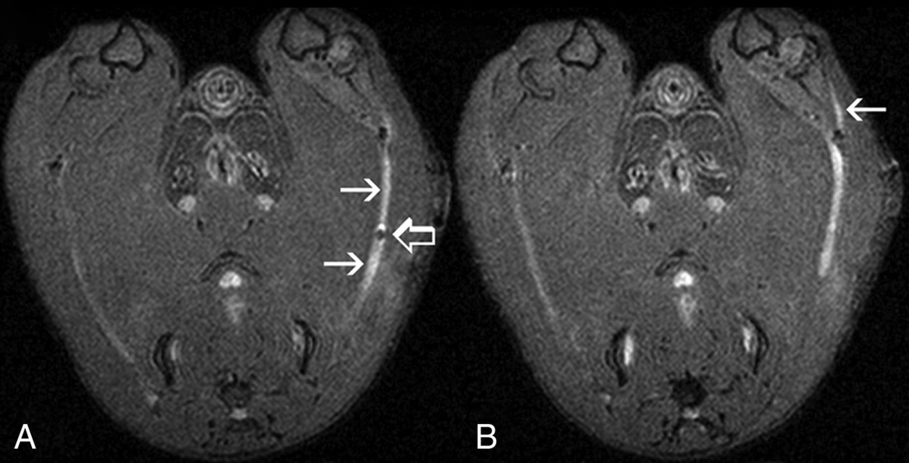
  • Fig 3.
    • Download figure
    • Open in new tab
    • Download powerpoint
    Fig 3.

    Axial T1WI with fat saturation, pre- (A) and post- (B–D) Gd-DTPA (same animal as in Fig 2). A, On precontrast images, both injured and nonoperative nerves are isointense to muscle. B, Intense Gd-DTPA enhancement (open arrows) is demonstrated a few millimeters proximal to, in the region of, and distal to the site of crush injury (arrow) (On-line Video 1). C, The nonoperative contralateral nerve (arrows) is well seen in this section and demonstrates a thin rim of peripheral enhancement but no internal enhancement (On-line Video 2). D, Enhancement is seen in a distal branch (arrow) of the injured sciatic nerve (On-line Video 3).
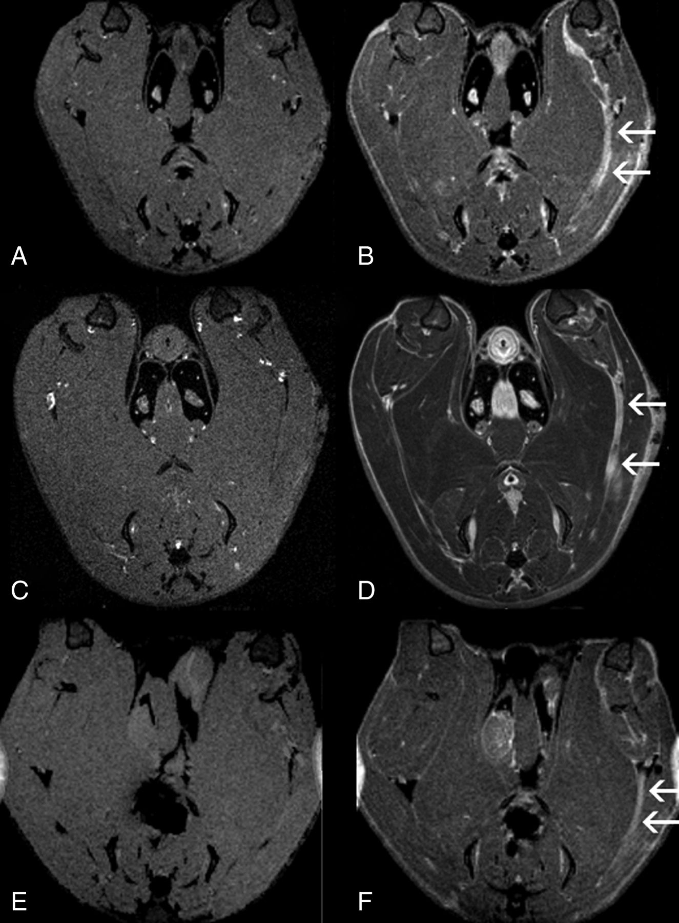
  • Fig 4.
    • Download figure
    • Open in new tab
    • Download powerpoint
    Fig 4.

    Axial T1WI pre- (left) and post- (right) Gd-DTPA with fat saturation acquired at days 7 (A and B), 13 (C and D), and 21 (E and F) after severe (forceps) crush injury. Robust enhancement of injured nerves is seen at all 3 time points (arrows).
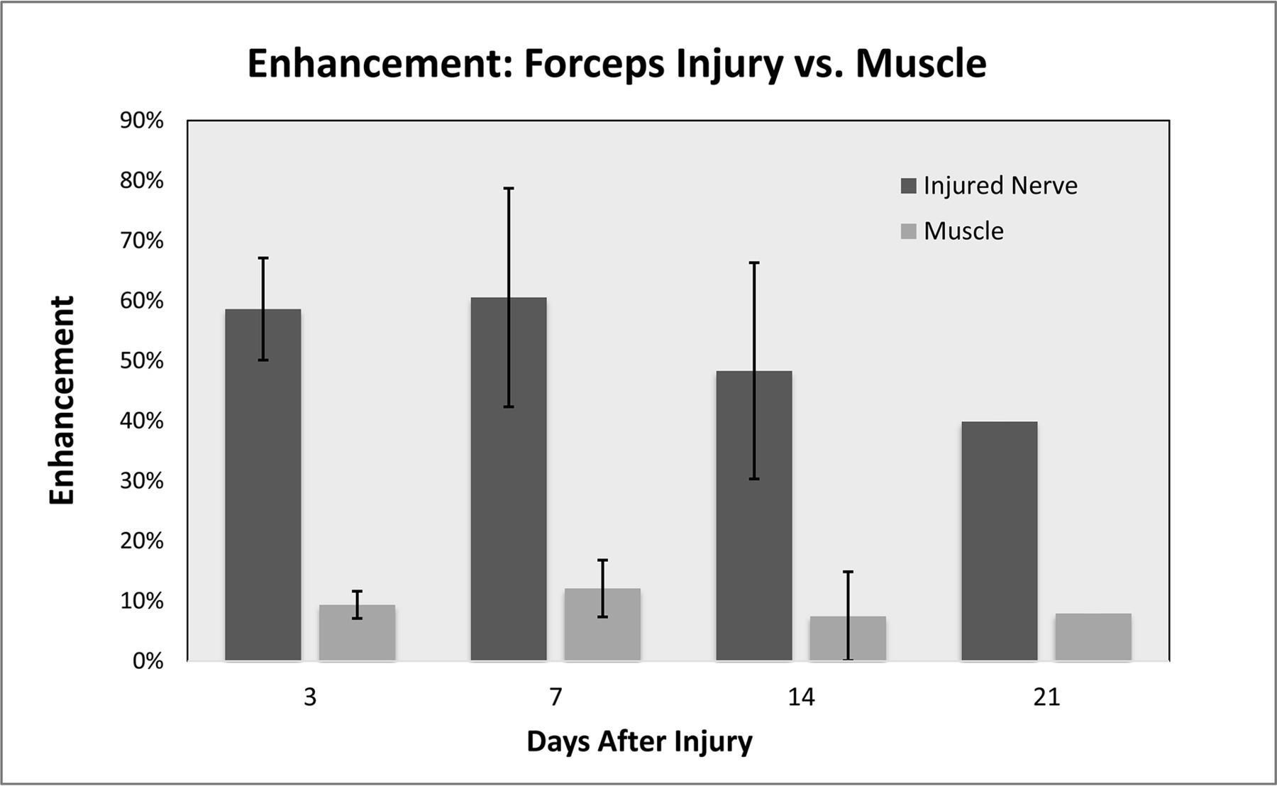
  • Fig 5.
    • Download figure
    • Open in new tab
    • Download powerpoint
    Fig 5.

    Peak enhancement after severe (forceps) crush injury compared with normal muscle between days 3 and 21. Enhancement is expressed as the percentage increase of the signal-to-noise ratio after Gd-DTPA administration. Error bars represent 1 SD.

  • Fig 6.
    • Download figure
    • Open in new tab
    • Download powerpoint
    Fig 6.

    ROI signal-to-noise ratio as a function of time after the intravenous administration of Gd-DTPA (same animal as in Figs 2 and 3). The presence of a delayed, higher peak suggests an enlarged extravascular extracellular space after severe (forceps) crush injury (On-line Video 4).13

  • Fig 7.
    • Download figure
    • Open in new tab
    • Download powerpoint
    Fig 7.

    ROI signal-to-noise ratio as a function of time after the intravenous administration of Gd-DTPA (same animal as in Figs 2, 3, and 6). The half-life of Gd-DTPA enhancement was approximately 1 hour after severe (forceps) crush injury.

Tables

  • Figures
    • View popup
    Table 1:

    Number of MRI sessions at each time point after forceps (#5 jeweler or toothed Adson) crush injury

    Forceps: Postop Day No.No. MRI Sessions (with Limited DCE)
    01 (0)
    14 (0)
    22 (0)
    33 (2)
    41 (0)
    51 (0)
    76 (5)
    122 (0)
    132 (1)
    145 (4)
    191 (1)
    211 (1)
    221 (1)
    301 (1)
    Total31 (16)
    • Note:—Postop indicates postoperative.

    • View popup
    Table 2:

    Number of MRI sessions at each time point after clip (10–60 g microvascular/microaneurysm) crush injury

    Clip: Postop Day No.No. MRI Sessions
    13
    24
    32
    43
    54
    61
    72
    Total19
PreviousNext
Back to top

In this issue

American Journal of Neuroradiology: 39 (1)
American Journal of Neuroradiology
Vol. 39, Issue 1
1 Jan 2018
  • Table of Contents
  • Index by author
  • Complete Issue (PDF)
Advertisement
Print
Download PDF
Email Article

Thank you for your interest in spreading the word on American Journal of Neuroradiology.

NOTE: We only request your email address so that the person you are recommending the page to knows that you wanted them to see it, and that it is not junk mail. We do not capture any email address.

Enter multiple addresses on separate lines or separate them with commas.
Gadolinium DTPA Enhancement Characteristics of the Rat Sciatic Nerve after Crush Injury at 4.7T
(Your Name) has sent you a message from American Journal of Neuroradiology
(Your Name) thought you would like to see the American Journal of Neuroradiology web site.
CAPTCHA
This question is for testing whether or not you are a human visitor and to prevent automated spam submissions.
Cite this article
B.J. Hill, K.R. Padgett, V. Kalra, A. Marcillo, B. Bowen, P. Pattany, D. Dietrich, R. Quencer
Gadolinium DTPA Enhancement Characteristics of the Rat Sciatic Nerve after Crush Injury at 4.7T
American Journal of Neuroradiology Jan 2018, 39 (1) 177-183; DOI: 10.3174/ajnr.A5437

Citation Manager Formats

  • BibTeX
  • Bookends
  • EasyBib
  • EndNote (tagged)
  • EndNote 8 (xml)
  • Medlars
  • Mendeley
  • Papers
  • RefWorks Tagged
  • Ref Manager
  • RIS
  • Zotero
0 Responses
Respond to this article
Share
Bookmark this article
Gadolinium DTPA Enhancement Characteristics of the Rat Sciatic Nerve after Crush Injury at 4.7T
B.J. Hill, K.R. Padgett, V. Kalra, A. Marcillo, B. Bowen, P. Pattany, D. Dietrich, R. Quencer
American Journal of Neuroradiology Jan 2018, 39 (1) 177-183; DOI: 10.3174/ajnr.A5437
del.icio.us logo Twitter logo Facebook logo Mendeley logo
  • Tweet Widget
  • Facebook Like
  • Google Plus One
Purchase

Jump to section

  • Article
    • Abstract
    • ABBREVIATIONS:
    • Materials and Methods
    • Results
    • Discussion
    • Conclusions
    • Acknowledgments
    • Footnotes
    • References
  • Figures & Data
  • Supplemental
  • Info & Metrics
  • Responses
  • References
  • PDF

Related Articles

  • PubMed
  • Google Scholar

Cited By...

  • Is Gd-DTPA Necessary in Clinical Practice for Peripheral Nerve Injury?
  • Crossref
  • Google Scholar

This article has not yet been cited by articles in journals that are participating in Crossref Cited-by Linking.

More in this TOC Section

Peripheral Nervous System

  • Dorsal Root Ganglion Volumetry by MR Gangliography
  • Role of MR Neurography for the Diagnosis of Peripheral Trigeminal Nerve Injuries in Patients with Prior Molar Tooth Extraction
Show more Peripheral Nervous System

Spine Imaging and Spine Image-Guided Interventions

  • Optimization of Photon Counting CT Myelography
  • Characteristics of SIH Type I Culprit Lesions
  • Management Outcomes For VO Spine Biopsy
Show more Spine Imaging and Spine Image-Guided Interventions

Similar Articles

Advertisement

Indexed Content

  • Current Issue
  • Accepted Manuscripts
  • Article Preview
  • Past Issues
  • Editorials
  • Editor's Choice
  • Fellows' Journal Club
  • Letters to the Editor
  • Video Articles

Cases

  • Case Collection
  • Archive - Case of the Week
  • Archive - Case of the Month
  • Archive - Classic Case

More from AJNR

  • Trainee Corner
  • Imaging Protocols
  • MRI Safety Corner
  • Book Reviews

Multimedia

  • AJNR Podcasts
  • AJNR Scantastics

Resources

  • Turnaround Time
  • Submit a Manuscript
  • Submit a Video Article
  • Submit an eLetter to the Editor/Response
  • Manuscript Submission Guidelines
  • Statistical Tips
  • Fast Publishing of Accepted Manuscripts
  • Graphical Abstract Preparation
  • Imaging Protocol Submission
  • Evidence-Based Medicine Level Guide
  • Publishing Checklists
  • Author Policies
  • Become a Reviewer/Academy of Reviewers
  • News and Updates

About Us

  • About AJNR
  • Editorial Board
  • Editorial Board Alumni
  • Alerts
  • Permissions
  • Not an AJNR Subscriber? Join Now
  • Advertise with Us
  • Librarian Resources
  • Feedback
  • Terms and Conditions
  • AJNR Editorial Board Alumni

American Society of Neuroradiology

  • Not an ASNR Member? Join Now

© 2025 by the American Society of Neuroradiology All rights, including for text and data mining, AI training, and similar technologies, are reserved.
Print ISSN: 0195-6108 Online ISSN: 1936-959X

Powered by HighWire