
Article Figures & Data
Figures
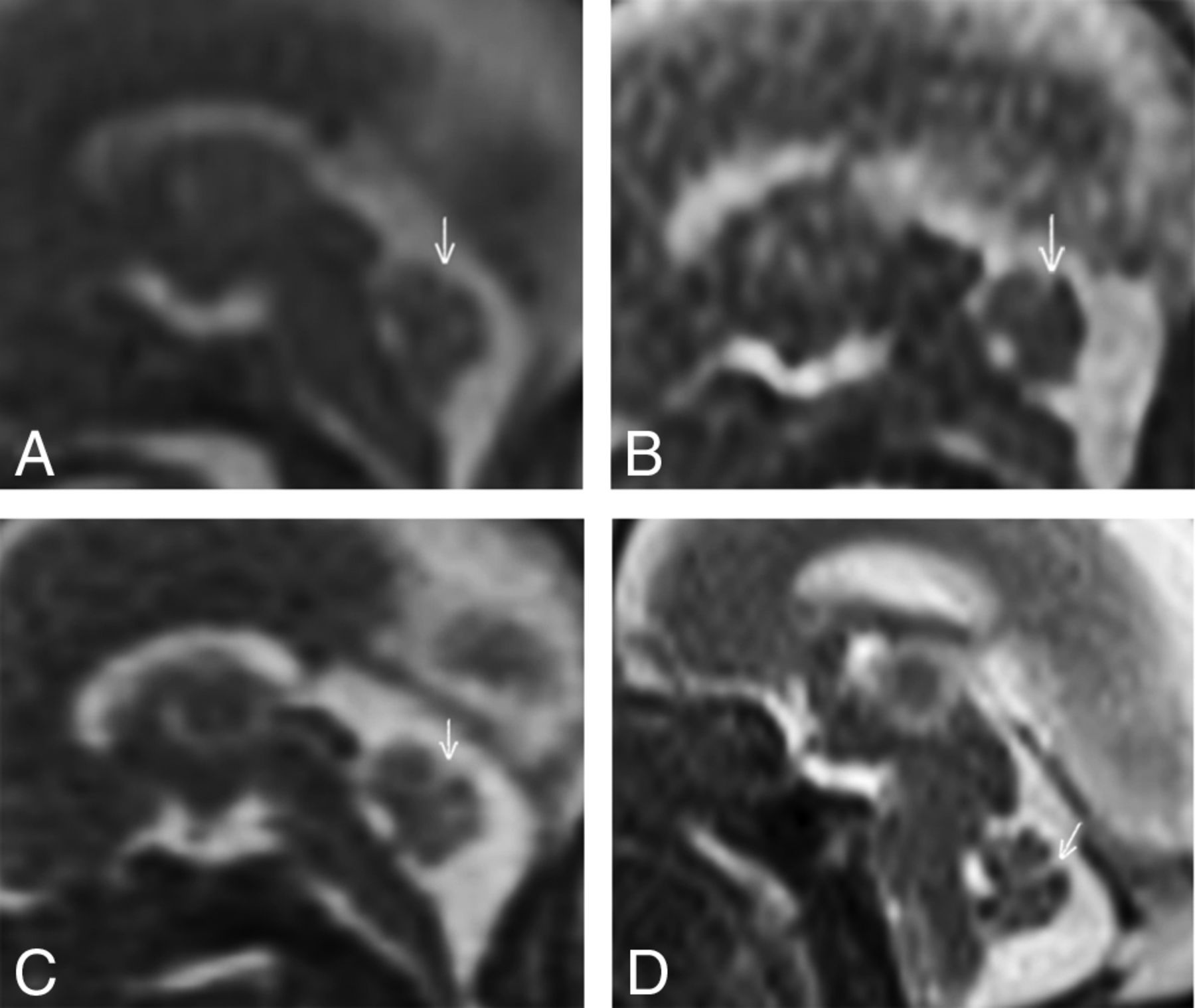
- Fig 1.
Sagittal T2 midline images of fetuses. Examples provide the qualitative assessment of the primary fissure of the cerebellum (arrows). A, Gestational age (GA), 21 weeks 1 day; 1.5T; score 0: no visible structure. B, GA, 21 weeks; 1.5T; score 1: partially visible structure. C, GA, 22 weeks; 1.5T; score 2: entirely visible structure, but ill-defined contour. D, GA, 20 weeks; 3T; score 3: entirely visible and sharp margins.
- Fig 2.
A coronal image was selected from each scan at the level of the third ventricle. One-millimeter ROIs were drawn in the developing brain layers: 1) germinal matrix, 2) periventricular, 3) subplate, and 4) cortical plate. These values were compared with an ROI outside the patient (air). This fetus had a GA of 20 weeks, scanned at 3T.
- Fig 3.
Estimated differences in the mean score between magnet types at each location from a linear mixed-effects model. Estimates are accompanied by 95% confidence intervals, adjusted for multiple testing.
- Fig 4.
The ratio between signal intensity and air at each location for each magnet. Each estimated ratio is shown as a small circle. Each superimposed box indicates the 25th percentile (lower part of the box), median (heavy horizontal line), and 75th percentile (upper part of the box). Note that the vertical axis has a logarithmic scale.


Tables
Fetal Sequences 1.5T, 3 planes, T2WI SSFSE Free-breathing TR/TE, 3000/87.8 ms, ST = 4 mm FOV, 340 × 340 mm2 Matrix, 320 × 224 px2 Time, 22.4 seconds 1.5T, 3 planes, steady-state FIESTA Free-breathing TR/TE, 3700/160 ms, ST = 4 mm FOV, 34 × 34 mm2 Matrix, 320 × 224 px2 Time, 25 seconds 3T, 3 planes, T2WI SSFSE Free-breathing TR/TE, 1400/96 ms, ST = 3 mm FOV, 280 × 280 mm2 Matrix, 320 × 288 px2 Time, 22.4 seconds Note:—SSFSE indicates single-shot FSE; px, pixel; ST, section thickness.
Magnet Score 0 1 2 3 1.5T 74 237 317 108 10.1% 32.2% 43.1% 14.7% 3T 20 71 152 309 3.6% 12.9% 27.5% 56.0% Location 1.5T 3T Germinal 7.3 (4.8–19.4) 72.3 (49.5–100.6) Periventricular layer 10.7 (6.2–21.1) 103.8 (54.9–181.7) Subplate 12.0 (8.4–25.4) 165.2 (97.2–270.1) Cortical 9.7 (5.5–19.5) 90.1 (61.7–119.4) Note:—IQR indicates interquartile range.


