
Article Figures & Data
Figures
- Fig 1.
Schematic representation of ascending and descending thoracic vertebral arteries. In its most typical form, a descending thoracic VA (right side) originates from the pretransverse segment (V1) of the cervical VA and curves sharply medially and caudally to enter the last transverse foramen or the first costotransverse space. It then continues caudally, passing through 1 or more costotransverse space, generally branching off a complete set of branches for the second and third thoracic ISAs (T2 and T3) and the medial branch of the first thoracic ISA (T1). In about 50% of cases, a descending thoracic VA provides an important contribution to the spinal cord vascularization. An ascending thoracic VA (left side) is the cranial prolongation of a thoracic ISA, which passes through 1 or more costotransverse space before continuing as a normal cervical VA. Strictly speaking, the thoracic VA segment only corresponds to the portion of the vessel delimited by costotransverse spaces (highlighted in green).
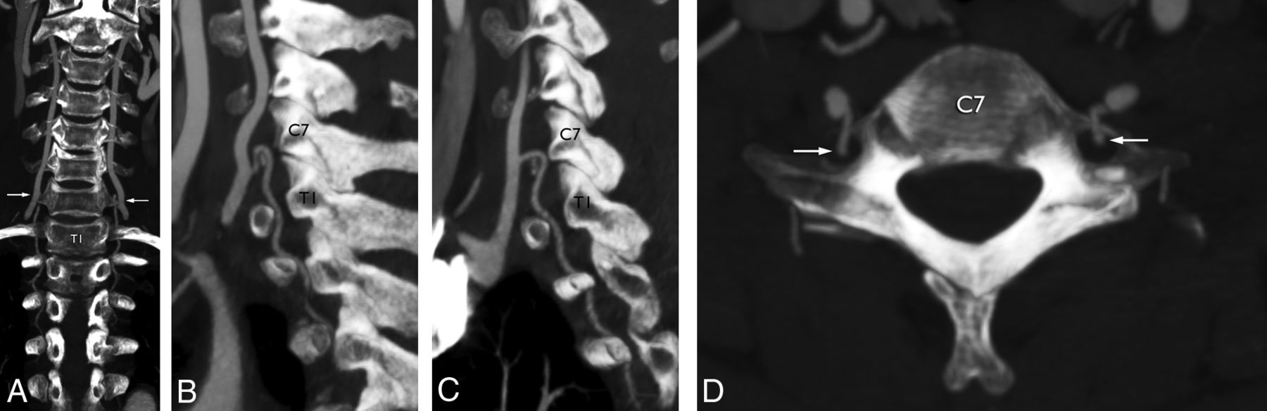
- Fig 2.
A 64-year-old man with a right descending thoracic vertebral artery. A, DSA, right VA injection, posteroanterior projection, shows a VA originating from the subclavian artery and dividing into ascending cervical (large white arrow) and descending thoracic (white arrowhead) VAs at the C7–T1 level. The descending thoracic VA provides the medial branches of C7 (small white arrow) and T1 (double small white arrow) and complete sets of branches for the T2 and T3 ISAs. The second thoracic ISA provides a prominent anterior radiculomedullary artery (small black arrow), which supplies the anterior spinal artery (black arrowheads). B, CTA, coronal reconstruction at the level of the transverse foramina. The ascending (large white arrow) and descending (white arrowhead) branches of the VA are identified. The small gray arrows point to the descending thoracic VA down to its termination at T3 (small black arrow). C, CTA, multilevel axial reconstructions, shows the course of the descending thoracic VA (small gray arrows) from its origin at C7–T1 (white arrowhead).
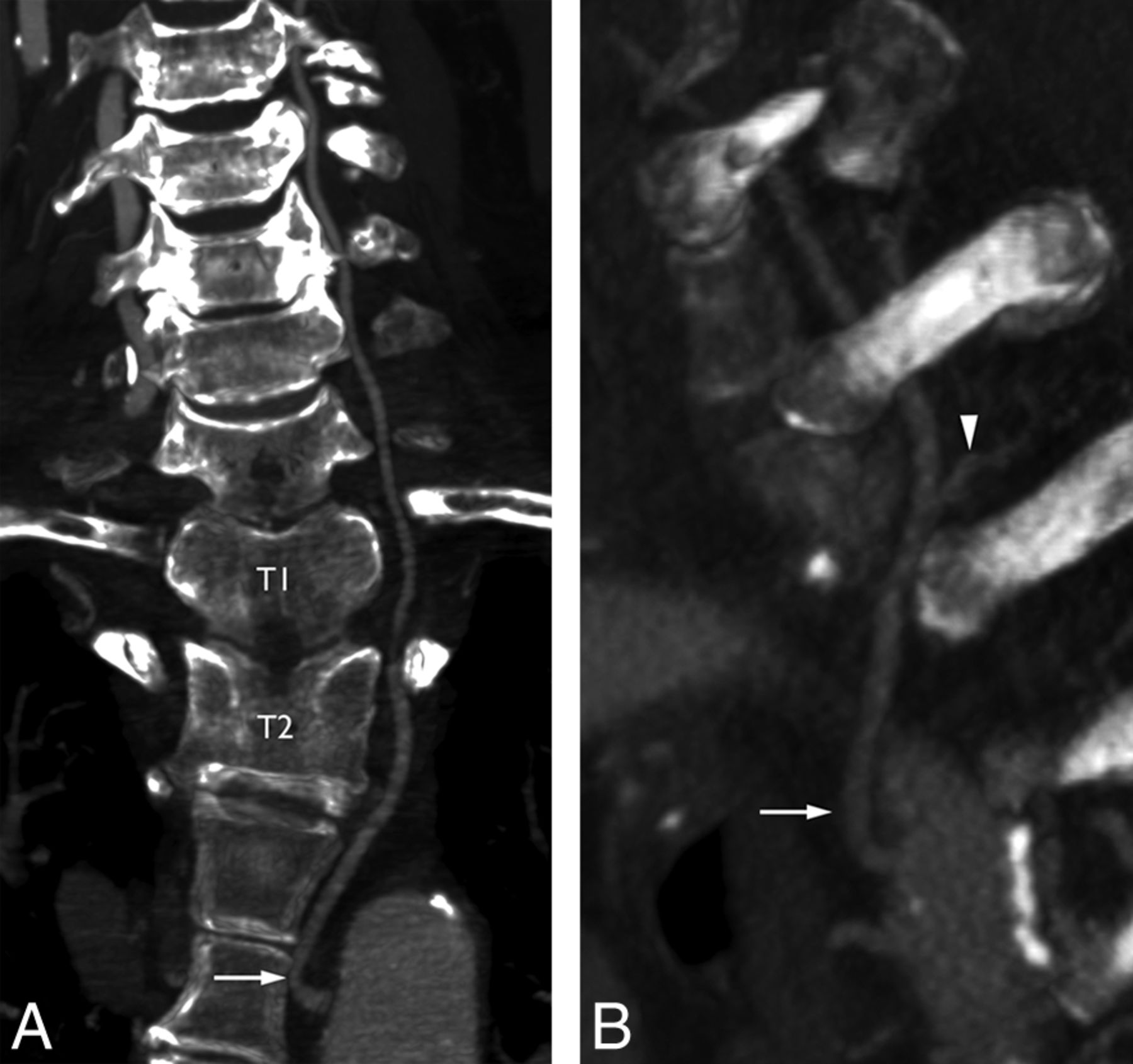
- Fig 3.
A 67-year-old woman with a right descending thoracic vertebral artery. A, DSA, right VA injection, posteroanterior projection. The VA divides into ascending cervical (large white arrow) and descending thoracic (white arrowhead) VAs at the C7–T1 level. The descending thoracic VA provides the medial branch of T1, which gives off a prominent anterior radiculomedullary artery (small black arrow). Note that the descending thoracic VA extends beyond the FOV and may provide additional ISAs. A prominent T2 dorsal (muscular) branch is seen coursing medially and caudally (black arrowhead) and should not be confused with a bronchial artery. B, DSA, right VA injection, nonsubtracted posteroanterior projection, documents the close relationship between the descending thoracic VA and the cervical pedicles (asterisks), notably at the level of the transverse foramina (small white arrows).
- Fig 4.
A 59-year-old woman with bilateral descending thoracic vertebral arteries. A, CTA, coronal reconstruction at the level of the transverse foramina, documents bilateral and symmetric descending thoracic VAs, both originating at C6–C7 (arrows) and ending at T4 on the right and T3 on the left. B, CTA, sagittal reconstruction at the level of the left transverse foramina. Note that the level of the variant (C6–C7) is indicated by its tight curve over the anterior margin of the transverse foramen rather than by the actual level of the bifurcation of the common VA trunk (C7–T1). C, CTA, sagittal reconstruction at the level of the right transverse foramina. D, CTA, axial reconstruction, shows the sharp posterior turn of the descending thoracic VA to pass through the C7 transverse foramina (white arrows).
- Fig 5.
A 30-year-old man with a right-sided descending thoracic vertebral artery. A, DSA, right VA injection, posteroanterior projection, subtracted view. Note the typical appearance of the descending thoracic VA, which provides, in this case, medial branches for C7 and T1 and a full set of intersegmental braches for T2 (the asterisks indicate the right T1 and T2 pedicles in all images). B, DSA, right VA injection, posteroanterior projection, nonsubtracted view. C, DSA, right costocervical trunk injection, posteroanterior projection, subtracted view. The existence of a supreme intercostal artery (white arrow) is independent of the presence of a descending thoracic VA. In this case, the supreme intercostal artery provides the C7 ISA (arrowhead) and the lateral branch of the T1 ISA (black arrow). D, DSA, right costocervical trunk injection, nonsubtracted posteroanterior projection. Contrary to a descending thoracic VA, the supreme intercostal artery lies away from the vertebral pedicles (hence outside the costotransverse spaces).
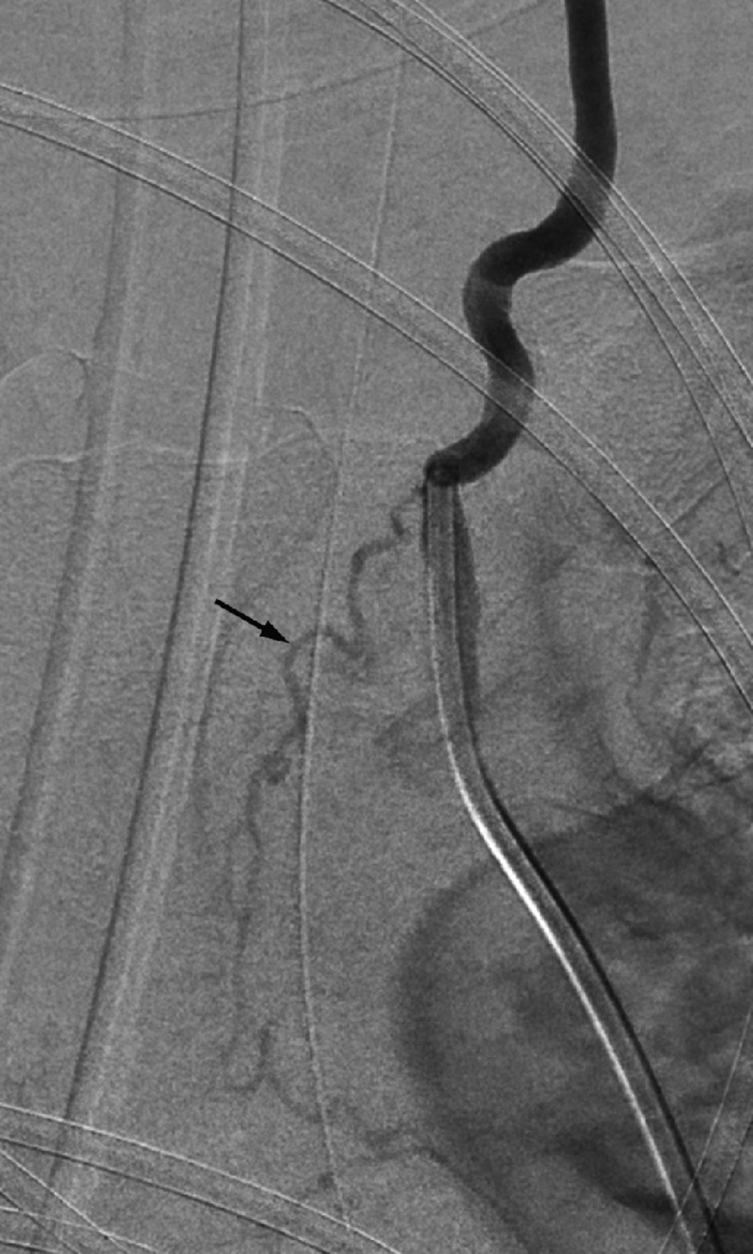
- Fig 6.
A 47-year-old woman with a right descending thoracic VA. This typical descending thoracic VA provides, in addition to the second thoracic ISA (white arrow), a prominent bronchial artery (black arrow).
- Fig 7.
A 20-year-old man with a left ascending thoracic VA. A, DSA, left T2 ISA injection, posteroanterior projection, subtracted view. The T2 ISA (white arrow) provides the second intercostal artery (c) before crossing through the T2 costotransverse space (white arrowhead). It then branches off contributions for the T1 (b) and C7 (a) ISAs and continues cranially as the cervical VA (black arrow). B, DSA, left T2 ISA injection, posteroanterior projection, nonsubtracted view. The asterisk indicates the approximate level of the origin of the left subclavian artery from the aortic arch, illustrating the distance separating the latter from the left T2 ostium. C, CT, axial view at the T2 level, shows a marked asymmetry of the size of the T2 costotransverse spaces. This sign should be taken into consideration when planning an upper thoracic spine procedure—for example, when choosing a needle path for a vertebral biopsy or a vertebroplasty.
- Fig 8.
A 64-year-old man with a left ascending thoracic VA. A, CTA, coronal MIP reconstruction, shows the origin of the left T2 ISA (white arrow) and its passage through the T2 and T1 costotransverse spaces. The vessel then continues cranially as the cervical VA. B, CTA, oblique MIP reconstruction, documents the T2 posterior intercostal artery (arrowhead), therefore identifying the ascending thoracic VA as being derived from the second thoracic ISA (arrow).
- Fig 9.
A 31-year-old woman with an atypical descending thoracic VA. A, DSA, left supreme intercostal artery injection, posteroanterior projection, subtracted view shows an example of a descending thoracic VA originating from the supreme intercostal artery (atypical form of Krassnig5); in this case, there is no connection with the cervical VA. The supreme intercostal artery provides the C7 (a) and T1 (b) ISAs as well as the lateral branch of the T2 ISA (c). A prominent ascending branch curves sharply downward to enter the first costotransverse space and continues caudally as a descending thoracic VA (black arrow) passing through the T2 and T3 the costotransverse spaces. This descending thoracic VA provides the T3 ISA (d) and the medial branch of the T2 ISA (gray arrowhead). Note the opacification of the T4 ISA (e) via a small paravertebral anastomotic connection (as shown in Fig 1, right side). The C7 ISA provides both an anterior (black arrowhead) and a posterior (white arrowhead) radiculomedullary artery. B, DSA, left supreme intercostal artery injection, posteroanterior projection, nonsubtracted view of same image.
- Fig 10.
Schematic representation of 2 types of descending thoracic VA variants. The descending thoracic VA (atypical form of Krassnig5) depicted on the right side is, as in Fig 9, connected with the supreme intercostal artery rather than the cervical VA. In the configuration shown on the left side (atypical form of Bühler21), the descending thoracic VA is the downward continuation of a thoracic ISA of aortic origin. a. indicates artery.
- Fig 11.
Examples of vertebral configurations similar to but distinct from thoracic VAs. A, CTA, sagittal MIP reconstruction. This case shows a descending branch with an appearance similar to that of a descending thoracic VA (arrow). However, the bifurcation is located at C5–C6, and the descending branch stops at C7, short of the first costotransverse space. There is, therefore, no thoracic segment. B, CTA, coronal MIP reconstruction, shows a vertebral artery originating from the aorta distal to the subclavian artery and entering the seventh transverse foramen (arrow). This vessel corresponds to a persistent seventh ISA and has no thoracic component.
- Fig 12.
First known representation of a descending thoracic vertebral artery by Joseph Maclise for Richard Quain in 1844 (Fig 5, Plate XXII).22 The figure accurately depicts a typical descending thoracic VA on the right side (12) and an atypical form on the left (12′), as later defined by Krassnig.5 While both vessels are clearly coursing within the costotransverse spaces, the distinction from the supreme (or superior) intercostal arteries has not yet been established. The original legend is the following: “The left vertebral enters the last cervical vertebra. On the right side the superior intercostal artery is derived from the vertebral, and passes downwards into the thorax through the foramen in the transverse process of the seventh cervical vertebra, and afterward between the necks of the ribs and the corresponding transverse processes of the dorsal vertebræ. It will be observed that the superior intercostal of the other side descends also between the ribs and the processes of the vertebræ; and that the first aortic intercostal branch occupies a similar position in reference to the bones.” 6 indicates right vertebral, 6′, left vertebral; 12, right superior intercostal; 12′, left superior intercostal; 12†, first aortic intercostal; 13, deep cervical (arteria profunda cervicis).
- Fig 13.
Reproduction of the original Fig 21 of Pensa,23 showing an example of a typical descending thoracic VA providing T1 partially and T2 entirely. The supreme intercostal artery (originating from an unlabeled costocervical trunk) is clearly distinguished from the thoracic VA, both in the illustration and in the Pensa's text, the latter emphasizing the respective precostal and retrocostal locations of the 2 vessels. However, the nomenclature remains uncertain; Pensa labeled the descending thoracic VA as an arteria intercostalis suprema (A.i.s). V indicates vertebral artery; T.t.c, thyrocervical trunk.
- Fig 14.
Vertebral artery variant in a 55-year-old woman. This left vertebral injection (posteroanterior projection, subtracted, A, and nonsubtracted, B, images) documents a vertebral artery that originates from the supreme intercostal artery (ie, a persistent seventh ISA) and enters the transverse canal at C7 (arrow). Because the vertebral artery becomes dominant in the adult stage, it falsely suggests an opposite relationship (ie, a supreme intercostal artery of vertebral origin).
- Fig 15.
Vertebral artery variant in a 58-year-old woman. Left vertebral injection (posteroanterior projection) documents a prominent aberrant bronchial branch (arrow) of vertebral origin.
- Fig 16.
Reproduction of the original Fig 16 (Vol. 1, page 36) of Adachi.19 Adachi presents a left ascending thoracic VA originating from the aorta at the T3–T4 level and passing behind the head of the first rib to enter the first costotransverse space before continuing cranially as the left cervical vertebral artery.
- Fig 17.
Examples of ascending and descending thoracic aortas in birds (from Pensa23). A, Bilateral ascending thoracic vertebral arteries in the chicken (Gallus domesticus); note a cranial anastomosis with the costocervical trunk. B, Bilateral descending thoracic vertebral arteries in the domestic pigeon (Columba livia). V indicates vertebral artery; Ai.c.d., arteria intercostalis communis descendens; A.cr., arteria cruralis.


Tables
Nine angiographic observations of the descending thoracic vertebral artery
Patient Sex Age (yr) Level Side ISA Branches Complete Partial ARMA Br 1 M 64 C7–T1 R T2–T3 Medial C7–T1 T2 Yes 2 W 67 C7–T1 R T2–T4?a Medial T1 T1 No 3 W 59 C6–C7 L T2–T3 Medial T1 T2 No 4 M 30 C6–C7 R T2 Medial C7 T2 No 5 W 47 C6–C7 R T3 Medial T2 – Yes 6 M 18 C7–T1 R T2–T3 Medial T1 T2 No 7 W 56 C7–T1 R T2–T3 Medial T1 – No 8 W 60 C7–T1 R T2–T4?a Medial T1 – No 9 W 31 C6–C7 R T1–T2 ?b – No Note:—ARMA indicates anterior radiculomedullary artery; Br, bronchial artery; R, right; L, left; –, none; M, man; W, woman.
↵a Question mark indicates that the caudal extension of the descending thoracic VA was beyond the FOV.
↵b Question mark indicates that motion artifacts prevented evaluating the presence of a proximal medial branch.


Jump to section
Related Articles
Cited By...
- No citing articles found.