
Article Figures & Data
Figures
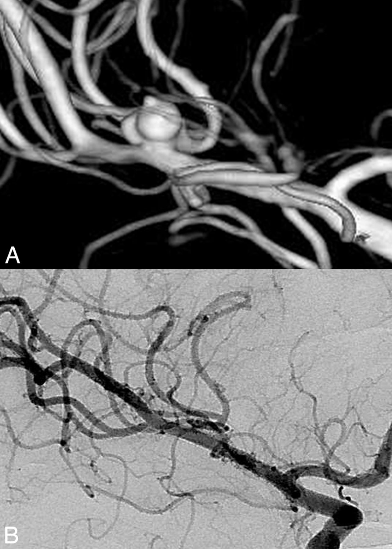
- Fig 1.
Pretreatment 3D image (A) shows a multilobulated right MCA aneurysm with a bleb. Note that the superior trunk originates from the sac. Control DSA 1 year after Pipeline Embolization Device placement. An image at a corresponding angle (B) demonstrates total occlusion of the aneurysm with the branch coming off the patent sac (Cekirge-Saatci class 1a occlusion).
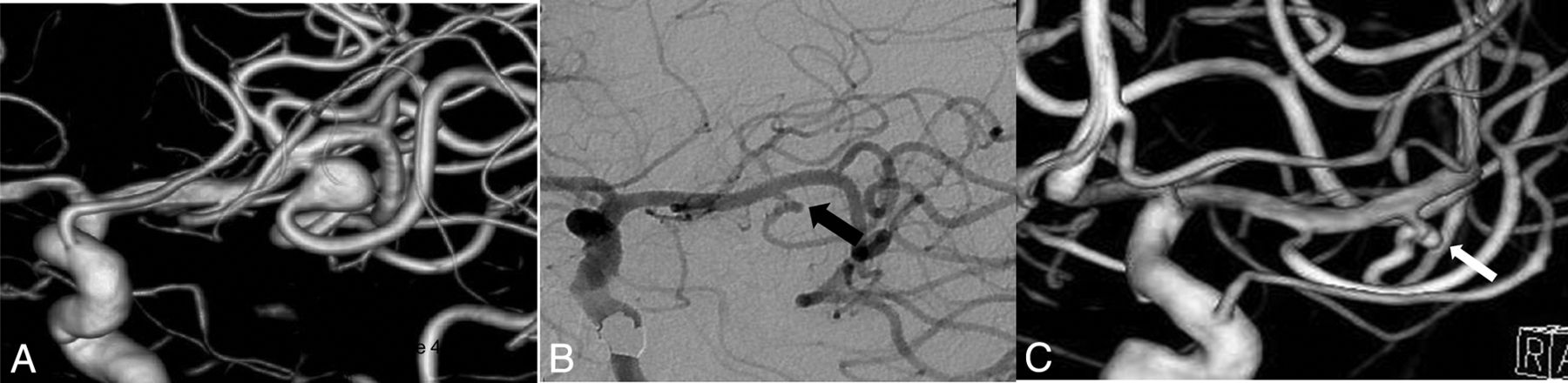
- Fig 2.
Pretreatment 3D (A) and 2D (B) images show a small, irregular right MCA aneurysm with an early bifurcating branch (arrow) originating from the sac. Corresponding DSA obtained 1 year after PED treatment (C) confirms the total occlusion of the aneurysm; the branch coming off the sac (arrow) is reduced in caliber (Cekirge-Saatci class 1b occlusion).
- Fig 3.
Pretreatment 3D image (A) shows a left MCA aneurysm with the inferior trunk coming off the sac. Posttreatment 1- (B) and 2-year (C) angiograms confirm the stable occlusion of the sac with the patent inferior trunk (arrow) having a tortuous origin (Cekirge-Saatci class 5 occlusion).
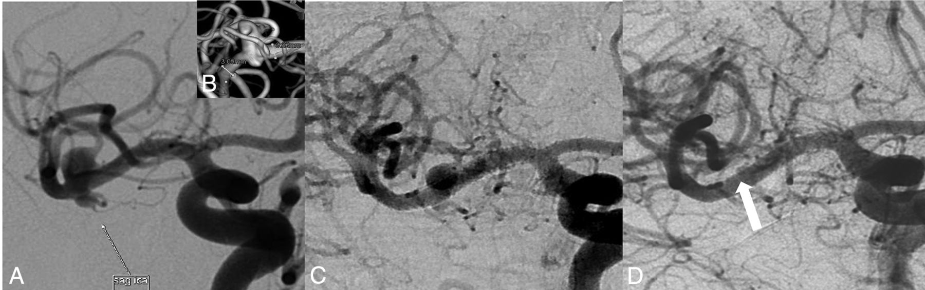
- Fig 4.
Pretreatment 2D (A) and 3D (B) images show an irregular right MCA aneurysm with a bleb. Note that the aneurysm has no neck, and a branch is originating from the sac. Six-month control DSA image at a corresponding angle (C) reveals the sac decreased in size, but still filling (class 2). Correlative 1-year control DSA image demonstrates total occlusion of the aneurysm sac with the relevant branch filling retrogradely (white arrow) (ie, Cekirge-Saatci class 1c occlusion).


Tables
PRE tx Discharge mRS (n = 58) 6-Month mRS (n = 58) ≥12-Month mRS (n = 58) mRS No. of Patients 0 1 2 3 0 1 2 3 0 1 2 0 12 12 0 0 0 12 0 0 0 12 0 0 1 41 0 37 1a 2a 26 11 4a 1a 30 9 3 2 5 0 4 1 1a 3 1 0 0a 3 1 0 Total 58 12 41 2 3 41 12 4 1 45 10 3 Note:—PRE tx indicates pretreatment values.
↵a Number of patients with clinical worsening in regard to pretreatment clinical status.
- Table 2:
Immediate postprocedure angiographic results and angiographic outcomes in controls
Cekirge-Saatci Evaluation Scale Immediate Angiographic Outcome ≥12-Month Angiographic Outcome Third (Midterm) Angiographic Outcome 1a 3 (4.8%) 18 (28.6%) 16 (40.0%) 1b 0 14 (22.2%) 10 (25.0%) 1c 0 13 (20.6%) 8 (20.0%) 2 0 2 (3.2%) 0 3 1 (1.6%) 1 (1.6%) 1 (2.5%) 4a 30 (47.6%) 0 0 4b 29 (46%) 0 0 5 0 15 (23.8%) 5 (12.5%) Total 63 (100%) 63 (100%) 40 (100%) χ2, 48.175; df, 3 χ2, 24.524; df, 5 χ2,15.750; df, 4 Significance level, P < .0001 Significance level, P = .0002 Significance level, P = .0034 Jailed Branch Fate 6-Month Follow-Up 12-Month Follow-Up 3rd (Midterm) Follow-Up Asymptomatic narrowing 18 28.6% 10 15.9% 5 12.5% Symptomatic narrowing 0 0 0 0 0 0 Asymptomatic occlusion 9 14.3% 0 0 1 2.5% Symptomatic occlusion 2 3.2% 0 0 0 0 Better opacification or “reopening” 0 0 5 7.9% 0 0 No change 34 54.0% 48 76.2% 34 85.0% Total 63 100.0% 63 100% 40 100% χ2, 36.365; df, 3 χ2, 52.667; df, 2 χ2, 48.650; df, 2 Significance level, P < .0001 Significance level, P < .0001 Significance level, P < .0001


Jump to section
Related Articles
Cited By...
- Two year follow-up of distal unruptured intracranial aneurysms treated with a surface modified flow diverter under prasugrel monotherapy
- Flow diversion for the treatment of intracranial bifurcation aneurysms: a systematic review and meta-analysis
- In vitro flow diversion effect of the ReSolv stent with the shelf technique in a bifurcation aneurysm model
- Magnetic resonance perfusion imaging findings following flow diversion in patients with complex middle cerebral artery bifurcation aneurysms: a single-center analysis regarding the jailed cortical branches
- In vitro flow diversion effect of the ReSolv stent with the shelf technique in a bifurcation aneurysm model
- Endoluminal flow diverting stents for middle cerebral artery bifurcation aneurysms: multicenter cohort
- Treatment of distal unruptured intracranial aneurysms using a surface-modified flow diverter under prasugrel monotherapy: a pilot safety trial
- Long-term safety and efficacy of distal aneurysm treatment with flow diversion in the M2 segment of the middle cerebral artery and beyond
- Aspirin monotherapy in the treatment of distal intracranial aneurysms with a surface modified flow diverter: a pilot study
- Outcome of intracranial flow diversion according to the antiplatelet regimen used: a systematic review and meta-analysis
- Treatment of Unruptured Distal Anterior Circulation Aneurysms with Flow-Diverter Stents: A Meta-Analysis
- Toward Better Understanding of Flow Diversion in Bifurcation Aneurysms
- Treatment of Middle Cerebral Artery Aneurysms with Flow-Diverter Stents: A Systematic Review and Meta-Analysis