Advertisement

Main menu

  • Home
  • Content
    • Current Issue
    • Accepted Manuscripts
    • Article Preview
    • Past Issue Archive
    • Video Articles
    • AJNR Case Collection
    • Case of the Week Archive
    • Case of the Month Archive
    • Classic Case Archive
  • Special Collections
    • AJNR Awards
    • Low-Field MRI
    • Alzheimer Disease
    • ASNR Foundation Special Collection
    • Photon-Counting CT
    • View All
  • Multimedia
    • AJNR Podcasts
    • AJNR SCANtastic
    • Trainee Corner
    • MRI Safety Corner
    • Imaging Protocols
  • For Authors
    • Submit a Manuscript
    • Submit a Video Article
    • Submit an eLetter to the Editor/Response
    • Manuscript Submission Guidelines
    • Statistical Tips
    • Fast Publishing of Accepted Manuscripts
    • Graphical Abstract Preparation
    • Imaging Protocol Submission
    • Author Policies
  • About Us
    • About AJNR
    • Editorial Board
    • Editorial Board Alumni
  • More
    • Become a Reviewer/Academy of Reviewers
    • Subscribers
    • Permissions
    • Alerts
    • Feedback
    • Advertisers
    • ASNR Home

User menu

  • Alerts
  • Log in

Search

  • Advanced search
American Journal of Neuroradiology

American Journal of Neuroradiology

ASHNR American Society of Functional Neuroradiology ASHNR American Society of Pediatric Neuroradiology ASSR
  • Alerts
  • Log in

Advanced Search

  • Home
  • Content
    • Current Issue
    • Accepted Manuscripts
    • Article Preview
    • Past Issue Archive
    • Video Articles
    • AJNR Case Collection
    • Case of the Week Archive
    • Case of the Month Archive
    • Classic Case Archive
  • Special Collections
    • AJNR Awards
    • Low-Field MRI
    • Alzheimer Disease
    • ASNR Foundation Special Collection
    • Photon-Counting CT
    • View All
  • Multimedia
    • AJNR Podcasts
    • AJNR SCANtastic
    • Trainee Corner
    • MRI Safety Corner
    • Imaging Protocols
  • For Authors
    • Submit a Manuscript
    • Submit a Video Article
    • Submit an eLetter to the Editor/Response
    • Manuscript Submission Guidelines
    • Statistical Tips
    • Fast Publishing of Accepted Manuscripts
    • Graphical Abstract Preparation
    • Imaging Protocol Submission
    • Author Policies
  • About Us
    • About AJNR
    • Editorial Board
    • Editorial Board Alumni
  • More
    • Become a Reviewer/Academy of Reviewers
    • Subscribers
    • Permissions
    • Alerts
    • Feedback
    • Advertisers
    • ASNR Home
  • Follow AJNR on Twitter
  • Visit AJNR on Facebook
  • Follow AJNR on Instagram
  • Join AJNR on LinkedIn
  • RSS Feeds

AJNR Awards, New Junior Editors, and more. Read the latest AJNR updates

Research ArticleNeurointervention

Treatment of Intra- and Extracranial Aneurysms Using the Flow-Redirection Endoluminal Device: Multicenter Experience and Follow-Up Results

F. Drescher, W. Weber, A. Berlis, S. Rohde, A. Carolus and S. Fischer
American Journal of Neuroradiology January 2017, 38 (1) 105-112; DOI: https://doi.org/10.3174/ajnr.A4964
F. Drescher
aFrom the Knappschaftskrankenhaus Bochum (F.D., W.W., S.F.), Universitätsklinik, Institut für Diagnostische und Interventionelle Radiologie, Neuroradiologie, Nuklearmedizin, Bochum, Germany
  • Find this author on Google Scholar
  • Find this author on PubMed
  • Search for this author on this site
  • ORCID record for F. Drescher
W. Weber
aFrom the Knappschaftskrankenhaus Bochum (F.D., W.W., S.F.), Universitätsklinik, Institut für Diagnostische und Interventionelle Radiologie, Neuroradiologie, Nuklearmedizin, Bochum, Germany
bKnappschaftskrankenhaus Recklinghausen Klinik für Radiologie Neuroradiologie und Nuklearmedizin (W.W.), Recklinghausen, Germany
  • Find this author on Google Scholar
  • Find this author on PubMed
  • Search for this author on this site
  • ORCID record for W. Weber
A. Berlis
cKlinikum Augsburg (A.B.), Klinik für Diagnostiche Radiologie und Neuroradiologie, Augsburg, Germany
  • Find this author on Google Scholar
  • Find this author on PubMed
  • Search for this author on this site
  • ORCID record for A. Berlis
S. Rohde
dKlinikum Dortmund (S.R.), Klinik für Radiologie und Neuroradiologie, Dortmund, Germany
  • Find this author on Google Scholar
  • Find this author on PubMed
  • Search for this author on this site
  • ORCID record for S. Rohde
A. Carolus
eKnappschaftskrankenhaus Bochum (A.C.), Universitätsklinik, Klinik für Neurochirurgie, Bochum, Germany.
  • Find this author on Google Scholar
  • Find this author on PubMed
  • Search for this author on this site
  • ORCID record for A. Carolus
S. Fischer
aFrom the Knappschaftskrankenhaus Bochum (F.D., W.W., S.F.), Universitätsklinik, Institut für Diagnostische und Interventionelle Radiologie, Neuroradiologie, Nuklearmedizin, Bochum, Germany
  • Find this author on Google Scholar
  • Find this author on PubMed
  • Search for this author on this site
  • ORCID record for S. Fischer
  • Article
  • Figures & Data
  • Info & Metrics
  • Responses
  • References
  • PDF
Loading

Article Figures & Data

Figures

  • Tables
  • Fig 1.
    • Download figure
    • Open in new tab
    • Download powerpoint
    Fig 1.

    Dual-layer structure of the FRED. The fluoroscopic visibility results from 2 interwoven helical marker strands delineating the dual-layer section (working length) and 4 radiopaque markers at the flared ends.

  • Fig 2.
    • Download figure
    • Open in new tab
    • Download powerpoint
    Fig 2.

    A, A male patient with multiple asymptomatic intracranial aneurysms with a small proximal aneurysm of the A1 segment status post stent-assisted coil occlusion of a distal ICA aneurysm, right oblique and cranial views. B, Placement of a 3.5-/13/7-mm FRED with its flared ends extending toward the ICA bifurcation to cover the aneurysm with the dual-layer part of the device, right oblique and cranial views. C, Stasis of contrast material up to the venous phase, right oblique and cranial views. D, 4-month follow-up angiography with complete occlusion of the aneurysm, right oblique and cranial views.
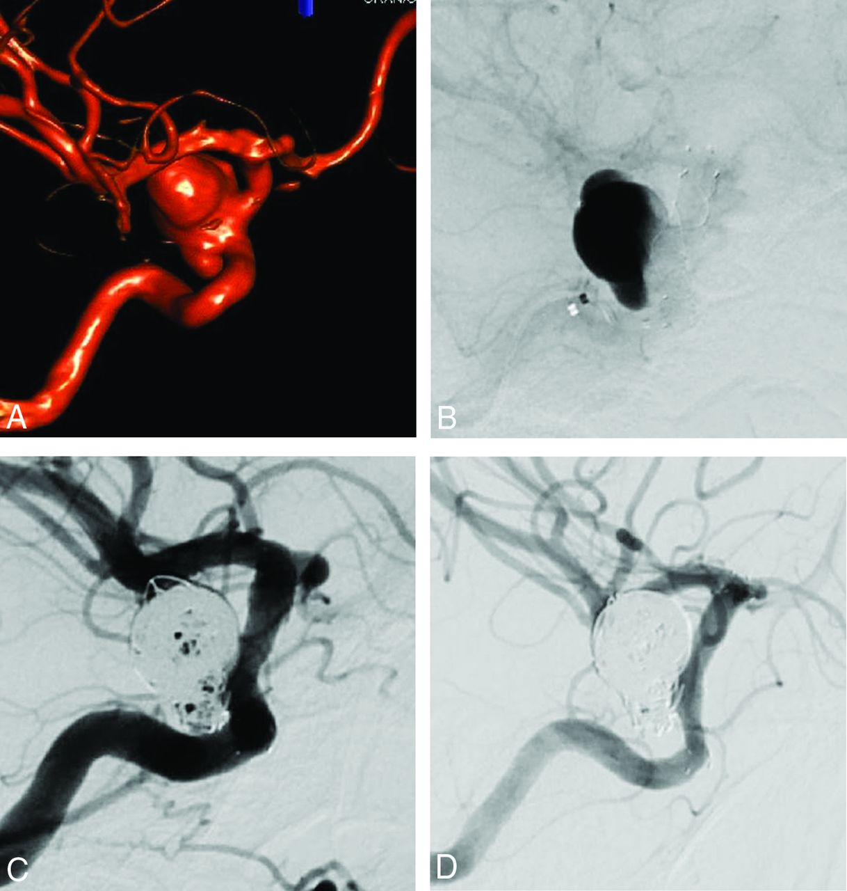
  • Fig 3.
    • Download figure
    • Open in new tab
    • Download powerpoint
    Fig 3.

    A, Finding of a large irregularly shaped aneurysm of the right ICA (posterior communicating segment) in a woman, causing symptoms of mass effect, 3D rotational angiography. B, Placement of a 4.0-/18/12-mm FRED after jailing of a microcatheter. Intra-aneurysmal stasis of contrast material, lateral view. C, Loose coil occlusion of the aneurysm, lateral view. D, Complete occlusion of the aneurysm on 3-month follow-up angiography, lateral view.
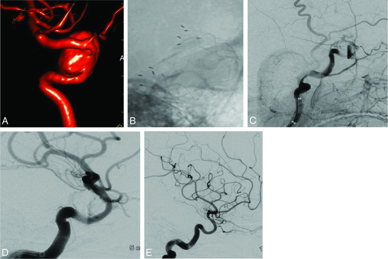
  • Fig 4.
    • Download figure
    • Open in new tab
    • Download powerpoint
    Fig 4.

    A, Incidental finding of a cavernous ICA aneurysm in a female patient, 3D rotational angiography. B, Placement of a 3.5-/22/16-mm FRED with incomplete expansion in the midsection of the flow diverter, recorded with fluoroscopy. C, Acute thrombotic occlusion of the ICA caused by the incompletely expanded flow diverter, right anterior oblique view. D, Status post dilation of the FRED with a coronary percutaneous transarterial angioplasty balloon (Sequent Medical, 2.75 × 10 mm), right anterior oblique view. E, 3-month follow-up angiography demonstrates complete occlusion of the cavernous aneurysm with regular opacification of the ICA, lateral view.

Tables

  • Figures
    • View popup
    Table 1:

    Inclusion and exclusion criteria for treatment with FRED

    Criteria
    In favor of treatment with FRED
        Intradural incidental aneurysm
        Intradural or extradural symptomatic aneurysm (mass effect)
        Supposed difficulty for coil treatment alone (dome-to-neck ratio of <1.2, broad-based aneurysm, fusiform morphology, blisterlike shape)
        Difficulty or impossibility of neurosurgical clip placement due to aneurysm morphology or anatomic location
        Acutely ruptured aneurysms without any alternative neurosurgical or endovascular treatment option
        Aneurysm remnant or reperfusion after endovascular or microsurgical treatment
        Documented response to medicamentous platelet function inhibition
    Exclusion for treatment with FRED
        Intradural aneurysm with a definable neck
        Intradural bifurcation aneurysm
        Documented nonresponse to medicamentous platelet function inhibition
        Patient preference for alternative treatment options
        Patient preference against any treatment
    • View popup
    Table 2:

    Summary of the locations of the treated aneurysms

    LocationAneurysms (No.)Ratio
    Anterior circulation (n = 39; 75.0%)
        ICA cervical815.4%
        ICA cavernous35.8%
        ICA paraophthalmic2140.5%
        ICA Pcom35.8%
        ACA23.8%
        MCA23.8%
    Posterior circulation (n = 13; 25.0%)
        BA23.8%
        VA V4917.3%
        PCA23.8%
    Total52100.0%
    • Note:—ACA indicates anterior cerebral artery; BA, basilar artery; VA, vertebral artery; PCA, posterior cerebral artery; Pcom, posterior communicating; MCA, middle cerebral artery.

    • View popup
    Table 3:

    Occlusion rates at 3- and 12-month follow-up

    Occlusion3-Month Follow-UpRatio12-Month Follow-UpRatio
    Complete occlusion2558.1%2775.0%
    Minor neck remnant1125.6%822.2%
    Major residual filling37.0%12.8%
    Unchanged filling49.3%00.0%
    Total43100.0%36100.0%
    • View popup
    Table 4:

    Summary of complications during the follow-up period

    Adverse EventsAt Discharge (n = 52 cases)Ratio3-Month Follow-Up (n = 43 cases)Ratio12-Month Follow-Up (n = 36 cases)Ratio
    Hemorrhagic00.0%12.3%00.0%
    Thromboembolic, symptomatic11.9%00.0%12.8%
    Thromboembolic, asymptomatic35.7%24.7%12.8%
    Total47.6%37.0%25.6%
    • View popup
    Table 5:

    Morbidity and mortality during the follow-up period

    At Discharge (n = 50 patients)Ratio3-Month Follow-Up (n = 41 patients)Ratio12-Month Follow-Up (n = 34 patients)Ratio
    Morbidity12.0%00.0%12.0%
    Mortality00.0%12.0%00.0
    • View popup
    Table 6:

    Complication rates with different flow diverters including the period under review

    AuthorYearFlow DiverterNo. of PatientsThromboembolic ComplicationsHemorrhagic ComplicationsMedian Follow-Up Time (mo)
    Möhlenbruch et al82015FRED2914.0%3.0%6
    Poncyljusz et al112013FRED617.0%0.0%3
    Briganti et al122016FRED200.0%0.0%12
    Lubicz et al132015Silk2623.1%11.5%6
    Briganti et al42012Silk + Pipeline2734.8%5.5%3
    Colby et al72016Pipeline Flex442.0%0.0%Not applicable
    De Vries et al142013Surpass3713.5%5.4%12
    Fischer et al152015p641303.0%0.0%9
    Our data2016FRED4815.4%1.9%12
PreviousNext
Back to top

In this issue

American Journal of Neuroradiology: 38 (1)
American Journal of Neuroradiology
Vol. 38, Issue 1
1 Jan 2017
  • Table of Contents
  • Index by author
  • Complete Issue (PDF)
Advertisement
Print
Download PDF
Email Article

Thank you for your interest in spreading the word on American Journal of Neuroradiology.

NOTE: We only request your email address so that the person you are recommending the page to knows that you wanted them to see it, and that it is not junk mail. We do not capture any email address.

Enter multiple addresses on separate lines or separate them with commas.
Treatment of Intra- and Extracranial Aneurysms Using the Flow-Redirection Endoluminal Device: Multicenter Experience and Follow-Up Results
(Your Name) has sent you a message from American Journal of Neuroradiology
(Your Name) thought you would like to see the American Journal of Neuroradiology web site.
CAPTCHA
This question is for testing whether or not you are a human visitor and to prevent automated spam submissions.
Cite this article
F. Drescher, W. Weber, A. Berlis, S. Rohde, A. Carolus, S. Fischer
Treatment of Intra- and Extracranial Aneurysms Using the Flow-Redirection Endoluminal Device: Multicenter Experience and Follow-Up Results
American Journal of Neuroradiology Jan 2017, 38 (1) 105-112; DOI: 10.3174/ajnr.A4964

Citation Manager Formats

  • BibTeX
  • Bookends
  • EasyBib
  • EndNote (tagged)
  • EndNote 8 (xml)
  • Medlars
  • Mendeley
  • Papers
  • RefWorks Tagged
  • Ref Manager
  • RIS
  • Zotero
0 Responses
Respond to this article
Share
Bookmark this article
Treatment of Intra- and Extracranial Aneurysms Using the Flow-Redirection Endoluminal Device: Multicenter Experience and Follow-Up Results
F. Drescher, W. Weber, A. Berlis, S. Rohde, A. Carolus, S. Fischer
American Journal of Neuroradiology Jan 2017, 38 (1) 105-112; DOI: 10.3174/ajnr.A4964
del.icio.us logo Twitter logo Facebook logo Mendeley logo
  • Tweet Widget
  • Facebook Like
  • Google Plus One
Purchase

Jump to section

  • Article
    • Abstract
    • ABBREVIATIONS:
    • Materials and Methods
    • Results
    • Discussion
    • Conclusions
    • Footnotes
    • References
  • Figures & Data
  • Info & Metrics
  • Responses
  • References
  • PDF

Related Articles

  • No related articles found.
  • PubMed
  • Google Scholar

Cited By...

  • Efficacy and safety of the dual-layer flow-diverting stent (FRED) for the treatment of intracranial aneurysms
  • On Flow Diversion: The Changing Landscape of Intracerebral Aneurysm Management
  • Transient in-stent stenosis at mid-term angiographic follow-up in patients treated with SILK flow diverter stents: incidence, clinical significance and long-term follow-up
  • SAFE study (Safety and efficacy Analysis of FRED Embolic device in aneurysm treatment): 1-year clinical and anatomical results
  • Feasibility, complications, morbidity, and mortality results at 6 months for aneurysm treatment with the Flow Re-Direction Endoluminal Device: report of SAFE study
  • European Multicenter Study for the Evaluation of a Dual-Layer Flow-Diverting Stent for Treatment of Wide-Neck Intracranial Aneurysms: The European Flow-Redirection Intraluminal Device Study
  • Multicenter Experience with FRED Jr Flow Re-Direction Endoluminal Device for Intracranial Aneurysms in Small Arteries
  • Crossref
  • Google Scholar

This article has not yet been cited by articles in journals that are participating in Crossref Cited-by Linking.

More in this TOC Section

Neurointervention

  • Rescue Reentry in Carotid Near-Occlusion
  • Contour Neurovascular System: Five Year Follow Up
  • Effect of SARS-CoV2 on Endovascular Thrombectomy
Show more Neurointervention

Extracranial Vascular

  • Proximal Vertebral Artery Variants and Embryology
  • High-Risk Plaque Features in Carotid MRI
  • Nonstenotic Carotid Plaques and Stroke Review
Show more Extracranial Vascular

Similar Articles

Advertisement

Indexed Content

  • Current Issue
  • Accepted Manuscripts
  • Article Preview
  • Past Issues
  • Editorials
  • Editor's Choice
  • Fellows' Journal Club
  • Letters to the Editor
  • Video Articles

Cases

  • Case Collection
  • Archive - Case of the Week
  • Archive - Case of the Month
  • Archive - Classic Case

More from AJNR

  • Trainee Corner
  • Imaging Protocols
  • MRI Safety Corner
  • Book Reviews

Multimedia

  • AJNR Podcasts
  • AJNR Scantastics

Resources

  • Turnaround Time
  • Submit a Manuscript
  • Submit a Video Article
  • Submit an eLetter to the Editor/Response
  • Manuscript Submission Guidelines
  • Statistical Tips
  • Fast Publishing of Accepted Manuscripts
  • Graphical Abstract Preparation
  • Imaging Protocol Submission
  • Evidence-Based Medicine Level Guide
  • Publishing Checklists
  • Author Policies
  • Become a Reviewer/Academy of Reviewers
  • News and Updates

About Us

  • About AJNR
  • Editorial Board
  • Editorial Board Alumni
  • Alerts
  • Permissions
  • Not an AJNR Subscriber? Join Now
  • Advertise with Us
  • Librarian Resources
  • Feedback
  • Terms and Conditions
  • AJNR Editorial Board Alumni

American Society of Neuroradiology

  • Not an ASNR Member? Join Now

© 2025 by the American Society of Neuroradiology All rights, including for text and data mining, AI training, and similar technologies, are reserved.
Print ISSN: 0195-6108 Online ISSN: 1936-959X

Powered by HighWire