
Article Figures & Data
Figures
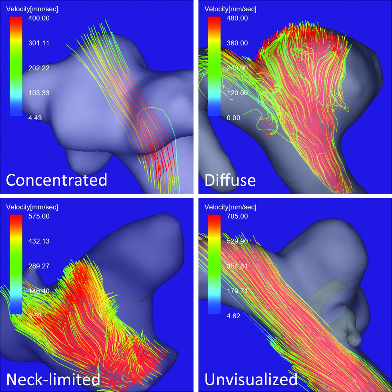
- Fig 1.
Classification of inflow jet patterns visualized on 4D flow MR images. Concentrated: An aneurysm on the anterior communicating artery with a concentrated inflow jet defined as a bundle of inflow streamlines intruding into the aneurysmal dome without dispersion in 20% of the width of the streamline bundle at the aneurysmal orifice and impacting the aneurysmal wall at a site more than half-way up the aneurysm height. Diffuse: A sidewall aneurysm on the ICA with a diffuse inflow jet defined as inflow streamlines intruding into the aneurysmal dome with dispersion in >20% of the width of the streamline bundle at the aneurysmal orifice and impacting the aneurysmal wall at a site more than half-way up the aneurysm height. Neck-limited: A sidewall aneurysm on the ICA with a neck-limited inflow jet defined as inflow streamlines impacting the aneurysmal wall between the neck and half-way down the aneurysm height. Unvisualized: A sidewall aneurysm on the ICA with unvisualized inflow streamlines defined as no inflow streams into the aneurysm.
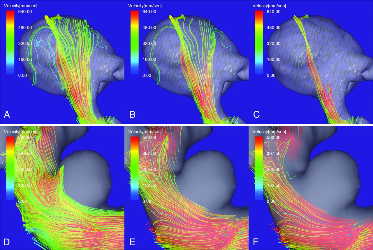
- Fig 2.
Inflow jet patterns on 4D flow MR images determined by observing the inflow streamline bundle with a velocity exceeding visualization thresholds corresponding to 60% (A and D), 75% (B and E), and 90% (C and F) of the maximum velocity in the parent artery. A–C, An aneurysm on the ICA segment branching the posterior communicating artery. D–F, An aneurysm on the paraclinoid segment of the ICA. A and B, A diffuse inflow jet intruding into the aneurysmal dome. The visualization thresholds are 60% (A) and 75% (B). C, A concentrated inflow jet intruding into the aneurysmal dome without dispersion (visualized at the 90% threshold). D, A neck-limited inflow jet visualized at the 60% threshold. E and F, Unvisualized inflow streams—that is, no inflow streams are observed at the 75% (E) and the 90% (F) thresholds.


Tables
- Table 1:
Inflow jet patterns visualized at the 60%, 75%, and 90% threshold of the maximum velocity in the parent arterya
Inflow Jet Pattern No. of Aneurysms (Threshold 60%) No. of Aneurysms (Threshold 75%) No. of Aneurysms (Threshold 90%) Concentrated inflow jet 8 (14.0%) 13 (22.8%) 16 (28.1%) Diffuse inflow jet 23 (40.4%) 18 (31.6%) 14 (24.6%) Neck-limited 15 (26.3%) 11 (19.3%) 9 (15.8%) Unvisualized 11 (19.3%) 15 (26.3%) 18 (31.6%) ↵a The distribution of the inflow jet patterns at the 60% and 90% threshold was significantly different (P = .0468).
- Table 2:
Maximum inflow velocity and maximum inflow rate in unruptured aneurysms with different inflow jet patterns visualized at the 75% threshold of the maximum velocity in the parent arterya
Inflow Jet Pattern Median (IQR) Concentrated Inflow Jet Diffuse Inflow Jet Neck-Limited Unvisualized Concentrated inflow jet Maximum inflow velocity (mm/s) 572 (206) .2539 (NS) .4689 (NS) .0002 (S) Maximum inflow rate (mL/s) 2610 (3080) .8414 (NS) .0049 (S) .0006 (S) Diffuse inflow jet Maximum inflow velocity (mm/s) 636 (289) .2539 (NS) .0963 (NS) <.0001 (S) Maximum inflow rate (mL/s) 2450 (3080) .8414 (NS) .0017 (S) <.0001 (S) Neck-limited Maximum inflow velocity (mm/s) 462 (380) .4689 (NS) .0963 (NS) .2645 (NS) Maximum inflow rate (mL/s) 890 (424) .0049 (S) .0017 (S) .3637 (NS) Unvisualized Maximum inflow velocity (mm/s) 382 (44.0) .0002 (S) <.0001 (S) .2645 (NS) Maximum inflow rate (mL/s) 696 (454) .0006 (S) <.0001 (S) .3637 (NS) Note:—IQR indicates interquartile range; S, significant; NS, not significant by the comparison test adjusted for the P value.
↵a Statistical analysis was performed between a variable in the left column and a variable in the headers.
- Table 3:
Inflow velocity ratio and inflow rate ratio in unruptured aneurysms with different inflow jet patterns visualized at the 75% threshold of the maximum velocity in the parent arterya
Inflow Jet Pattern and Ratio Median % (IQR) Concentrated Inflow Jet Diffuse Inflow Jet Neck-Limited Unvisualized Concentrated inflow jet Inflow velocity 94.1 (15.5) .5222 (NS) .0138 (S) <.0001 (S) Inflow rate 89.9 (52.3) .1735 (NS) .0059 (S) <.0001 (S) Diffuse inflow jet Inflow velocity 101 (31.8) .5222 (NS) .0053 (S) <.0001 (S) Inflow rate 59.3 (59.9) .1735 (NS) .0194 (S) <.0001 (S) Neck-limited Inflow velocity 69.4 (28.8) .0138 (S) .0053 (S) .0516 (NS) Inflow rate 29.8 (33.3) .0059 (S) .0194 (S) .1021 (NS) Unvisualized Inflow velocity 59.1 (7.62) <.0001 (S) <.0001 (S) .0516 (NS) Inflow rate 21.6 (16.0) <.0001 (S) <.0001 (S) .1021 (NS) Note:—IQR indicates interquartile range; S, significant; NS, not significant by the comparison test adjusted for the P value; inflow velocity ratio, the ratio of the maximum inflow velocity to the maximum flow velocity in the parent artery; inflow rate ratio, the ratio of the maximum inflow rate to the maximum flow rate in the parent artery.
↵a Statistical analysis was performed between a variable shown in the left column and a variable in the headers.


