
Article Figures & Data
Figures
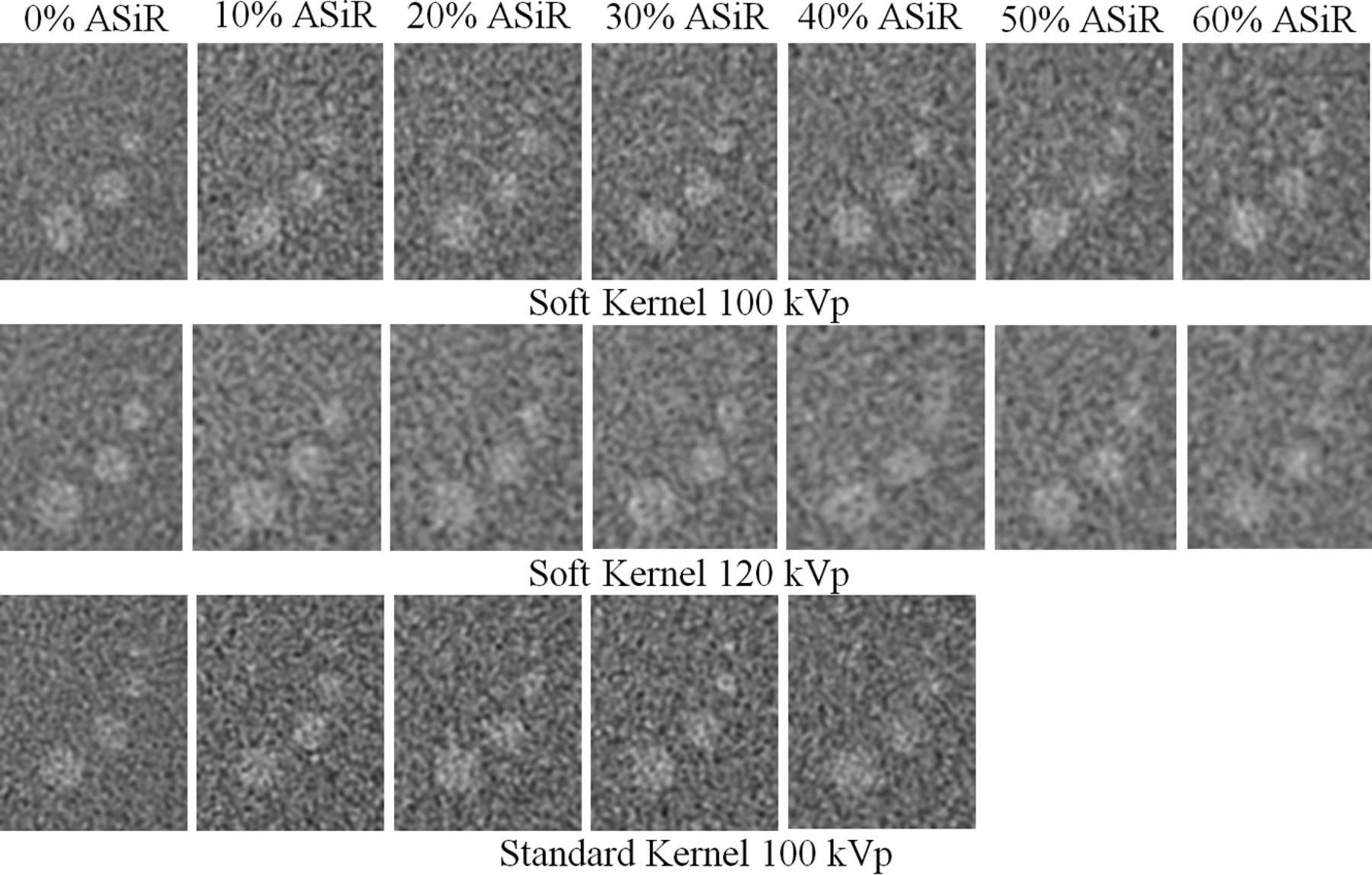
- Fig 1.
Texture of image noise as it appears in reconstructed images changes as the mean of the NPS curve shifts along the abscissa; shifts in mean NPS frequency are associated with changes in the appearance of image noise texture. A, NPS curves of the standard reconstruction kernel are reconstructed at 3 levels of ASIR. B, A corresponding ROI of 128 × 128 pixels from the center of a water phantom shows the appearance of the noise texture as it correlates with a 32% shift in NPS mean frequency along the abscissa from curve A to B and a 52% shift in curve A to C.
- Fig 2.
Dose-reduced ASIR protocols compare the mean NPS frequency shift (A) as a function of the level of ASIR reconstruction. An acceptable tolerance for the appearance of noise texture in the reconstructed image is reported in the literature,4,5,20,21 based on a 25% shift of NPS noise frequency (dashed line). B, Corresponding reductions of CTDIvol for the protocols by using ASIR are plotted and fit by using a log-regression function.
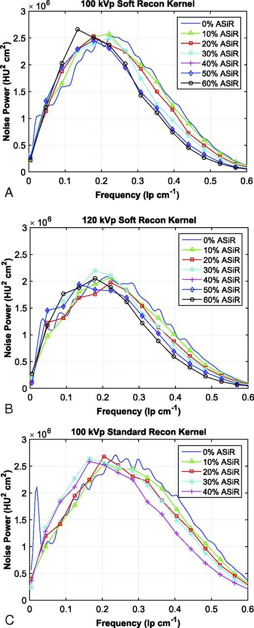
- Fig 3.
Noise power spectra acquired with the soft reconstruction kernel at 100 kVp from 240 to 120 mA (A), with the soft reconstruction kernel at 120 kVp from 200 to 110 mA (B), and the standard reconstruction kernel at 100 kVp from 250 to 140 mA (C). The calculated spectra are reconstructed at 0%–60% ASIR (soft reconstruction kernel) and at 0%–40% ASIR (standard reconstruction kernel).
- Fig 4.
Images of the 3-, 5-, 7-, and 9-mm low-contrast targets in the Catphan 700 phantom are acquired with FBP and dose-reduced ASIR reconstruction up to 60% ASIR for the soft reconstruction kernels at both 100 and 120 kVp and up to 40% for the standard reconstruction kernel at 100 kVp.
- Fig 5.
A 3-year-old boy with scans 6 months apart. A, The original brain protocol is acquired at 200 mA and 100 kVp with a CTDIvol of 25.1 mGy by using FBP. B, The patient is re-examined postsurgery with the dose-reduced brain protocol by using 60% ASIR, 120 mA, and 100 kVp, with a CTDIvol of 15.0 mGy. Both examinations were acquired by using the GE Healthcare soft reconstruction kernel.


Tables
Patient Age (yr) A or H Rotation (sec) Collimation (mm) Section (mm) Reconstruction Kernel kVp Pre-ASIR (mA) Post-ASIR (mA) Brain 0–2b A 0.5 20 5 Soft and bone 100 280 150 2–5 1 200 120 6–10 220 130 11–18 240 140 ≥19 120 200 105 Sinus ≥19 H 0.5 40 2.5 Soft and bone 120 NI = 7.5 155 0–18 100 220 130 Orbits 0–18c Hd 0.5 20 1.25 Standard and bone 100 240 155 Temporal bone ≥19 H 1 20 1.25 Standard and bone 120 250 150 2–18 0.5 120 400 230 Maxillary bone ≥19 Hd 0.5 20 2.5 Standard and bone 120 NI = 7.5 NI = 9.25 0–18 H 100 300 180 Note:—A indicates axial; H, helical; NI, Noise Index; SFOV, scan FOV.
↵a All protocols were imaged with a SFOV using “Head” unless otherwise indicated. All helical acquisitions were scanned with a pitch of 0.984 unless otherwise indicated.
↵b SFOV used “Ped Head.”
↵c SFOV used “Small Head.”
↵d Pitch = 0.516.
Patient Age Category Protocol CTDIvol (mGy) Noise (HU) Original Dose-Reduced Difference Original Dose-Reduced Difference 0–23 mo Brain 15.0 ± 0.7 8.0 ± 0.4 −47% 4.4 ± 1.0 4.2 ± 0.7 −3% 2–5 yr Brain 24.1 ± 0.9 14.6 ± 0.6 −39% 4.2 ± 0.7 4.1 ± 0.7 −3% 6–10 yr Brain 26.3 ± 1.3 15.9 ± 0.4 −40% 4.1 ± 0.5 4.2 ± 0.6 4% 11–18 yr Brain 29.1 ± 0.9 17.0 ± 0.5 −42% 4.4 ± 0.6 4.5 ± 0.6 3% ≥ 19 yr Brain 36.6 ± 0.8 18.9 ± 0.5 −48% 4.3 ± 0.6 4.4 ± 0.4 3% 0–18 yr Maxilla 19.4 ± 0.0 11.5 ± 0.1 −41% 11.2 ± 2.8 11.6 ± 1.6 3% ≥19 yr Maxilla 22.8 ± 0.0 18.7 ± 0.2 −18% 9.6 ± 1.4 9.2 ± 1.7 −3% 0–18 yr Orbits 26.9 ± 8.0 15.8 ± 0.5 −41% 7.5 ± 1.2 7.2 ± 0.1 −4% 0–18 yr Sinus 13.1 ± 0.0 7.2 ± 0.3 −45% 8.5 ± 1.2 8.9 ± 1.1 5% ≥19 yr Sinus 22.8 ± 0.0 13.7 ± 0.1 −40% 8.3 ± 0.9 8.2 ± 0.6 −1% 2–18 yr Temporal 40.7 ± 0.0 22.8 ± 0.0 −44% 9.3 ± 1.4 9.2 ± 1.2 −2% ≥19 yra Temporal 49.9 ± 0.0 29.7 ± 0.0 −40% 9.3 ± 1.0 ↵a No dose-reduced patient examinations were available for comparison. Dose-reduced CTDIvol value is calculated on the basis of scan parameters. Dose difference is a theoretic calculation.


