Advertisement

Main menu

  • Home
  • Content
    • Current Issue
    • Accepted Manuscripts
    • Article Preview
    • Past Issue Archive
    • Video Articles
    • AJNR Case Collection
    • Case of the Week Archive
    • Case of the Month Archive
    • Classic Case Archive
  • Special Collections
    • AJNR Awards
    • Low-Field MRI
    • Alzheimer Disease
    • ASNR Foundation Special Collection
    • Photon-Counting CT
    • View All
  • Multimedia
    • AJNR Podcasts
    • AJNR SCANtastic
    • Trainee Corner
    • MRI Safety Corner
    • Imaging Protocols
  • For Authors
    • Submit a Manuscript
    • Submit a Video Article
    • Submit an eLetter to the Editor/Response
    • Manuscript Submission Guidelines
    • Statistical Tips
    • Fast Publishing of Accepted Manuscripts
    • Graphical Abstract Preparation
    • Imaging Protocol Submission
    • Author Policies
  • About Us
    • About AJNR
    • Editorial Board
    • Editorial Board Alumni
  • More
    • Become a Reviewer/Academy of Reviewers
    • Subscribers
    • Permissions
    • Alerts
    • Feedback
    • Advertisers
    • ASNR Home

User menu

  • Alerts
  • Log in

Search

  • Advanced search
American Journal of Neuroradiology

American Journal of Neuroradiology

ASHNR American Society of Functional Neuroradiology ASHNR American Society of Pediatric Neuroradiology ASSR
  • Alerts
  • Log in

Advanced Search

  • Home
  • Content
    • Current Issue
    • Accepted Manuscripts
    • Article Preview
    • Past Issue Archive
    • Video Articles
    • AJNR Case Collection
    • Case of the Week Archive
    • Case of the Month Archive
    • Classic Case Archive
  • Special Collections
    • AJNR Awards
    • Low-Field MRI
    • Alzheimer Disease
    • ASNR Foundation Special Collection
    • Photon-Counting CT
    • View All
  • Multimedia
    • AJNR Podcasts
    • AJNR SCANtastic
    • Trainee Corner
    • MRI Safety Corner
    • Imaging Protocols
  • For Authors
    • Submit a Manuscript
    • Submit a Video Article
    • Submit an eLetter to the Editor/Response
    • Manuscript Submission Guidelines
    • Statistical Tips
    • Fast Publishing of Accepted Manuscripts
    • Graphical Abstract Preparation
    • Imaging Protocol Submission
    • Author Policies
  • About Us
    • About AJNR
    • Editorial Board
    • Editorial Board Alumni
  • More
    • Become a Reviewer/Academy of Reviewers
    • Subscribers
    • Permissions
    • Alerts
    • Feedback
    • Advertisers
    • ASNR Home
  • Follow AJNR on Twitter
  • Visit AJNR on Facebook
  • Follow AJNR on Instagram
  • Join AJNR on LinkedIn
  • RSS Feeds

AJNR Awards, New Junior Editors, and more. Read the latest AJNR updates

Research ArticleSpine Imaging and Spine Image-Guided Interventions

Radiographic Local Control of Spinal Metastases with Percutaneous Radiofrequency Ablation and Vertebral Augmentation

A.N. Wallace, A. Tomasian, D. Vaswani, R. Vyhmeister, R.O. Chang and J.W. Jennings
American Journal of Neuroradiology April 2016, 37 (4) 759-765; DOI: https://doi.org/10.3174/ajnr.A4595
A.N. Wallace
aFrom the Mallinckrodt Institute of Radiology (A.N.W., A.T., D.V., J.W.J.), Siteman Cancer Center, Washington University School of Medicine, St. Louis, Missouri
  • Find this author on Google Scholar
  • Find this author on PubMed
  • Search for this author on this site
  • ORCID record for A.N. Wallace
A. Tomasian
aFrom the Mallinckrodt Institute of Radiology (A.N.W., A.T., D.V., J.W.J.), Siteman Cancer Center, Washington University School of Medicine, St. Louis, Missouri
  • Find this author on Google Scholar
  • Find this author on PubMed
  • Search for this author on this site
  • ORCID record for A. Tomasian
D. Vaswani
aFrom the Mallinckrodt Institute of Radiology (A.N.W., A.T., D.V., J.W.J.), Siteman Cancer Center, Washington University School of Medicine, St. Louis, Missouri
  • Find this author on Google Scholar
  • Find this author on PubMed
  • Search for this author on this site
  • ORCID record for D. Vaswani
R. Vyhmeister
bWashington University School of Medicine (R.V., R.O.C.), St. Louis, Missouri.
  • Find this author on Google Scholar
  • Find this author on PubMed
  • Search for this author on this site
  • ORCID record for R. Vyhmeister
R.O. Chang
bWashington University School of Medicine (R.V., R.O.C.), St. Louis, Missouri.
  • Find this author on Google Scholar
  • Find this author on PubMed
  • Search for this author on this site
  • ORCID record for R.O. Chang
J.W. Jennings
aFrom the Mallinckrodt Institute of Radiology (A.N.W., A.T., D.V., J.W.J.), Siteman Cancer Center, Washington University School of Medicine, St. Louis, Missouri
  • Find this author on Google Scholar
  • Find this author on PubMed
  • Search for this author on this site
  • ORCID record for J.W. Jennings
  • Article
  • Figures & Data
  • Info & Metrics
  • Responses
  • References
  • PDF
Loading

Article Figures & Data

Figures

  • Fig 1.
    • Download figure
    • Open in new tab
    • Download powerpoint
    Fig 1.

    A 29-year-old woman with stage IV (T1b, N1, M1) cardiac angiosarcoma who presented with low back pain. Sagittal T1-weighted (A) and STIR (B) MR imaging show diffuse T1 hypointensity and heterogeneous T2 hyperintensity of the lumbar vertebral body marrow, consistent with marrow-replacing tumor. She was treated with conventional external-beam radiation therapy (30 Gy in 10 fractions); however, her back pain persisted. Sagittal STIR (C) and T1-weighted, fat-suppressed, postcontrast (D) MR imaging performed 5 months later show interval progression of multiple spinal metastases with new epidural extension of tumor at T11, L1, L2, and L3 (black block arrows) and pathologic fractures of the L2 and L3 vertebral bodies. She could not receive additional radiation therapy due to the cumulative dose to the spinal cord. Consequently, she underwent radiofrequency ablation and vertebral augmentation of T11, L1, L2, and L3. Anteroposterior (E) and lateral (F) fluoroscopic images show percutaneous cannulae in both pedicles of T11 and the ablation probe curving into the left posteroinferior vertebral body (black arrowheads). Sagittal STIR (G) and T1-weighted, fat-suppressed, postcontrast (H) MR imaging performed 6 months later show interval retraction of the epidural tumor at T11, L1, L2, and L3. Signal void corresponding to cement (white asterisks) with surrounding T2-hyperintense, enhancing granulation tissue is noted at the treated levels (white block arrows).

  • Fig 2.
    • Download figure
    • Open in new tab
    • Download powerpoint
    Fig 2.

    Examples of ablation failure. A, Axial CT image shows a T12 lytic squamous cell carcinoma metastasis. B, CT scan obtained 4 months after radiofrequency ablation and vertebral augmentation shows new osteolysis medial to the cement and extending across the midline (white arrow), consistent with progression of residual tumor. C, Axial postcontrast, T1-weighted, fat-suppressed MR imaging performed 6 months after radiofrequency ablation and vertebral augmentation of an L3 non-small cell carcinoma metastasis shows signal void corresponding to cement in the ablation cavity (white asterisk), with residual enhancing tumor in the right lateral and posterior vertebral body that extends into the epidural space (black asterisks). D, Axial [18F] fluorodeoxyglucose PET/CT scan obtained 1 month after radiofrequency ablation and vertebral augmentation of an L5 liposarcoma metastasis shows residual hypermetabolic tumor along the right anterolateral aspect of the vertebral body (white dashed arrow).
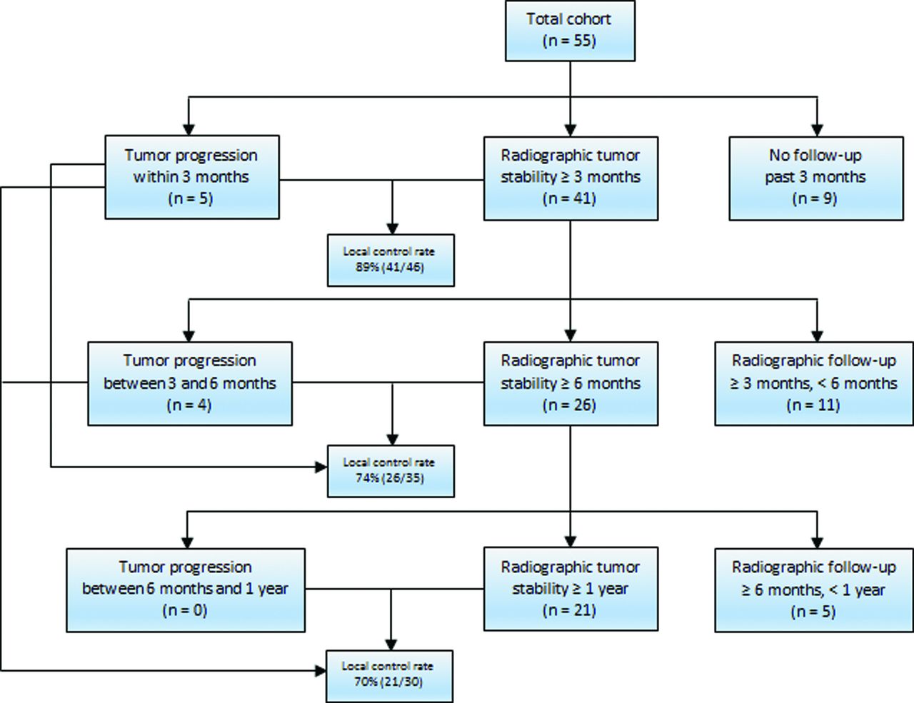
  • Fig 3.
    • Download figure
    • Open in new tab
    • Download powerpoint
    Fig 3.

    Flowchart summarizing overall radiographic local tumor control results at 3-month, 6-month, and 1-year follow-ups.

PreviousNext
Back to top

In this issue

American Journal of Neuroradiology: 37 (4)
American Journal of Neuroradiology
Vol. 37, Issue 4
1 Apr 2016
  • Table of Contents
  • Index by author
  • Complete Issue (PDF)
Advertisement
Print
Download PDF
Email Article

Thank you for your interest in spreading the word on American Journal of Neuroradiology.

NOTE: We only request your email address so that the person you are recommending the page to knows that you wanted them to see it, and that it is not junk mail. We do not capture any email address.

Enter multiple addresses on separate lines or separate them with commas.
Radiographic Local Control of Spinal Metastases with Percutaneous Radiofrequency Ablation and Vertebral Augmentation
(Your Name) has sent you a message from American Journal of Neuroradiology
(Your Name) thought you would like to see the American Journal of Neuroradiology web site.
CAPTCHA
This question is for testing whether or not you are a human visitor and to prevent automated spam submissions.
Cite this article
A.N. Wallace, A. Tomasian, D. Vaswani, R. Vyhmeister, R.O. Chang, J.W. Jennings
Radiographic Local Control of Spinal Metastases with Percutaneous Radiofrequency Ablation and Vertebral Augmentation
American Journal of Neuroradiology Apr 2016, 37 (4) 759-765; DOI: 10.3174/ajnr.A4595

Citation Manager Formats

  • BibTeX
  • Bookends
  • EasyBib
  • EndNote (tagged)
  • EndNote 8 (xml)
  • Medlars
  • Mendeley
  • Papers
  • RefWorks Tagged
  • Ref Manager
  • RIS
  • Zotero
0 Responses
Respond to this article
Share
Bookmark this article
Radiographic Local Control of Spinal Metastases with Percutaneous Radiofrequency Ablation and Vertebral Augmentation
A.N. Wallace, A. Tomasian, D. Vaswani, R. Vyhmeister, R.O. Chang, J.W. Jennings
American Journal of Neuroradiology Apr 2016, 37 (4) 759-765; DOI: 10.3174/ajnr.A4595
del.icio.us logo Twitter logo Facebook logo Mendeley logo
  • Tweet Widget
  • Facebook Like
  • Google Plus One
Purchase

Jump to section

  • Article
    • Abstract
    • ABBREVIATION:
    • Materials and Methods
    • Results
    • Discussion
    • Conclusions
    • Footnotes
    • REFERENCES
  • Figures & Data
  • Info & Metrics
  • Responses
  • References
  • PDF

Related Articles

  • No related articles found.
  • PubMed
  • Google Scholar

Cited By...

  • Photodynamic Therapy for the Treatment of Vertebral Metastases: A Phase I Clinical Trial
  • Percutaneous Radiofrequency Ablation of Spinal Osteoid Osteomas Using a Targeted Navigational Bipolar Electrode System
  • Simultaneous Bipedicular Radiofrequency Ablation Combined with Vertebral Augmentation for Local Tumor Control of Spinal Metastases
  • Republished: Renal cell carcinoma metastasis involving vertebral hemangioma: dual percutaneous treatment by navigational bipolar radiofrequency ablation and high viscosity cement vertebroplasty
  • Is an Intact Posterior Vertebral Body Cortex Protective for Percutaneous Ablation?
  • Percutaneous Spinal Ablation in a Sheep Model: Protective Capacity of an Intact Cortex, Correlation of Ablation Parameters with Ablation Zone Size, and Correlation of Postablation MRI and Pathologic Findings
  • Renal cell carcinoma metastasis involving vertebral hemangioma: dual percutaneous treatment by navigational bipolar radiofrequency ablation and high viscosity cement vertebroplasty
  • Crossref
  • Google Scholar

This article has not yet been cited by articles in journals that are participating in Crossref Cited-by Linking.

More in this TOC Section

Spine Imaging and Spine Image-Guided Interventions

  • Optimization of Photon Counting CT Myelography
  • Characteristics of SIH Type I Culprit Lesions
  • Management Outcomes For VO Spine Biopsy
Show more Spine Imaging and Spine Image-Guided Interventions

Neurointervention

  • Rescue Reentry in Carotid Near-Occlusion
  • Contour Neurovascular System: Five Year Follow Up
  • Effect of SARS-CoV2 on Endovascular Thrombectomy
Show more Neurointervention

Similar Articles

Advertisement

Indexed Content

  • Current Issue
  • Accepted Manuscripts
  • Article Preview
  • Past Issues
  • Editorials
  • Editor's Choice
  • Fellows' Journal Club
  • Letters to the Editor
  • Video Articles

Cases

  • Case Collection
  • Archive - Case of the Week
  • Archive - Case of the Month
  • Archive - Classic Case

More from AJNR

  • Trainee Corner
  • Imaging Protocols
  • MRI Safety Corner
  • Book Reviews

Multimedia

  • AJNR Podcasts
  • AJNR Scantastics

Resources

  • Turnaround Time
  • Submit a Manuscript
  • Submit a Video Article
  • Submit an eLetter to the Editor/Response
  • Manuscript Submission Guidelines
  • Statistical Tips
  • Fast Publishing of Accepted Manuscripts
  • Graphical Abstract Preparation
  • Imaging Protocol Submission
  • Evidence-Based Medicine Level Guide
  • Publishing Checklists
  • Author Policies
  • Become a Reviewer/Academy of Reviewers
  • News and Updates

About Us

  • About AJNR
  • Editorial Board
  • Editorial Board Alumni
  • Alerts
  • Permissions
  • Not an AJNR Subscriber? Join Now
  • Advertise with Us
  • Librarian Resources
  • Feedback
  • Terms and Conditions
  • AJNR Editorial Board Alumni

American Society of Neuroradiology

  • Not an ASNR Member? Join Now

© 2025 by the American Society of Neuroradiology All rights, including for text and data mining, AI training, and similar technologies, are reserved.
Print ISSN: 0195-6108 Online ISSN: 1936-959X

Powered by HighWire