Advertisement

Main menu

  • Home
  • Content
    • Current Issue
    • Accepted Manuscripts
    • Article Preview
    • Past Issue Archive
    • Video Articles
    • AJNR Case Collection
    • Case of the Week Archive
    • Case of the Month Archive
    • Classic Case Archive
  • Special Collections
    • AJNR Awards
    • Low-Field MRI
    • Alzheimer Disease
    • ASNR Foundation Special Collection
    • Photon-Counting CT
    • View All
  • Multimedia
    • AJNR Podcasts
    • AJNR SCANtastic
    • Trainee Corner
    • MRI Safety Corner
    • Imaging Protocols
  • For Authors
    • Submit a Manuscript
    • Submit a Video Article
    • Submit an eLetter to the Editor/Response
    • Manuscript Submission Guidelines
    • Statistical Tips
    • Fast Publishing of Accepted Manuscripts
    • Graphical Abstract Preparation
    • Imaging Protocol Submission
    • Author Policies
  • About Us
    • About AJNR
    • Editorial Board
    • Editorial Board Alumni
  • More
    • Become a Reviewer/Academy of Reviewers
    • Subscribers
    • Permissions
    • Alerts
    • Feedback
    • Advertisers
    • ASNR Home

User menu

  • Alerts
  • Log in

Search

  • Advanced search
American Journal of Neuroradiology

American Journal of Neuroradiology

ASHNR American Society of Functional Neuroradiology ASHNR American Society of Pediatric Neuroradiology ASSR
  • Alerts
  • Log in

Advanced Search

  • Home
  • Content
    • Current Issue
    • Accepted Manuscripts
    • Article Preview
    • Past Issue Archive
    • Video Articles
    • AJNR Case Collection
    • Case of the Week Archive
    • Case of the Month Archive
    • Classic Case Archive
  • Special Collections
    • AJNR Awards
    • Low-Field MRI
    • Alzheimer Disease
    • ASNR Foundation Special Collection
    • Photon-Counting CT
    • View All
  • Multimedia
    • AJNR Podcasts
    • AJNR SCANtastic
    • Trainee Corner
    • MRI Safety Corner
    • Imaging Protocols
  • For Authors
    • Submit a Manuscript
    • Submit a Video Article
    • Submit an eLetter to the Editor/Response
    • Manuscript Submission Guidelines
    • Statistical Tips
    • Fast Publishing of Accepted Manuscripts
    • Graphical Abstract Preparation
    • Imaging Protocol Submission
    • Author Policies
  • About Us
    • About AJNR
    • Editorial Board
    • Editorial Board Alumni
  • More
    • Become a Reviewer/Academy of Reviewers
    • Subscribers
    • Permissions
    • Alerts
    • Feedback
    • Advertisers
    • ASNR Home
  • Follow AJNR on Twitter
  • Visit AJNR on Facebook
  • Follow AJNR on Instagram
  • Join AJNR on LinkedIn
  • RSS Feeds

AJNR Awards, New Junior Editors, and more. Read the latest AJNR updates

Research ArticleADULT BRAIN
Open Access

On the Use of DSC-MRI for Measuring Vascular Permeability

J.T. Skinner, P.L. Moots, G.D. Ayers and C.C. Quarles
American Journal of Neuroradiology January 2016, 37 (1) 80-87; DOI: https://doi.org/10.3174/ajnr.A4478
J.T. Skinner
aFrom the Vanderbilt University Institute of Imaging Science (J.T.S., C.C.Q.)
dDepartments of Radiology and Radiological Sciences (J.T.S., C.C.Q.)
  • Find this author on Google Scholar
  • Find this author on PubMed
  • Search for this author on this site
P.L. Moots
eNeurology (P.L.M.)
  • Find this author on Google Scholar
  • Find this author on PubMed
  • Search for this author on this site
G.D. Ayers
fBiostatistics (G.D.A.), Vanderbilt University School of Medicine, Nashville, Tennessee.
  • Find this author on Google Scholar
  • Find this author on PubMed
  • Search for this author on this site
C.C. Quarles
aFrom the Vanderbilt University Institute of Imaging Science (J.T.S., C.C.Q.)
bDepartments of Cancer Biology (C.C.Q.)
cBiomedical Engineering (C.C.Q.), Vanderbilt University, Nashville, Tennessee
dDepartments of Radiology and Radiological Sciences (J.T.S., C.C.Q.)
  • Find this author on Google Scholar
  • Find this author on PubMed
  • Search for this author on this site
  • Article
  • Figures & Data
  • Info & Metrics
  • Responses
  • References
  • PDF
Loading

Article Figures & Data

Figures

  • Tables
  • Fig 1.
    • Download figure
    • Open in new tab
    • Download powerpoint
    Fig 1.

    A, Representative uncorrected tumor ΔR2* time course and the associated Weisskoff model fit (solid) used to compute K2 at TE1 (square), TE2 (dot), and dual-echo (diamond). B, Corresponding tissue residue functions used to compute Ka at TE1, TE2, and dual-echo.

  • Fig 2.
    • Download figure
    • Open in new tab
    • Download powerpoint
    Fig 2.

    A, T1-weighted post-Gd anatomic image showing a high-grade brain tumor. Sample computed permeability maps (units in minute−1), Ktrans (B), K2 (C), and Ka (D).

  • Fig 3.
    • Download figure
    • Open in new tab
    • Download powerpoint
    Fig 3.

    A, Sample voxelwise comparison between K2 at TE2 and Ktrans. B, Sample voxelwise comparison between Ka at TE2 and Ktrans. C, Voxelwise comparison between K2 (y-axis) and Ka (x-axis). Linear regression line shown in black.
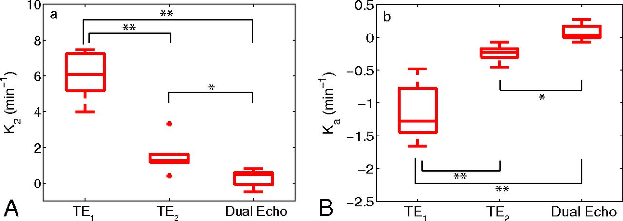
  • Fig 4.
    • Download figure
    • Open in new tab
    • Download powerpoint
    Fig 4.

    Boxplots of median parameter estimates (from all patients) calculated at various TEs for K2 (A) and Ka (B). Boxplots display the median, 25th, and 75th percentiles (edges of box) and extreme data points (whiskers). Outliers are plotted individually (plus sign). Significance was determined by the Mann-Whitney U test. * indicates P < .01; **, P < .001. Note: Positive outlier for K2 at TE1 not pictured.

  • Fig 5.
    • Download figure
    • Open in new tab
    • Download powerpoint
    Fig 5.

    Sample mean ΔR2* time course (TE = 31 ms) for a tumor ROI (A) and the resulting ΔR1 time course (B). Mean ΔR2* (C) and ΔR1 (D) time courses from the same tumor with voxels separated by whether they predominately exhibit T2* leakage effects (T2* voxels) or T1 leakage effects (T1 voxels).

Tables

  • Figures
    • View popup
    Table 1:

    Patient demographics

    PatientAge (yr)SexPrior ResectionPathologyOS (mo)
    161FemaleYesGrade IV glioblastoma17.9
    266MaleYesGrade IV glioblastoma18.2
    365MaleYesGrade III anaplastic astrocytomaNA
    451MaleYesGrade IV glioblastoma4.3
    555MaleNoGrade III oligodendroglioma13.1
    640MaleYesGrade IV glioblastoma11.0
    742FemaleYesGrade IV glioblastomaNA
    • Note:—OS indicates overall survival after radiologically confirmed tumor recurrence/progression; NA, not applicable.

    • View popup
    Table 2:

    Patient-specific estimates of DSC-MRI and DCE-MRI parameters separated by the predominant leakage effect

    Patient No.No. of Voxels (%)K2 (min−1)Ka (min−1)Ktrans (min−1)ve
    T1T2*T1T2*T1T2*T1T2*T1T2*
    144 (79%)12 (21%)1.8071.205−0.373−0.2500.2230.0660.2210.072
    2214 (45%)265 (55%)1.229−0.815−0.3420.0260.1690.1630.3590.258
    3126 (61%)79 (39%)2.3740.822−0.372−0.1170.0890.0380.3280.150
    4368 (47%)417 (53%)1.7670.700−0.536−0.4690.1040.0780.2280.140
    5187 (56%)147 (44%)1.9750.787−0.149−0.0250.0690.0440.2840.107
    6734 (93%)52 (7%)3.7260.240−0.2560.0040.0990.0500.2900.138
    716 (64%)9 (36%)2.5910.025−0.4180.0240.2000.1790.2030.107
    Meanw2.6270.289−0.329−0.2080.1090.0920.2850.167
    • Note:—Meanw indicates weighted mean.

PreviousNext
Back to top

In this issue

American Journal of Neuroradiology: 37 (1)
American Journal of Neuroradiology
Vol. 37, Issue 1
1 Jan 2016
  • Table of Contents
  • Index by author
  • Complete Issue (PDF)
Advertisement
Print
Download PDF
Email Article

Thank you for your interest in spreading the word on American Journal of Neuroradiology.

NOTE: We only request your email address so that the person you are recommending the page to knows that you wanted them to see it, and that it is not junk mail. We do not capture any email address.

Enter multiple addresses on separate lines or separate them with commas.
On the Use of DSC-MRI for Measuring Vascular Permeability
(Your Name) has sent you a message from American Journal of Neuroradiology
(Your Name) thought you would like to see the American Journal of Neuroradiology web site.
CAPTCHA
This question is for testing whether or not you are a human visitor and to prevent automated spam submissions.
Cite this article
J.T. Skinner, P.L. Moots, G.D. Ayers, C.C. Quarles
On the Use of DSC-MRI for Measuring Vascular Permeability
American Journal of Neuroradiology Jan 2016, 37 (1) 80-87; DOI: 10.3174/ajnr.A4478

Citation Manager Formats

  • BibTeX
  • Bookends
  • EasyBib
  • EndNote (tagged)
  • EndNote 8 (xml)
  • Medlars
  • Mendeley
  • Papers
  • RefWorks Tagged
  • Ref Manager
  • RIS
  • Zotero
0 Responses
Respond to this article
Share
Bookmark this article
On the Use of DSC-MRI for Measuring Vascular Permeability
J.T. Skinner, P.L. Moots, G.D. Ayers, C.C. Quarles
American Journal of Neuroradiology Jan 2016, 37 (1) 80-87; DOI: 10.3174/ajnr.A4478
del.icio.us logo Twitter logo Facebook logo Mendeley logo
  • Tweet Widget
  • Facebook Like
  • Google Plus One
Purchase

Jump to section

  • Article
    • Abstract
    • ABBREVIATIONS:
    • Materials and Methods
    • Results
    • Discussion
    • Conclusions
    • Footnotes
    • References
  • Figures & Data
  • Info & Metrics
  • Responses
  • References
  • PDF

Related Articles

  • No related articles found.
  • PubMed
  • Google Scholar

Cited By...

  • Blood-Brain Barrier Permeability and Kinetics of Inflammatory Markers in Acute Stroke Patients Treated With Thrombectomy
  • Sex-specific differences in white matter microvascular integrity after ischaemic stroke
  • Prognostic Predictions for Patients with Glioblastoma after Standard Treatment: Application of Contrast Leakage Information from DSC-MRI within Nonenhancing FLAIR High-Signal-Intensity Lesions
  • Clinical Value of Vascular Permeability Estimates Using Dynamic Susceptibility Contrast MRI: Improved Diagnostic Performance in Distinguishing Hypervascular Primary CNS Lymphoma from Glioblastoma
  • Crossref
  • Google Scholar

This article has not yet been cited by articles in journals that are participating in Crossref Cited-by Linking.

More in this TOC Section

  • Diagnostic Neuroradiology of Monoclonal Antibodies
  • NCCT vs. MRI for Brain Atrophy in Acute Stroke
  • Clinical Outcomes After Chiari I Decompression
Show more Adult Brain

Similar Articles

Advertisement

Indexed Content

  • Current Issue
  • Accepted Manuscripts
  • Article Preview
  • Past Issues
  • Editorials
  • Editor's Choice
  • Fellows' Journal Club
  • Letters to the Editor
  • Video Articles

Cases

  • Case Collection
  • Archive - Case of the Week
  • Archive - Case of the Month
  • Archive - Classic Case

More from AJNR

  • Trainee Corner
  • Imaging Protocols
  • MRI Safety Corner
  • Book Reviews

Multimedia

  • AJNR Podcasts
  • AJNR Scantastics

Resources

  • Turnaround Time
  • Submit a Manuscript
  • Submit a Video Article
  • Submit an eLetter to the Editor/Response
  • Manuscript Submission Guidelines
  • Statistical Tips
  • Fast Publishing of Accepted Manuscripts
  • Graphical Abstract Preparation
  • Imaging Protocol Submission
  • Evidence-Based Medicine Level Guide
  • Publishing Checklists
  • Author Policies
  • Become a Reviewer/Academy of Reviewers
  • News and Updates

About Us

  • About AJNR
  • Editorial Board
  • Editorial Board Alumni
  • Alerts
  • Permissions
  • Not an AJNR Subscriber? Join Now
  • Advertise with Us
  • Librarian Resources
  • Feedback
  • Terms and Conditions
  • AJNR Editorial Board Alumni

American Society of Neuroradiology

  • Not an ASNR Member? Join Now

© 2025 by the American Society of Neuroradiology All rights, including for text and data mining, AI training, and similar technologies, are reserved.
Print ISSN: 0195-6108 Online ISSN: 1936-959X

Powered by HighWire