
Article Figures & Data
Figures
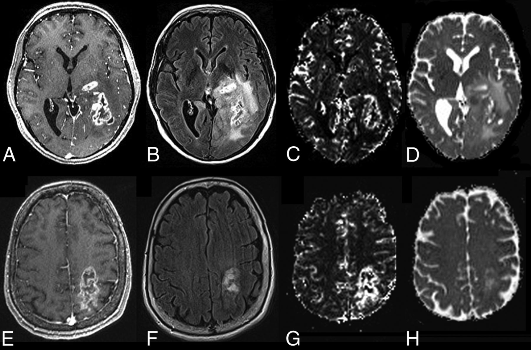
- Fig 1.
Representative MR images and parametric maps from patients with GBM. The top row (A–D) shows images from a 66-year-old woman with GBM who survived for >58 months after gross total resection of her GBM in the left temporo-occipital lobe. Representative MR images from a 75-year-old man with a GBM in the left parietal lobe with only a 5-month survival are shown (E–H). Images of both patients demonstrate heterogeneous peripheral enhancement (A and E) and extensive surrounding FLAIR signal abnormality (B and F) on axial contrast-enhanced T1-weighted and FLAIR images, respectively. The patient with short survival has a higher CBV (G) (rCBVmax = 12.22) in comparison with the patient with long survival (C) (rCBVmax = 3.57). The MD maps from these patients (D and H) do not show any difference (6.50 × 10⁻⁴ mm2/s versus 6.80 × 10⁻⁴ mm2/s).
- Fig 2.
Receiver operative characteristic curves of rCBVmax (solid line) by using a rCBVmax cutoff value of 5.79. The area under the curve was 0.93. The receiver operating curve of MDmin (dotted line) by using a cutoff value of 8.35 × 10⁻⁴mm2/s demonstrated only a modest area under the curve of 0.55.
- Fig 3.
Kaplan-Meier curves for patients with low (<5.79, solid line) and high (≥5.79, dotted line) rCBVmax. GBMs with low rCBVmax had a median survival time of 23 ± 3.4 months, whereas GBMs with high rCBVmax had a median survival time of 5 ± 1.9 months (P < .001). Cum indicates cumulative.
- Fig 4.
Kaplan-Meier curves for patients with GBM with low (<0.835 × 10⁻³mm2/s, solid line) and high (≥0.835 × 10⁻³mm2/s, dotted line) MDmin. GBMs with a low MDmin have a median survival time of 14 ± 2.0 months, whereas GBMs with a high MDmin have a median survival time of 18 ± 1.4 months. There is no significant difference between the 2 groups (P > .05). Cum indicates cumulative.


Tables
Average ± SD, sensitivity, specificity, PPV, and NPV of pretreatment rCBVmax and MDmin in patients with GBM demonstrating long (≥15 mo) survival and short (<15 mo) survival
Long Survival (n = 30) Short Survival (n = 28) Cutoff AUC Sensitivity Specificity PPV NPV Mean ± SD Mean ± SD rCBVmax 4.78 ± 1.30 9.90 ± 4.01a 5.79 0.93 0.89 0.90 0.89 0.90 MDmin (10−3mm2/s) 0.80 ± 0.17 0.75 ± 0.15 0.83 0.55 0.71 0.47 0.56 0.64 Note:—NPV indicates negative predictive value; PPV, positive predictive value; AUC, area under the curve.
↵a P < .01.


Jump to section
Related Articles
Cited By...
- No citing articles found.