Advertisement

Main menu

  • Home
  • Content
    • Current Issue
    • Accepted Manuscripts
    • Article Preview
    • Past Issue Archive
    • Video Articles
    • AJNR Case Collection
    • Case of the Week Archive
    • Case of the Month Archive
    • Classic Case Archive
  • Special Collections
    • AJNR Awards
    • Low-Field MRI
    • Alzheimer Disease
    • ASNR Foundation Special Collection
    • Photon-Counting CT
    • View All
  • Multimedia
    • AJNR Podcasts
    • AJNR SCANtastic
    • Trainee Corner
    • MRI Safety Corner
    • Imaging Protocols
  • For Authors
    • Submit a Manuscript
    • Submit a Video Article
    • Submit an eLetter to the Editor/Response
    • Manuscript Submission Guidelines
    • Statistical Tips
    • Fast Publishing of Accepted Manuscripts
    • Graphical Abstract Preparation
    • Imaging Protocol Submission
    • Author Policies
  • About Us
    • About AJNR
    • Editorial Board
    • Editorial Board Alumni
  • More
    • Become a Reviewer/Academy of Reviewers
    • Subscribers
    • Permissions
    • Alerts
    • Feedback
    • Advertisers
    • ASNR Home

User menu

  • Alerts
  • Log in

Search

  • Advanced search
American Journal of Neuroradiology

American Journal of Neuroradiology

ASHNR American Society of Functional Neuroradiology ASHNR American Society of Pediatric Neuroradiology ASSR
  • Alerts
  • Log in

Advanced Search

  • Home
  • Content
    • Current Issue
    • Accepted Manuscripts
    • Article Preview
    • Past Issue Archive
    • Video Articles
    • AJNR Case Collection
    • Case of the Week Archive
    • Case of the Month Archive
    • Classic Case Archive
  • Special Collections
    • AJNR Awards
    • Low-Field MRI
    • Alzheimer Disease
    • ASNR Foundation Special Collection
    • Photon-Counting CT
    • View All
  • Multimedia
    • AJNR Podcasts
    • AJNR SCANtastic
    • Trainee Corner
    • MRI Safety Corner
    • Imaging Protocols
  • For Authors
    • Submit a Manuscript
    • Submit a Video Article
    • Submit an eLetter to the Editor/Response
    • Manuscript Submission Guidelines
    • Statistical Tips
    • Fast Publishing of Accepted Manuscripts
    • Graphical Abstract Preparation
    • Imaging Protocol Submission
    • Author Policies
  • About Us
    • About AJNR
    • Editorial Board
    • Editorial Board Alumni
  • More
    • Become a Reviewer/Academy of Reviewers
    • Subscribers
    • Permissions
    • Alerts
    • Feedback
    • Advertisers
    • ASNR Home
  • Follow AJNR on Twitter
  • Visit AJNR on Facebook
  • Follow AJNR on Instagram
  • Join AJNR on LinkedIn
  • RSS Feeds

AJNR Awards, New Junior Editors, and more. Read the latest AJNR updates

Getting new auth cookie, if you see this message a lot, tell someone!
Research ArticleHead and Neck Imaging

Radiology Reports for Incidental Thyroid Nodules on CT and MRI: High Variability across Subspecialties

A.T. Grady, J.A. Sosa, T.P. Tanpitukpongse, K.R. Choudhury, R.T. Gupta and J.K. Hoang
American Journal of Neuroradiology February 2015, 36 (2) 397-402; DOI: https://doi.org/10.3174/ajnr.A4089
A.T. Grady
aFrom the Departments of Radiology (A.T.G., T.P.T., K.R.C., R.T.G., J.K.H.)
  • Find this author on Google Scholar
  • Find this author on PubMed
  • Search for this author on this site
J.A. Sosa
bSurgery (J.A.S.)
dDuke Cancer Institute (J.A.S.)
eDuke Clinical Research Institute (J.A.S.), Duke University Medical Center, Durham, North Carolina.
  • Find this author on Google Scholar
  • Find this author on PubMed
  • Search for this author on this site
T.P. Tanpitukpongse
aFrom the Departments of Radiology (A.T.G., T.P.T., K.R.C., R.T.G., J.K.H.)
  • Find this author on Google Scholar
  • Find this author on PubMed
  • Search for this author on this site
K.R. Choudhury
aFrom the Departments of Radiology (A.T.G., T.P.T., K.R.C., R.T.G., J.K.H.)
  • Find this author on Google Scholar
  • Find this author on PubMed
  • Search for this author on this site
R.T. Gupta
aFrom the Departments of Radiology (A.T.G., T.P.T., K.R.C., R.T.G., J.K.H.)
  • Find this author on Google Scholar
  • Find this author on PubMed
  • Search for this author on this site
J.K. Hoang
aFrom the Departments of Radiology (A.T.G., T.P.T., K.R.C., R.T.G., J.K.H.)
cRadiation Oncology (J.K.H.)
  • Find this author on Google Scholar
  • Find this author on PubMed
  • Search for this author on this site
  • Article
  • Figures & Data
  • Supplemental
  • Info & Metrics
  • Responses
  • References
  • PDF
Loading

Article Figures & Data

Figures

  • Tables
  • Fig 1.
    • Download figure
    • Open in new tab
    • Download powerpoint
    Fig 1.

    Flowchart of study patients. FNA indicates fine needle aspiration.
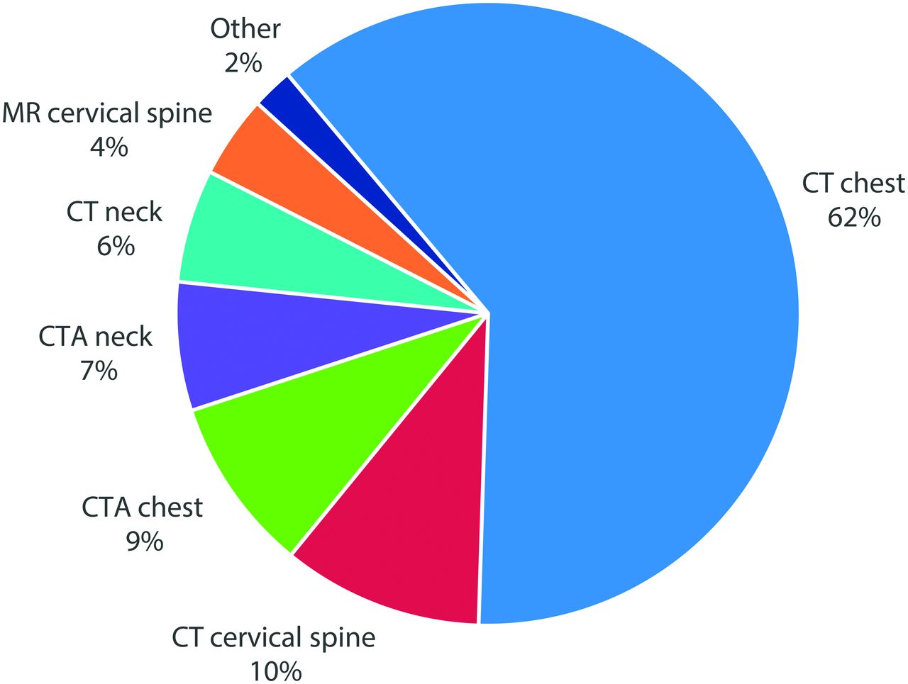
  • Fig 2.
    • Download figure
    • Open in new tab
    • Download powerpoint
    Fig 2.

    Source of imaging studies with reported incidental thyroid nodules.

  • Fig 3.
    • Download figure
    • Open in new tab
    • Download powerpoint
    Fig 3.

    Three patients with incidental thyroid nodules that were similar in size but were reported differently. A, A 46-year-old man with a 12-mm incidental nodule in the left thyroid lobe detected on chest CTA performed to evaluate an abdominal aortic aneurysm. The nodule was reported only in the “Findings” section of the report without a recommendation. B, A 47-year-old woman with a 10-mm incidental nodule in the right thyroid lobe detected on chest CTA performed to evaluate chest pain. The nodule was reported in the “Impression” section without a recommendation. C, A 63-year-old man with several incidental thyroid nodules detected on cervical spine CT performed to evaluate neck injury. The largest was in the left thyroid lobe and measured 10 mm. The nodule was reported in the “Impression” section with a recommendation for sonography.

Tables

  • Figures
  • The Duke 3-tiered system for CT, MRI, or PET-detected thyroid nodules1,13,14,a

    CategoryCriteria for CategoriesRecommendations
    Risk category 1: highly suspicious for malignancyPET avid thyroid noduleStrongly consider work-up with ultrasound for any size nodule
    Suspicious lymphadenopathy;b extrathyroid spread with or without signs of vocal cord palsy on side of nodule; lung metastases
    Risk category 2: indeterminate with risk factor of young ageAge younger than 35 yearsConsider work-up with ultrasound if ≥1 cm in adults
    Consider work-up with ultrasound for any size in pediatric patients
    Risk category 3: indeterminate without risk factorsAge 35 years or olderConsider work-up with ultrasound if ≥1.5 cm
    • ↵a Intended for management of incidental thyroid nodules in low-risk patients.

    • ↵b Suspicious lymph nodes are defined as nodes >10 mm in the short axis (with the exception of jugulodigastric lymph nodes, which are permitted to be up to 15 mm in the short axis) or nodes that contain either calcifications, cystic components, or irregular margins.

PreviousNext
Back to top

In this issue

American Journal of Neuroradiology: 36 (2)
American Journal of Neuroradiology
Vol. 36, Issue 2
1 Feb 2015
  • Table of Contents
  • Index by author
  • Complete Issue (PDF)
Advertisement
Print
Download PDF
Email Article

Thank you for your interest in spreading the word on American Journal of Neuroradiology.

NOTE: We only request your email address so that the person you are recommending the page to knows that you wanted them to see it, and that it is not junk mail. We do not capture any email address.

Enter multiple addresses on separate lines or separate them with commas.
Radiology Reports for Incidental Thyroid Nodules on CT and MRI: High Variability across Subspecialties
(Your Name) has sent you a message from American Journal of Neuroradiology
(Your Name) thought you would like to see the American Journal of Neuroradiology web site.
CAPTCHA
This question is for testing whether or not you are a human visitor and to prevent automated spam submissions.
Cite this article
A.T. Grady, J.A. Sosa, T.P. Tanpitukpongse, K.R. Choudhury, R.T. Gupta, J.K. Hoang
Radiology Reports for Incidental Thyroid Nodules on CT and MRI: High Variability across Subspecialties
American Journal of Neuroradiology Feb 2015, 36 (2) 397-402; DOI: 10.3174/ajnr.A4089

Citation Manager Formats

  • BibTeX
  • Bookends
  • EasyBib
  • EndNote (tagged)
  • EndNote 8 (xml)
  • Medlars
  • Mendeley
  • Papers
  • RefWorks Tagged
  • Ref Manager
  • RIS
  • Zotero
0 Responses
Respond to this article
Share
Bookmark this article
Radiology Reports for Incidental Thyroid Nodules on CT and MRI: High Variability across Subspecialties
A.T. Grady, J.A. Sosa, T.P. Tanpitukpongse, K.R. Choudhury, R.T. Gupta, J.K. Hoang
American Journal of Neuroradiology Feb 2015, 36 (2) 397-402; DOI: 10.3174/ajnr.A4089
del.icio.us logo Twitter logo Facebook logo Mendeley logo
  • Tweet Widget
  • Facebook Like
  • Google Plus One
Purchase

Jump to section

  • Article
    • Abstract
    • ABBREVIATION:
    • Materials and Methods
    • Results
    • Discussion
    • Conclusions
    • Footnotes
    • REFERENCES
  • Figures & Data
  • Supplemental
  • Info & Metrics
  • Responses
  • References
  • PDF

Related Articles

  • PubMed
  • Google Scholar

Cited By...

  • No citing articles found.
  • Crossref
  • Google Scholar

This article has not yet been cited by articles in journals that are participating in Crossref Cited-by Linking.

More in this TOC Section

  • MRI of the Parasellar Ligaments
  • ASL Sensitivity for Head and Neck Paraganglioma
  • Post SRS Peritumoral Hyperintense Signal of VSs
Show more Head and Neck Imaging

Similar Articles

Advertisement

Indexed Content

  • Current Issue
  • Accepted Manuscripts
  • Article Preview
  • Past Issues
  • Editorials
  • Editor's Choice
  • Fellows' Journal Club
  • Letters to the Editor
  • Video Articles

Cases

  • Case Collection
  • Archive - Case of the Week
  • Archive - Case of the Month
  • Archive - Classic Case

More from AJNR

  • Trainee Corner
  • Imaging Protocols
  • MRI Safety Corner
  • Book Reviews

Multimedia

  • AJNR Podcasts
  • AJNR Scantastics

Resources

  • Turnaround Time
  • Submit a Manuscript
  • Submit a Video Article
  • Submit an eLetter to the Editor/Response
  • Manuscript Submission Guidelines
  • Statistical Tips
  • Fast Publishing of Accepted Manuscripts
  • Graphical Abstract Preparation
  • Imaging Protocol Submission
  • Evidence-Based Medicine Level Guide
  • Publishing Checklists
  • Author Policies
  • Become a Reviewer/Academy of Reviewers
  • News and Updates

About Us

  • About AJNR
  • Editorial Board
  • Editorial Board Alumni
  • Alerts
  • Permissions
  • Not an AJNR Subscriber? Join Now
  • Advertise with Us
  • Librarian Resources
  • Feedback
  • Terms and Conditions
  • AJNR Editorial Board Alumni

American Society of Neuroradiology

  • Not an ASNR Member? Join Now

© 2025 by the American Society of Neuroradiology All rights, including for text and data mining, AI training, and similar technologies, are reserved.
Print ISSN: 0195-6108 Online ISSN: 1936-959X

Powered by HighWire