
Article Figures & Data
Figures
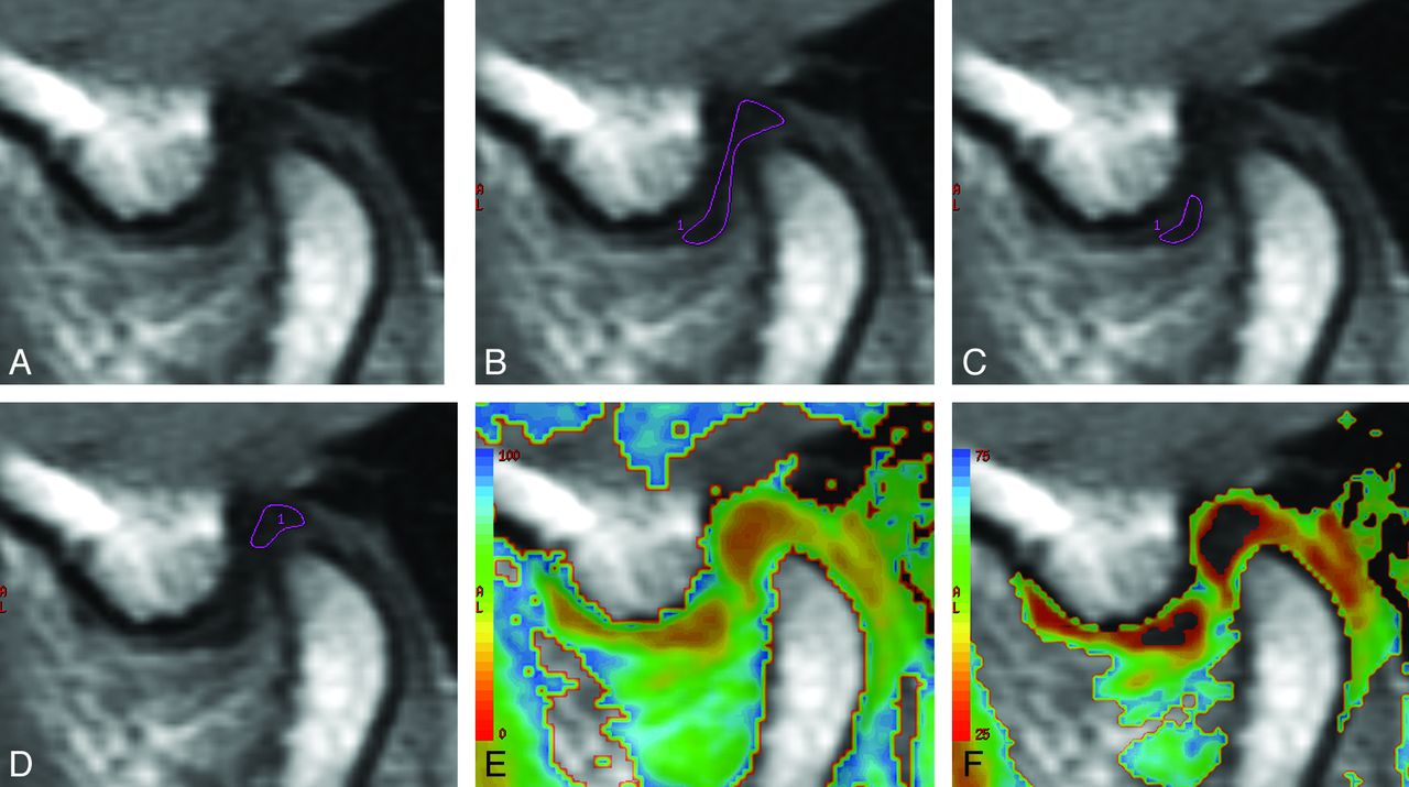
- Fig 1.
MR images of the articular disk of the TMJ, in a volunteer, used to measure the T2 relaxation time. A, The source MR image of the articular disk. The ROIs used for the measurement of the T2 relaxation time of the entire articular disk (B), the anterior band of the articular disk (C), and the posterior band of the articular disk (D). The T2 relaxation times on a color map ranging from 0 to 100 ms (E) and 25 to 75 ms (F).
- Fig 2.
The T2 relaxation times according to the MR image interpretations. A, The T2 relaxation times according to the articular disk position and function categories. The asterisk indicates P < .05/5 in the Mann-Whitney test with a Bonferroni correction. B, The T2 relaxation times according to the articular disk configuration categories. “Thickening” indicates thickening of the posterior band. C, The T2 relaxation times according to the joint effusion categories. “None” indicates no or minimal fluid; “Moderate,” moderate fluid; “Marked,” marked fluid; and “Extensive,” extensive fluid. The asterisk indicates P < .05/5 in the Mann-Whitney test with a Bonferroni correction. D, The T2 relaxation times according to the osteoarthritis categories. The asterisk indicates P < .05/3 in the Mann-Whitney test with a Bonferroni correction. E, The T2 relaxation times according to the bone marrow abnormality categories. The asterisk indicates P < .05/3 in the Mann-Whitney test with a Bonferroni correction. NorVol indicates healthy volunteers.


Tables
Volunteers Patients Cases 17 144 Male 12 34 Female 5 110 Age (yr) Range 23–32 11–80 Median 26 36 Articular disk position and function (joints) Normal superior 34 113 PADDWR 0 18 PADDWOR 0 2 ADDWR 0 48 ADDWOR 0 107 Articular disk configuration (joints) Biconcave 32 143 Biplanar 2 20 Hemiconvex 0 37 Thickening of the posterior band 0 36 Biconvex 0 7 Folded 0 45 Joint effusion (joints) None or minimal fluid 34 169 Moderate fluid 0 80 Marked fluid 0 24 Extensive fluid 0 15 Osteoarthritis (joints) Negative 34 237 Positive 0 51 Bone marrow abnormality (joints) Negative 34 260 Positive 0 28 T2 Relaxation Time (ms) P Volunteers Left TMJ 29.6 ± 3.9 Right TMJ 28.9 ± 3.8 .585 All TMJs 29.3 ± 3.8 Anterior band of all TMJs 29.0 ± 5.4 Posterior band of all TMJs 26.5 ± 3.6 .007 Male 29.0 ± 3.6 Female 29.8 ± 4.3 .620 Age ≤26 years 29.4 ± 3.9 Age >26 years 29.1 ± 3.8 .814 Patients All TMJs 30.7 ± 5.1 .177a Male 29.5 ± 4.0 Female 31.1 ± 5.4 .890 Age ≤36 years 29.5 ± 4.4 Age >36 years 31.9 ± 5.5 <.001 ↵a The T2 relaxation time of all TMJs in patients was compared with that of all TMJs in volunteers.
Variable Unstandardized Coefficient (ß) Unstandardized Coefficient (SE) Standardized Coefficient (ß) T P 95% Confidence Interval Disk position and function 0.652 1.116 0.052 0.585 .559 −1.543 2.848 Disk configuration −1.658 1.014 −0.12 −1.635 .103 −3.653 0.337 Joint effusion 2.467 0.838 0.188 2.944 .003 0.819 4.116 Osteoarthritis 3.986 1.221 0.273 3.264 .001 1.583 6.389 Bone marrow abnormalities 2.896 1.243 0.19 2.331 .02 0.451 5.341


Jump to section
Related Articles
Cited By...
- No citing articles found.