Advertisement

Main menu

  • Home
  • Content
    • Current Issue
    • Accepted Manuscripts
    • Article Preview
    • Past Issue Archive
    • Video Articles
    • AJNR Case Collection
    • Case of the Week Archive
    • Case of the Month Archive
    • Classic Case Archive
  • Special Collections
    • AJNR Awards
    • Low-Field MRI
    • Alzheimer Disease
    • ASNR Foundation Special Collection
    • Photon-Counting CT
    • View All
  • Multimedia
    • AJNR Podcasts
    • AJNR SCANtastic
    • Trainee Corner
    • MRI Safety Corner
    • Imaging Protocols
  • For Authors
    • Submit a Manuscript
    • Submit a Video Article
    • Submit an eLetter to the Editor/Response
    • Manuscript Submission Guidelines
    • Statistical Tips
    • Fast Publishing of Accepted Manuscripts
    • Graphical Abstract Preparation
    • Imaging Protocol Submission
    • Author Policies
  • About Us
    • About AJNR
    • Editorial Board
    • Editorial Board Alumni
  • More
    • Become a Reviewer/Academy of Reviewers
    • Subscribers
    • Permissions
    • Alerts
    • Feedback
    • Advertisers
    • ASNR Home

User menu

  • Alerts
  • Log in

Search

  • Advanced search
American Journal of Neuroradiology

American Journal of Neuroradiology

ASHNR American Society of Functional Neuroradiology ASHNR American Society of Pediatric Neuroradiology ASSR
  • Alerts
  • Log in

Advanced Search

  • Home
  • Content
    • Current Issue
    • Accepted Manuscripts
    • Article Preview
    • Past Issue Archive
    • Video Articles
    • AJNR Case Collection
    • Case of the Week Archive
    • Case of the Month Archive
    • Classic Case Archive
  • Special Collections
    • AJNR Awards
    • Low-Field MRI
    • Alzheimer Disease
    • ASNR Foundation Special Collection
    • Photon-Counting CT
    • View All
  • Multimedia
    • AJNR Podcasts
    • AJNR SCANtastic
    • Trainee Corner
    • MRI Safety Corner
    • Imaging Protocols
  • For Authors
    • Submit a Manuscript
    • Submit a Video Article
    • Submit an eLetter to the Editor/Response
    • Manuscript Submission Guidelines
    • Statistical Tips
    • Fast Publishing of Accepted Manuscripts
    • Graphical Abstract Preparation
    • Imaging Protocol Submission
    • Author Policies
  • About Us
    • About AJNR
    • Editorial Board
    • Editorial Board Alumni
  • More
    • Become a Reviewer/Academy of Reviewers
    • Subscribers
    • Permissions
    • Alerts
    • Feedback
    • Advertisers
    • ASNR Home
  • Follow AJNR on Twitter
  • Visit AJNR on Facebook
  • Follow AJNR on Instagram
  • Join AJNR on LinkedIn
  • RSS Feeds

AJNR Awards, New Junior Editors, and more. Read the latest AJNR updates

Getting new auth cookie, if you see this message a lot, tell someone!
Research ArticleBrain

Preoperative Prognostic Value of MRI Findings in 108 Patients with Idiopathic Normal Pressure Hydrocephalus

J. Virhammar, K. Laurell, K.G. Cesarini and E.-M. Larsson
American Journal of Neuroradiology December 2014, 35 (12) 2311-2318; DOI: https://doi.org/10.3174/ajnr.A4046
J. Virhammar
aFrom the Departments of Neuroscience and Neurology (J.V., K.L.)
  • Find this author on Google Scholar
  • Find this author on PubMed
  • Search for this author on this site
K. Laurell
aFrom the Departments of Neuroscience and Neurology (J.V., K.L.)
dDepartment of Pharmacology and Clinical Neuroscience (K.L.), Östersund, Umeå University, Umeå, Sweden.
  • Find this author on Google Scholar
  • Find this author on PubMed
  • Search for this author on this site
K.G. Cesarini
bNeuroscience and Neurosurgery (K.G.C.)
  • Find this author on Google Scholar
  • Find this author on PubMed
  • Search for this author on this site
E.-M. Larsson
cRadiology (E.-M.L.), Uppsala University, Uppsala, Sweden
  • Find this author on Google Scholar
  • Find this author on PubMed
  • Search for this author on this site
  • Article
  • Figures & Data
  • Info & Metrics
  • Responses
  • References
  • PDF
Loading

Article Figures & Data

Figures

  • Tables
  • Fig 1.
    • Download figure
    • Open in new tab
    • Download powerpoint
    Fig 1.

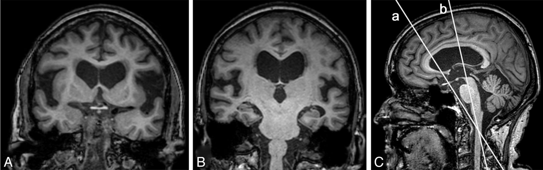
    Nine different patients with iNPH. A, Evans index = x/y. B, Callosal angle. C, Narrow medial sulci and 2 focally dilated sulci on the left side. D, DESH. E, A flow void in the aqueduct and fourth ventricle graded as 2. In addition, a flow void in the foramina of Monro. F, Large diameter of the third ventricle. G, Dilated temporal horns. H, DWMH graded as 3 in a patient who also has PVH. I, PVH graded as 2.

  • Fig 2.
    • Download figure
    • Open in new tab
    • Download powerpoint
    Fig 2.

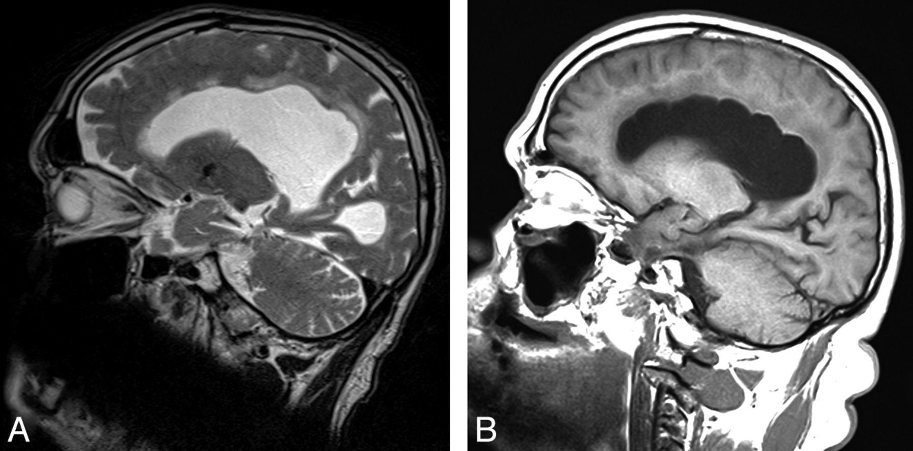
    T1-weighted 3D images of a patient with iNPH. A and B, Coronal images illustrate the different heights of the Sylvian fissure that can be achieved depending on the angulation of the section. B, The Sylvian fissure ordinal. C, Sagittal image with orientation lines represented by the coronal images A and B.

  • Fig 3.
    • Download figure
    • Open in new tab
    • Download powerpoint
    Fig 3.

    Two patients with focal bulging of the roof of the lateral ventricles. Sagittal images include the most cranial portions of the lateral ventricles. A, T2-weighted image. B, T1-weighted image.

  • Fig 4.
    • Download figure
    • Open in new tab
    • Download powerpoint
    Fig 4.

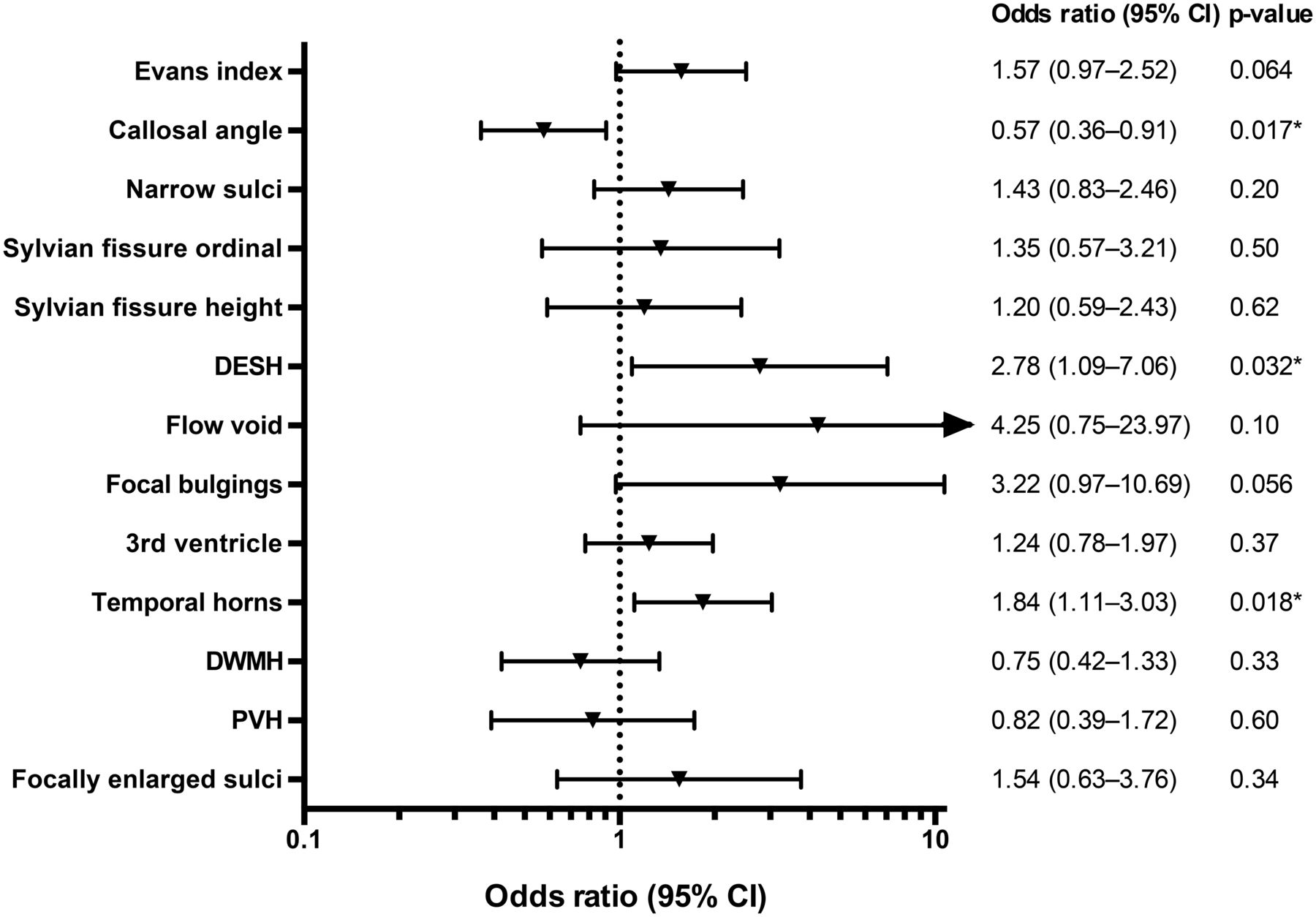
    Forest plot with sex-adjusted odds ratios for all imaging features. OR with a 95% CI of 1-SD increase for continuous variables and a 1-U increase for dichotomous and ordinal variables. An arrow indicates that the confidence interval extends beyond the range of the plot. The Sylvian fissure ordinal is the ordinal scale 0–2; the Sylvian fissure height is measured in millimeters. The asterisk indicates P < .05.

Tables

  • Figures
    • View popup
    Table 1:

    Interrater reliability between 2 independent investigators for all imaging findings

    Imaging FeatureReliability
    Evans index (ICC)0.93
    Callosal angle (ICC)0.95
    Narrow sulci (κ)0.64
    Sylvian fissure original (κ)a0.36
    Sylvian fissure ordinal (κ)b0.62
    Sylvian fissure height (ICC) (mm)0.89
    DESHcNA
    Flow void (κ)0.33
    Focal bulging (κ)0.28
    Third ventricle (ICC)0.96
    Temporal horns (ICC)0.80
    DWMH (κ)0.67
    PVH (κ)0.72
    Focally enlarged sulci (κ)0.54
    Aqueductal stenosis (κ)0.32
    • Note:—NA indicates not applicable.

    • ↵a The original method to measure the Sylvian fissure.7

    • ↵b Ordinal scale of 0–2.

    • ↵c Calculated from Sylvian fissure ordinal and narrow sulci.

    • View popup
    Table 2:

    Prevalence and grading of imaging findings measured on dichotomous and ordinal scales

    Imaging Feature (Grading Range)Frequency of Different Grading (No.) (%)aSample (No.)
    0123
    Sylvian fissure ordinal (0–2)23 (21)77 (71)8 (8)NA108
    DESH (0–1)36 (33)72 (67)NANA108
    Flow void (0–3)b0 (0)0 (0)9 (25)27 (75)36
    Narrow sulci (0–2)24 (22)22 (20)62 (58)NA108
    Focal bulging (0–1)13 (12)95 (88)NANA108
    DWMH (0–3)5 (5)11 (10)31 (29)60 (56)107
    PVH (0–2)27 (25)66 (62)14 (13)NA107
    Focally enlarged sulci (0–1)45 (42)63 (58)NANA108
    Aqueductal stenosis (0–1)105 (97)3 (3)NANA108
    • Note:—NA indicates not applicable.

    • ↵a Dichotomous variables: 0 = not present, 1 = present.

    • ↵b Flow void in the aqueduct and fourth ventricle.

    • View popup
    Table 3:

    Correlations among different imaging markers at baselinea

    Imaging FeatureSignificant Correlations
    Evans indexThird ventricle (r = 0.39b)
    Callosal angleDESH (r = −0.27c), focal bulging (r = −0.28c), temporal horns (r = −0.33d)
    DESHFocal bulging (r = 0.34d), focally enlarged sulci (r = 0.32d), callosal angle (r = −0.27c)
    Focal bulgingTemporal horns (r = 0.26c), callosal angle (r = −0.28c), DESH (r = 0.34d)
    Third ventricleTemporal horns (r = 0.38b)
    DWMHPVH (r = 0.68b)
    • Note:—r indicates Spearman correlation coefficient.

    • ↵a Measurements of Sylvian fissure and narrow sulci at the high convexity were not included because they are components of DESH.

    • ↵b P < .0001.

    • ↵c P < .01.

    • ↵d P < .001.

PreviousNext
Back to top

In this issue

American Journal of Neuroradiology: 35 (12)
American Journal of Neuroradiology
Vol. 35, Issue 12
1 Dec 2014
  • Table of Contents
  • Index by author
  • Complete Issue (PDF)
Advertisement
Print
Download PDF
Email Article

Thank you for your interest in spreading the word on American Journal of Neuroradiology.

NOTE: We only request your email address so that the person you are recommending the page to knows that you wanted them to see it, and that it is not junk mail. We do not capture any email address.

Enter multiple addresses on separate lines or separate them with commas.
Preoperative Prognostic Value of MRI Findings in 108 Patients with Idiopathic Normal Pressure Hydrocephalus
(Your Name) has sent you a message from American Journal of Neuroradiology
(Your Name) thought you would like to see the American Journal of Neuroradiology web site.
CAPTCHA
This question is for testing whether or not you are a human visitor and to prevent automated spam submissions.
Cite this article
J. Virhammar, K. Laurell, K.G. Cesarini, E.-M. Larsson
Preoperative Prognostic Value of MRI Findings in 108 Patients with Idiopathic Normal Pressure Hydrocephalus
American Journal of Neuroradiology Dec 2014, 35 (12) 2311-2318; DOI: 10.3174/ajnr.A4046

Citation Manager Formats

  • BibTeX
  • Bookends
  • EasyBib
  • EndNote (tagged)
  • EndNote 8 (xml)
  • Medlars
  • Mendeley
  • Papers
  • RefWorks Tagged
  • Ref Manager
  • RIS
  • Zotero
0 Responses
Respond to this article
Share
Bookmark this article
Preoperative Prognostic Value of MRI Findings in 108 Patients with Idiopathic Normal Pressure Hydrocephalus
J. Virhammar, K. Laurell, K.G. Cesarini, E.-M. Larsson
American Journal of Neuroradiology Dec 2014, 35 (12) 2311-2318; DOI: 10.3174/ajnr.A4046
del.icio.us logo Twitter logo Facebook logo Mendeley logo
  • Tweet Widget
  • Facebook Like
  • Google Plus One
Purchase

Jump to section

  • Article
    • Abstract
    • ABBREVIATIONS:
    • Materials and Methods
    • Results
    • Discussion
    • Conclusions
    • Acknowledgments
    • Footnotes
    • References
  • Figures & Data
  • Info & Metrics
  • Responses
  • References
  • PDF

Related Articles

  • No related articles found.
  • PubMed
  • Google Scholar

Cited By...

  • Can Shunt Response in Patients with Idiopathic Normal Pressure Hydrocephalus Be Predicted from Preoperative Brain Imaging? A Retrospective Study of the Diagnostic Use of the Normal Pressure Hydrocephalus Radscale in 119 Patients
  • Prognostic Utility of Disproportionately Enlarged Subarachnoid Space Hydrocephalus in Idiopathic Normal Pressure Hydrocephalus Treated with Ventriculoperitoneal Shunt Surgery: A Systematic Review and Meta-analysis
  • Aqueductal CSF Stroke Volume Is Increased in Patients with Idiopathic Normal Pressure Hydrocephalus and Decreases after Shunt Surgery
  • Absence of Disproportionately Enlarged Subarachnoid Space Hydrocephalus, a Sharp Callosal Angle, or Other Morphologic MRI Markers Should Not Be Used to Exclude Patients with Idiopathic Normal Pressure Hydrocephalus from Shunt Surgery
  • High-Convexity Tightness Predicts the Shunt Response in Idiopathic Normal Pressure Hydrocephalus
  • Quantitative MRI for Rapid and User-Independent Monitoring of Intracranial CSF Volume in Hydrocephalus
  • Optimal Diagnostic Indices for Idiopathic Normal Pressure Hydrocephalus Based on the 3D Quantitative Volumetric Analysis for the Cerebral Ventricle and Subarachnoid Space
  • Crossref
  • Google Scholar

This article has not yet been cited by articles in journals that are participating in Crossref Cited-by Linking.

More in this TOC Section

  • Multimodal CT Provides Improved Performance for Lacunar Infarct Detection
  • Optimal MRI Sequence for Identifying Occlusion Location in Acute Stroke: Which Value of Time-Resolved Contrast-Enhanced MRA?
  • Evaluating the Effects of White Matter Multiple Sclerosis Lesions on the Volume Estimation of 6 Brain Tissue Segmentation Methods
Show more Brain

Similar Articles

Advertisement

Indexed Content

  • Current Issue
  • Accepted Manuscripts
  • Article Preview
  • Past Issues
  • Editorials
  • Editor's Choice
  • Fellows' Journal Club
  • Letters to the Editor
  • Video Articles

Cases

  • Case Collection
  • Archive - Case of the Week
  • Archive - Case of the Month
  • Archive - Classic Case

More from AJNR

  • Trainee Corner
  • Imaging Protocols
  • MRI Safety Corner
  • Book Reviews

Multimedia

  • AJNR Podcasts
  • AJNR Scantastics

Resources

  • Turnaround Time
  • Submit a Manuscript
  • Submit a Video Article
  • Submit an eLetter to the Editor/Response
  • Manuscript Submission Guidelines
  • Statistical Tips
  • Fast Publishing of Accepted Manuscripts
  • Graphical Abstract Preparation
  • Imaging Protocol Submission
  • Evidence-Based Medicine Level Guide
  • Publishing Checklists
  • Author Policies
  • Become a Reviewer/Academy of Reviewers
  • News and Updates

About Us

  • About AJNR
  • Editorial Board
  • Editorial Board Alumni
  • Alerts
  • Permissions
  • Not an AJNR Subscriber? Join Now
  • Advertise with Us
  • Librarian Resources
  • Feedback
  • Terms and Conditions
  • AJNR Editorial Board Alumni

American Society of Neuroradiology

  • Not an ASNR Member? Join Now

© 2025 by the American Society of Neuroradiology All rights, including for text and data mining, AI training, and similar technologies, are reserved.
Print ISSN: 0195-6108 Online ISSN: 1936-959X

Powered by HighWire