Advertisement

Main menu

  • Home
  • Content
    • Current Issue
    • Accepted Manuscripts
    • Article Preview
    • Past Issue Archive
    • Video Articles
    • AJNR Case Collection
    • Case of the Week Archive
    • Case of the Month Archive
    • Classic Case Archive
  • Special Collections
    • AJNR Awards
    • Low-Field MRI
    • Alzheimer Disease
    • ASNR Foundation Special Collection
    • Photon-Counting CT
    • View All
  • Multimedia
    • AJNR Podcasts
    • AJNR SCANtastic
    • Trainee Corner
    • MRI Safety Corner
    • Imaging Protocols
  • For Authors
    • Submit a Manuscript
    • Submit a Video Article
    • Submit an eLetter to the Editor/Response
    • Manuscript Submission Guidelines
    • Statistical Tips
    • Fast Publishing of Accepted Manuscripts
    • Graphical Abstract Preparation
    • Imaging Protocol Submission
    • Author Policies
  • About Us
    • About AJNR
    • Editorial Board
    • Editorial Board Alumni
  • More
    • Become a Reviewer/Academy of Reviewers
    • Subscribers
    • Permissions
    • Alerts
    • Feedback
    • Advertisers
    • ASNR Home

User menu

  • Alerts
  • Log in

Search

  • Advanced search
American Journal of Neuroradiology

American Journal of Neuroradiology

ASHNR American Society of Functional Neuroradiology ASHNR American Society of Pediatric Neuroradiology ASSR
  • Alerts
  • Log in

Advanced Search

  • Home
  • Content
    • Current Issue
    • Accepted Manuscripts
    • Article Preview
    • Past Issue Archive
    • Video Articles
    • AJNR Case Collection
    • Case of the Week Archive
    • Case of the Month Archive
    • Classic Case Archive
  • Special Collections
    • AJNR Awards
    • Low-Field MRI
    • Alzheimer Disease
    • ASNR Foundation Special Collection
    • Photon-Counting CT
    • View All
  • Multimedia
    • AJNR Podcasts
    • AJNR SCANtastic
    • Trainee Corner
    • MRI Safety Corner
    • Imaging Protocols
  • For Authors
    • Submit a Manuscript
    • Submit a Video Article
    • Submit an eLetter to the Editor/Response
    • Manuscript Submission Guidelines
    • Statistical Tips
    • Fast Publishing of Accepted Manuscripts
    • Graphical Abstract Preparation
    • Imaging Protocol Submission
    • Author Policies
  • About Us
    • About AJNR
    • Editorial Board
    • Editorial Board Alumni
  • More
    • Become a Reviewer/Academy of Reviewers
    • Subscribers
    • Permissions
    • Alerts
    • Feedback
    • Advertisers
    • ASNR Home
  • Follow AJNR on Twitter
  • Visit AJNR on Facebook
  • Follow AJNR on Instagram
  • Join AJNR on LinkedIn
  • RSS Feeds

AJNR Awards, New Junior Editors, and more. Read the latest AJNR updates

Research ArticleBrain
Open Access

Automatic Lesion Incidence Estimation and Detection in Multiple Sclerosis Using Multisequence Longitudinal MRI

E.M. Sweeney, R.T. Shinohara, C.D. Shea, D.S. Reich and C.M. Crainiceanu
American Journal of Neuroradiology January 2013, 34 (1) 68-73; DOI: https://doi.org/10.3174/ajnr.A3172
E.M. Sweeney
aFrom the Department of Biostatistics (E.M.S., R.T.S., D.S.R., C.M.C.), Johns Hopkins University, Baltimore, Maryland
bTranslational Neuroradiology Unit, Neuroimmunology Branch, National Institute of Neurological Disease and Stroke (E.M.S., R.T.S., C.D.S., D.S.R.)
  • Find this author on Google Scholar
  • Find this author on PubMed
  • Search for this author on this site
R.T. Shinohara
aFrom the Department of Biostatistics (E.M.S., R.T.S., D.S.R., C.M.C.), Johns Hopkins University, Baltimore, Maryland
bTranslational Neuroradiology Unit, Neuroimmunology Branch, National Institute of Neurological Disease and Stroke (E.M.S., R.T.S., C.D.S., D.S.R.)
  • Find this author on Google Scholar
  • Find this author on PubMed
  • Search for this author on this site
C.D. Shea
bTranslational Neuroradiology Unit, Neuroimmunology Branch, National Institute of Neurological Disease and Stroke (E.M.S., R.T.S., C.D.S., D.S.R.)
  • Find this author on Google Scholar
  • Find this author on PubMed
  • Search for this author on this site
D.S. Reich
aFrom the Department of Biostatistics (E.M.S., R.T.S., D.S.R., C.M.C.), Johns Hopkins University, Baltimore, Maryland
bTranslational Neuroradiology Unit, Neuroimmunology Branch, National Institute of Neurological Disease and Stroke (E.M.S., R.T.S., C.D.S., D.S.R.)
cDiagnostic Radiology Department, Clinical Center (D.S.R.), National Institutes of Health, Bethesda, Maryland.
  • Find this author on Google Scholar
  • Find this author on PubMed
  • Search for this author on this site
C.M. Crainiceanu
aFrom the Department of Biostatistics (E.M.S., R.T.S., D.S.R., C.M.C.), Johns Hopkins University, Baltimore, Maryland
  • Find this author on Google Scholar
  • Find this author on PubMed
  • Search for this author on this site
  • Article
  • Figures & Data
  • Supplemental
  • Info & Metrics
  • Responses
  • References
  • PDF
Loading

Article Figures & Data

Figures

  • Tables
  • Fig 1.
    • Download figure
    • Open in new tab
    • Download powerpoint
    Fig 1.

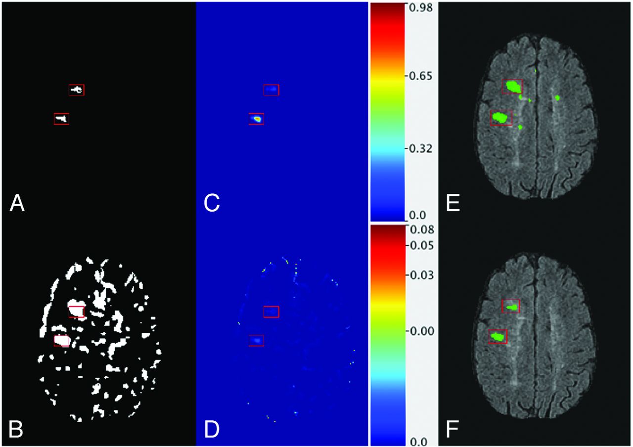
    Areas with lesion incidence are indicated with red boxes. A, Neuroradiologist manual segmentation of an axial section of the brain. B, Selected voxels for SuBLIME modeling. C, Axial section of the probability map from the full model. D, Axial section of the probability map from the SuBLIME model fit with only the T2-weighted image. E, Binary segmentation of the probability map from the full model with false-positive rate of 0.01 overlaid on the FLAIR image. F, Binary segmentation of the probability map from the full model with false-positive rate of 0.001 overlaid on the FLAIR image.

  • Fig 2.
    • Download figure
    • Open in new tab
    • Download powerpoint
    Fig 2.

    A, ROC curves for the voxel-level detection of incident and enlarging lesions for different thresholds of the probability maps produced from SuBLIME, as well as different thresholds of the T2-weighted subtraction without the model and voxel selection procedure. The red ROC curve is for the full model and has an AUC of 99% (95% CI [97%, 100%]). The blue ROC curve is for the model fit with only the T2-weighted image and has an AUC of 97% (95% CI [88%, 99%]). The green ROC curve is for thresholding the T2-weighted subtraction image without the model and voxel selection procedure and has an AUC of 92% (95% CI [83%, 95%]). B, Partial ROC curves for false-positive rates up to 0.01. The red curve is for the full model and the blue curve is for the model fit with only the T2-weighted image. The green curve is for thresholding the T2-weighted subtraction image without the model and voxel selection procedure.

Tables

  • Figures
    • View popup
    Table 1:

    Subject demographic, diagnosis, and treatment information

    SetSubtypeAgeSexEDSSTreatment with Disease-Modifying Therapy (Baseline)
    ValidationRRMS37Male1.5Yes
    ValidationRRMS38Female2.0Yes
    ValidationRRMS48Male3.0No
    TrainingRRMS38Female1.5No
    TrainingRRMS30Female1.0Yes
    TrainingRRMS43Female1.5Yes
    ValidationRRMS35Female1.5Yes
    TrainingRRMS37Female1.0Yes
    TrainingRRMS47Female3.0Yes
    ValidationRRMS56Female1.0No
    • Note:—EDSS indicates Expanded Disability Status Scale; RRMS, relaping-remitting multiple sclerosis.

    • View popup
    Table 2:

    Study scanning parameters

    FA (degrees)TR (ms)TE (ms)TI (ms)
    FLAIR(90, 90)(10,000, 10,000)(77.8, 159.5)(2200, 2500)
    T2-weighted(90, 90)(3400, 6500)(94.6, 112.0)NA
    PD(90, 90)(3400, 6500)(11.8, 15.0)NA
    T1-weighted(13, 20)(7.68, 10.3)(1.88, 4.05)(450, 750)a
    • Note:—FA indicates flip angle; NA, not available.

    • ↵a 106 of the T1-weighted scans did not have an inversion time.

    • View popup
    Table 3:

    Binary segmentations

    False- Positive RateThreshold ValueSpecificitySensitivityVolume Change (Actual 625 mm3)
    0.010.00220.990.9530454
    0.0010.02290.9990.833520
    0.000250.08150.999750.541082
    0.00010.13960.99990.35509
PreviousNext
Back to top

In this issue

American Journal of Neuroradiology: 34 (1)
American Journal of Neuroradiology
Vol. 34, Issue 1
1 Jan 2013
  • Table of Contents
  • Index by author
Advertisement
Print
Download PDF
Email Article

Thank you for your interest in spreading the word on American Journal of Neuroradiology.

NOTE: We only request your email address so that the person you are recommending the page to knows that you wanted them to see it, and that it is not junk mail. We do not capture any email address.

Enter multiple addresses on separate lines or separate them with commas.
Automatic Lesion Incidence Estimation and Detection in Multiple Sclerosis Using Multisequence Longitudinal MRI
(Your Name) has sent you a message from American Journal of Neuroradiology
(Your Name) thought you would like to see the American Journal of Neuroradiology web site.
CAPTCHA
This question is for testing whether or not you are a human visitor and to prevent automated spam submissions.
Cite this article
E.M. Sweeney, R.T. Shinohara, C.D. Shea, D.S. Reich, C.M. Crainiceanu
Automatic Lesion Incidence Estimation and Detection in Multiple Sclerosis Using Multisequence Longitudinal MRI
American Journal of Neuroradiology Jan 2013, 34 (1) 68-73; DOI: 10.3174/ajnr.A3172

Citation Manager Formats

  • BibTeX
  • Bookends
  • EasyBib
  • EndNote (tagged)
  • EndNote 8 (xml)
  • Medlars
  • Mendeley
  • Papers
  • RefWorks Tagged
  • Ref Manager
  • RIS
  • Zotero
0 Responses
Respond to this article
Share
Bookmark this article
Automatic Lesion Incidence Estimation and Detection in Multiple Sclerosis Using Multisequence Longitudinal MRI
E.M. Sweeney, R.T. Shinohara, C.D. Shea, D.S. Reich, C.M. Crainiceanu
American Journal of Neuroradiology Jan 2013, 34 (1) 68-73; DOI: 10.3174/ajnr.A3172
del.icio.us logo Twitter logo Facebook logo Mendeley logo
  • Tweet Widget
  • Facebook Like
  • Google Plus One
Purchase

Jump to section

  • Article
    • Abstract
    • ABBREVIATIONS:
    • Materials and Methods
    • Results
    • Discussion
    • Conclusions
    • Acknowledgments
    • Footnotes
    • References
  • Figures & Data
  • Supplemental
  • Info & Metrics
  • Responses
  • References
  • PDF

Related Articles

  • PubMed
  • Google Scholar

Cited By...

  • Evaluation of the Statistical Detection of Change Algorithm for Screening Patients with MS with New Lesion Activity on Longitudinal Brain MRI
  • Evaluation of the Statistical Detection of Change Algorithm for Screening Patients with MS with New Lesion Activity on Longitudinal Brain MRI
  • Evaluation of statistical detection of change algorithm for triaging multiple sclerosis patients with new lesion activity on longitudinal brain MRI
  • Automatic brain lesion segmentation on standard magnetic resonance images: a scoping review
  • Intensity warping for multisite MRI harmonization
  • A New Approach to Symmetric Registration of Longitudinal Structural MRI of the Human Brain
  • An automated statistical technique for counting distinct multiple sclerosis lesions can recover aspects of lesion history and provide relevant disease information
  • Improved Detection of New MS Lesions during Follow-Up Using an Automated MR Coregistration-Fusion Method
  • An Automated Statistical Technique for Counting Distinct Multiple Sclerosis Lesions
  • Improved Automatic Detection of New T2 Lesions in Multiple Sclerosis Using Deformation Fields
  • Crossref
  • Google Scholar

This article has not yet been cited by articles in journals that are participating in Crossref Cited-by Linking.

More in this TOC Section

  • Evaluating the Effects of White Matter Multiple Sclerosis Lesions on the Volume Estimation of 6 Brain Tissue Segmentation Methods
  • Quiet PROPELLER MRI Techniques Match the Quality of Conventional PROPELLER Brain Imaging Techniques
  • Predictors of Reperfusion in Patients with Acute Ischemic Stroke
Show more Brain

Similar Articles

Advertisement

Indexed Content

  • Current Issue
  • Accepted Manuscripts
  • Article Preview
  • Past Issues
  • Editorials
  • Editor's Choice
  • Fellows' Journal Club
  • Letters to the Editor
  • Video Articles

Cases

  • Case Collection
  • Archive - Case of the Week
  • Archive - Case of the Month
  • Archive - Classic Case

More from AJNR

  • Trainee Corner
  • Imaging Protocols
  • MRI Safety Corner
  • Book Reviews

Multimedia

  • AJNR Podcasts
  • AJNR Scantastics

Resources

  • Turnaround Time
  • Submit a Manuscript
  • Submit a Video Article
  • Submit an eLetter to the Editor/Response
  • Manuscript Submission Guidelines
  • Statistical Tips
  • Fast Publishing of Accepted Manuscripts
  • Graphical Abstract Preparation
  • Imaging Protocol Submission
  • Evidence-Based Medicine Level Guide
  • Publishing Checklists
  • Author Policies
  • Become a Reviewer/Academy of Reviewers
  • News and Updates

About Us

  • About AJNR
  • Editorial Board
  • Editorial Board Alumni
  • Alerts
  • Permissions
  • Not an AJNR Subscriber? Join Now
  • Advertise with Us
  • Librarian Resources
  • Feedback
  • Terms and Conditions
  • AJNR Editorial Board Alumni

American Society of Neuroradiology

  • Not an ASNR Member? Join Now

© 2025 by the American Society of Neuroradiology All rights, including for text and data mining, AI training, and similar technologies, are reserved.
Print ISSN: 0195-6108 Online ISSN: 1936-959X

Powered by HighWire