
Article Figures & Data
Figures
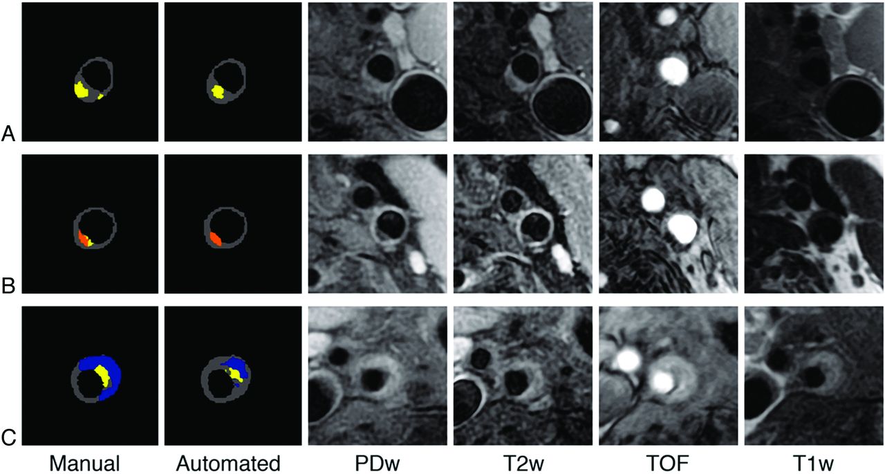
- Fig 1.
Three illustrative examples of manual and automated segmentation. A, Lipid core (yellow) corresponds to an area in which the signal intensity dropped on T2WI compared with PDw images. B, Calcifications (orange) correspond to an area of hypointensity on all 4 sequences. C, Recent hemorrhage (blue) corresponds to an area of hyperintensities on T1WI and TOF images. Note that the automated classifier underestimated the hemorrhage area.
- Fig 2.
Graphs showing scatterplots of the volumes measured with the manual and automated methods for each component. Each dot corresponds to 1 patient.
- Fig 3.
Bland-Altman graphics showing the differences between volumes obtained by manual and automated segmentation plotted against the mean of the 2 measurements. Volumes per patient are expressed in mm3. The dotted lines indicate the average bias and the dashed lines show the 95% CI (mean bias ± 1.96 SD). For each component, a negative bias indicates that the automated segmentation overestimates the volumes. A positive bias indicates that the automated segmentation underestimates the volumes.


Tables
- Table 1:
Percentage of agreement and κ statistic of the plaque components per patient (n = 40)
Plaque Component Manual Automated Agreement κ [95% CI] Absence Presence Calcification Absence 19 3 80.0% 0.59 [0.36–0.82] Presence 5 13 Hemorrhage Absence 15 4 82.5% 0.65 [0.42–0.88] Presence 3 18 Lipid core Absence 1 0 97.5% 0.65 [0.03–1.27] Presence 1 38 Plaque Component Volume (mm3) Mean SD Median IQR P value* Hemorrhage (n = 18) Manual 112 103 86 21–199 0.16 Automated 90 104 58 13–124 Calcification (n = 13) Manual 20 18 18 11–26 0.5 Automated 15 17 7 2–23 Lipid core (n = 38) Manual 73 66 51 33–91 0.01 Automated 125 149 82 19–196 Fibrous tissue (n = 40) Manual 272 176 229 163–344 0.27 Automated 237 162 216 122–340 Note:—IQR indicates interquartile range; n = number of patients for whom a given component was detected by both methods.
↵* Paired Wilcoxon test.


