
Article Figures & Data
Figures
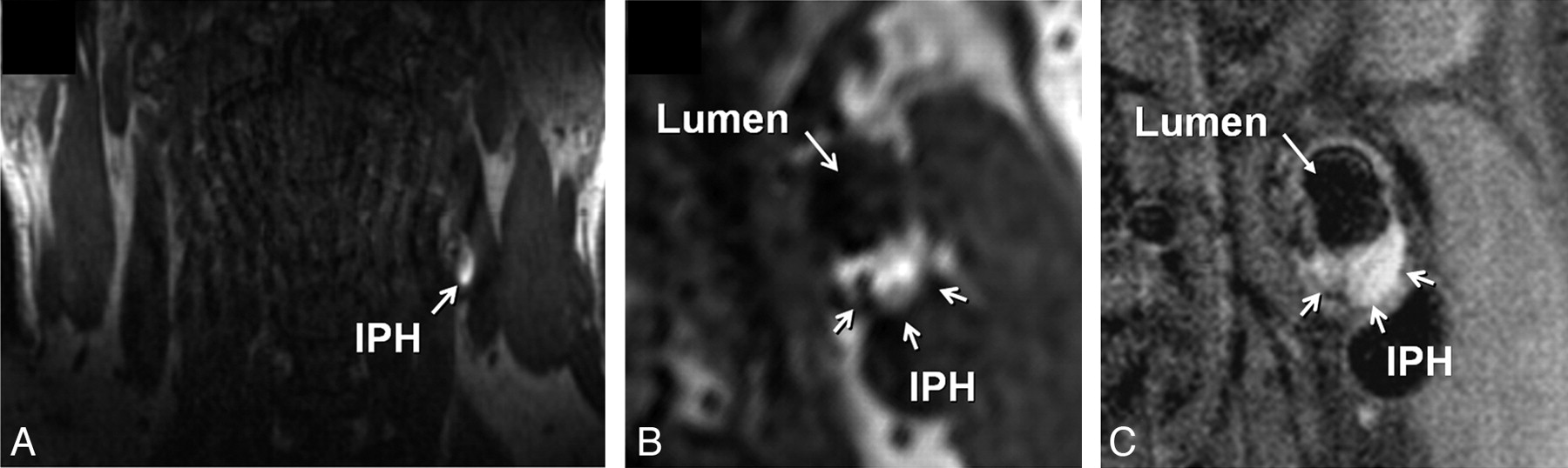
- Fig 1.
Identification of IPH in the left carotid artery of a 60-year-old man with an ipsilateral cerebrovascular ischemic event. A, Source image from the coronally oriented CE-MRA mask sequence shows hyperintense signal intensity in the left carotid artery wall corresponding to IPH. The high signal intensity is seen on the transverse view of the mask image (B) reconstructed in the same plane as the T1-weighted precontrast BBMRI (C).
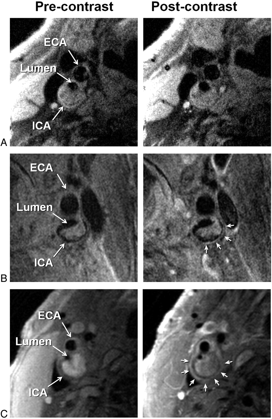
- Fig 2.
Examples of adventitial enhancement categories based on BBMRI acquired after the intravenous administration of gadolinium-based contrast (right column) by using the corresponding precontrast images for reference (left column). Categories 0, 1, and 2 are shown with no enhancement (A), <50% enhancement (B), and ≥50% enhancement (C) of the adventitia (arrows), respectively. ICA and ECA indicate internal carotid artery and external carotid artery, respectively.
- Fig 3.
The relation between adventitial enhancement and prior cerebrovascular ischemic events stratified for patients with and without IPH present.


Tables
Patient Characteristics Age (yr) 72.5 (10.0, 45–89) Female 11 (23%) Active smoker 7 (15%) Diabetes mellitus 7 (15%) Hypertension 26 (55%) Hyperlipidemia 20 (43%) Statin use 37 (78%) Aspirin use 29 (61%) Clinical presentation Asymptomatic 23 (49%) TIA 9 (19%) Stroke 15 (32%) -
↵a Data are expressed as mean (SD, range) or number of participants (% of 47).
-
- Table 2:
Characteristics of participants grouped by adventitial enhancement category and IPH presencea
Adventitial Enhancement IPH Category 0 (n = 7) Category 1 (n = 16) Category 2 (n = 24) P No. (n = 25) Yes (n = 22) P Age (yr) 68.7 ± 11 71.2 ± 9.6 74.4 ± 9.7 .34 70.5 ± 11 74.8 ± 8.3 .15 Female 2 3 6 .84 9 2 .03 Active smoker 0 5 2 .07 5 2 .27 Diabetes mellitus 2 3 2 .36 4 3 .57 Hypertension 3 9 14 .76 14 12 .58 Hyperlipidemia 4 5 11 .46 11 9 .53 Statin use 5 14 18 .56 21 16 .28 Aspirin use 4 12 13 .40 15 14 .52 TIA/stroke (%) 14 48 63 .02 9 15 .03 Stenosis (%) 53 ± 25 54 ± 30 62 ± 26 .62 47 ± 29 71 ± 19 .002 Maximum wall thickness (mm) 4.63 ± 1.84 4.75 ± 1.55 4.82 ± 1.14 .95 4.42 ± 1.60 5.13 ± 0.99 .04 -
↵a Data are expressed as mean ± SD or number of participants. Based on data from reader 1.
-
- Table 3:
Multivariable analysis for participants with previous cerebrovascular ischemic eventsa
Multivariable OR 95% CI P Age (yr) 0.99 0.91–1.08 .88 Female 0.43 0.07–2.72 .37 Active smoker 7.50 1.10–51.47 .04 Diabetes mellitus 10.25 0.91–115.92 .06 Hypertension 0.58 0.10–3.34 .54 Hyperlipidemia 0.51 0.08–3.14 .47 Statin use 0.81 0.06–9.84 .87 Aspirin use 0.99 0.10–9.70 .99 Stenosis (%) 0.05 0.02–1.50 .09 Maximum wall thickness (mm) 0.77 0.40–1.45 .42 Adventitial enhancement (category 1 vs category 0) 14.90 0.98–225.93 .05 Adventitial enhancement (category 2 vs category 0) 51.17 3.40–469.80 .004 IPH presence 10.18 1.42–72.21 .02 -
↵a Adjusting for coils and gadolinium dose.
-
In this issue


Jump to section
Related Articles
Cited By...
- Characterization of Restenosis following Carotid Endarterectomy Using Contrast-Enhanced Vessel Wall MR Imaging
- Imaging of the vulnerable carotid plaque: Role of imaging techniques and a research agenda
- Gadolinium Enhancement of the Aneurysm Wall in Extracranial Carotid Artery Aneurysms
- Carotid Vessel Wall Imaging on CTA
- Differential Features of Culprit Intracranial Atherosclerotic Lesions: A Whole-Brain Vessel Wall Imaging Study in Patients With Acute Ischemic Stroke
- Carotid Artery Wall Imaging: Perspective and Guidelines from the ASNR Vessel Wall Imaging Study Group and Expert Consensus Recommendations of the American Society of Neuroradiology
- Intracranial Vessel Wall MRI: Principles and Expert Consensus Recommendations of the American Society of Neuroradiology
- Magnetic Resonance Imaging of Plaque Morphology, Burden, and Distribution in Patients With Symptomatic Middle Cerebral Artery Stenosis
- Detection of Carotid Artery Stenosis: A Comparison between 2 Unenhanced MRAs and Dual-Source CTA
- Identifying Which Patients With Asymptomatic Carotid Stenosis Could Benefit From Intervention
- Contrast-Enhanced Ultrasound for the Evaluation of Neovascularization in Atherosclerotic Carotid Artery Plaques
- Imaging Intracranial Vessel Wall Pathology With Magnetic Resonance Imaging: Current Prospects and Future Directions
- Is Carotid Intima-Media Thickness as Predictive as Other Noninvasive Techniques for the Detection of Coronary Artery Disease?
- Intraplaque High-Intensity Signal on 3D Time-of-Flight MR Angiography Is Strongly Associated with Symptomatic Carotid Artery Stenosis
- Matrix metalloproteinase-9 expression in carotid atherosclerotic plaque and contrast-enhanced MRI in a swine model
- Vasa Vasorum Enhancement on Computerized Tomographic Angiography Correlates With Symptomatic Patients With 50% to 70% Carotid Artery Stenosis
- Adventitial Perfusion and Intraplaque Hemorrhage: A Dynamic Contrast-Enhanced MRI Study in the Carotid Artery