Advertisement

Main menu

  • Home
  • Content
    • Current Issue
    • Accepted Manuscripts
    • Article Preview
    • Past Issue Archive
    • Video Articles
    • AJNR Case Collection
    • Case of the Week Archive
    • Case of the Month Archive
    • Classic Case Archive
  • Special Collections
    • AJNR Awards
    • Low-Field MRI
    • Alzheimer Disease
    • ASNR Foundation Special Collection
    • Photon-Counting CT
    • View All
  • Multimedia
    • AJNR Podcasts
    • AJNR SCANtastic
    • Trainee Corner
    • MRI Safety Corner
    • Imaging Protocols
  • For Authors
    • Submit a Manuscript
    • Submit a Video Article
    • Submit an eLetter to the Editor/Response
    • Manuscript Submission Guidelines
    • Statistical Tips
    • Fast Publishing of Accepted Manuscripts
    • Graphical Abstract Preparation
    • Imaging Protocol Submission
    • Author Policies
  • About Us
    • About AJNR
    • Editorial Board
    • Editorial Board Alumni
  • More
    • Become a Reviewer/Academy of Reviewers
    • Subscribers
    • Permissions
    • Alerts
    • Feedback
    • Advertisers
    • ASNR Home

User menu

  • Alerts
  • Log in

Search

  • Advanced search
American Journal of Neuroradiology

American Journal of Neuroradiology

ASHNR American Society of Functional Neuroradiology ASHNR American Society of Pediatric Neuroradiology ASSR
  • Alerts
  • Log in

Advanced Search

  • Home
  • Content
    • Current Issue
    • Accepted Manuscripts
    • Article Preview
    • Past Issue Archive
    • Video Articles
    • AJNR Case Collection
    • Case of the Week Archive
    • Case of the Month Archive
    • Classic Case Archive
  • Special Collections
    • AJNR Awards
    • Low-Field MRI
    • Alzheimer Disease
    • ASNR Foundation Special Collection
    • Photon-Counting CT
    • View All
  • Multimedia
    • AJNR Podcasts
    • AJNR SCANtastic
    • Trainee Corner
    • MRI Safety Corner
    • Imaging Protocols
  • For Authors
    • Submit a Manuscript
    • Submit a Video Article
    • Submit an eLetter to the Editor/Response
    • Manuscript Submission Guidelines
    • Statistical Tips
    • Fast Publishing of Accepted Manuscripts
    • Graphical Abstract Preparation
    • Imaging Protocol Submission
    • Author Policies
  • About Us
    • About AJNR
    • Editorial Board
    • Editorial Board Alumni
  • More
    • Become a Reviewer/Academy of Reviewers
    • Subscribers
    • Permissions
    • Alerts
    • Feedback
    • Advertisers
    • ASNR Home
  • Follow AJNR on Twitter
  • Visit AJNR on Facebook
  • Follow AJNR on Instagram
  • Join AJNR on LinkedIn
  • RSS Feeds

AJNR Awards, New Junior Editors, and more. Read the latest AJNR updates

Research ArticleBrainE

Effect of Stenting on Cerebral CT Perfusion in Symptomatic and Asymptomatic Patients with Carotid Artery Stenosis

L.G. Merckel, J. Van der Heijden, L.M. Jongen, H.W. van Es, M. Prokop and A. Waaijer
American Journal of Neuroradiology February 2012, 33 (2) 280-285; DOI: https://doi.org/10.3174/ajnr.A2757
L.G. Merckel
  • Find this author on Google Scholar
  • Find this author on PubMed
  • Search for this author on this site
J. Van der Heijden
  • Find this author on Google Scholar
  • Find this author on PubMed
  • Search for this author on this site
L.M. Jongen
  • Find this author on Google Scholar
  • Find this author on PubMed
  • Search for this author on this site
H.W. van Es
  • Find this author on Google Scholar
  • Find this author on PubMed
  • Search for this author on this site
M. Prokop
  • Find this author on Google Scholar
  • Find this author on PubMed
  • Search for this author on this site
A. Waaijer
  • Find this author on Google Scholar
  • Find this author on PubMed
  • Search for this author on this site
  • Article
  • Figures & Data
  • Info & Metrics
  • Responses
  • References
  • PDF
Loading

Article Figures & Data

Figures

  • Tables
  • Fig 1.
    • Download figure
    • Open in new tab
    • Download powerpoint
    Fig 1.

    Manual outlining of middle cerebral artery territory on transverse CT sections, according to the maps of Damasio.20

  • Fig 2.
    • Download figure
    • Open in new tab
    • Download powerpoint
    Fig 2.

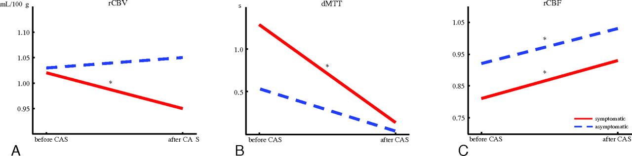
    Relative CTP values measured before and after treatment in symptomatic and asymptomatic patients: rCBV (A), dMTT (B), and rCBF (C).

  • Fig 3.
    • Download figure
    • Open in new tab
    • Download powerpoint
    Fig 3.

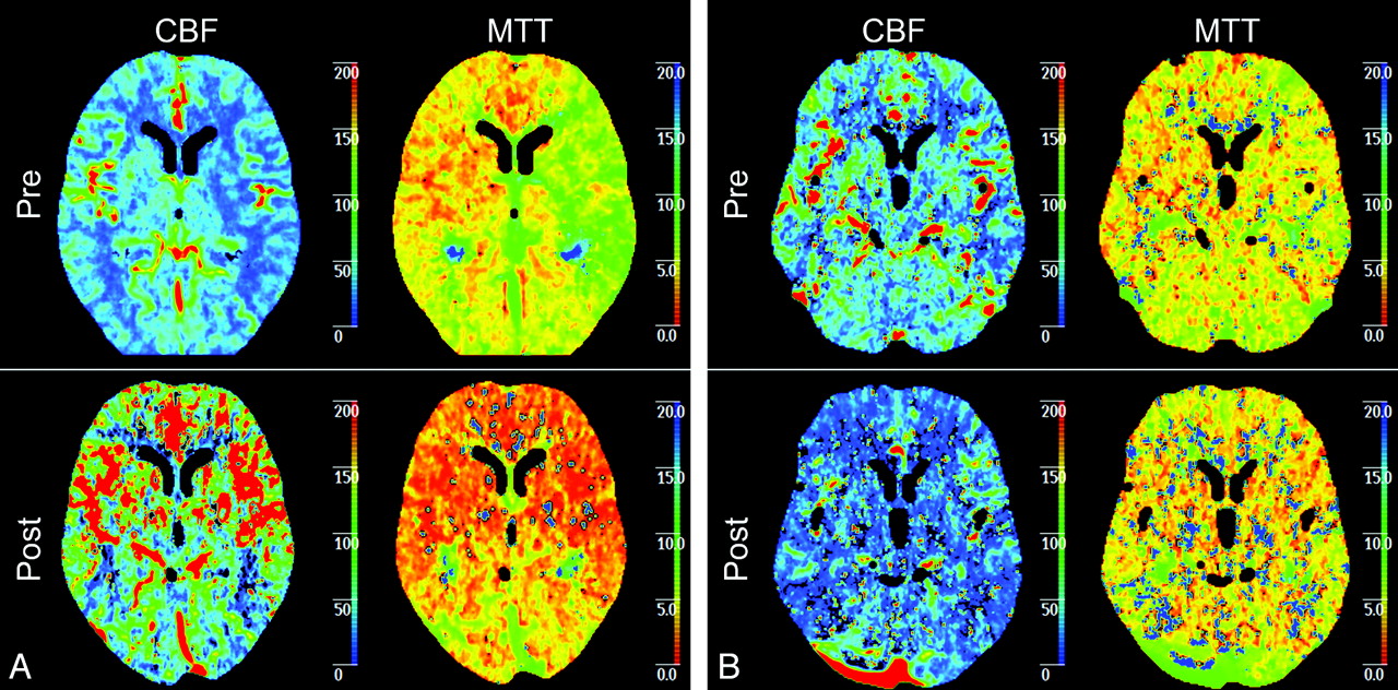
    A, Example of symptomatic patient with a left-sided carotid artery stenosis of 99%. Before treatment, CBF and MTT show explicit differences between the right and left hemispheres with a higher CBF and a higher MTT in the right hemisphere in comparison with the left hemisphere. After CAS, both CBF and MTT show a symmetric pattern of perfusion. B, An example of a asymptomatic patient with a left-sided carotid artery stenosis of 95%. Before treatment, CBF and MTT show differences between the right and left hemisphere with a higher CBF and a higher MTT in the right hemisphere in comparison with the left hemisphere. However, this difference is not as clear as in the symptomatic patient. After CAS, both CBF and MTT show a symmetric pattern of perfusion.

Tables

  • Figures
    • View popup
    Table 1:

    Patient characteristics (n = 45)

    Patient DemographicsSymptomatic Patients (n = 31)Asymptomatic Patients (n= 14)P Valuea
    Age (yr) (mean) (range)67.0 ± 10.4 (43–82)69.6 ± 9.0 (56–82).43b
    Male (No.) (%)21 (67.7)13 (92.9).13c
    Carotid arteries
    Left side treated (No.) (%)15 (48.4)9 (64.3).32
    Stenosis grade (%), mean ± SD (range)89.5 ± 9.4 (65–99)88.9 ± 5.8 (75–99).32b
    Symptoms
    Stroke (No.) (%)11 (35.5)NoneN.A.
    TIA (No.) (%)13 (41.9)NoneN.A.
    AF (No.) (%)7 (22.6)NoneN.A.
    Interval in days
    Pretreatment CTP to treatment (mean) (range)3.2 ± 6.4 (0–35)3.5 ± 1.9 (3–10).03b
    Treatment to posttreatment CTP (mean) (range)33.9 ± 4.8 (22–44)71.9 ± 82.6 (8–263).84b
    Medical history
    Hypertension (No.) (%)25 (80.6)13 (92.9).41c
    Diabetes mellitus (No.) (%)3 (9.7)3 (21.4).36c
    Hypercholesterolemia (No.) (%)12 (38.7)12 (85.7).008
    Smoking (No.) (%)10 (32.3)6 (42.9).52c
    Previous myocardial infarction (No.) (%)4 (12.9)3 (21.4).66c
    Previous CABG (No.) (%)2 (6.5)1 (7.1)1.00c
    • Note:—N.A. indicates not applicable; AE, atrial fibrillation.

    • ↵a P values analyzed with the Pearson χ2 test.

    • ↵b P values analyzed with Mann-Whitney U test.

    • ↵c P values analyzed with Fisher exact test.

    • View popup
    Table 2:

    Comparison of pre- and posttreatment CTP data for symptomatic and asymptomatic patientsa

    Pre- and PosttreatmentrCBV (mL/100 g) (mean)P ValuedMTT (s) (mean)P ValuerCBF (mL/100 g/min) (mean)P Value
    Symptomatic patients before treatment (n = 31)1.02 ± 0.131.29 ± 1.210.81 ± 0.14
    Symptomatic patients after treatment (n = 31)0.95 ± 0.14.036b0.14 ± 1.08<.001b0.93 ± 0.17<.001b
    Asymptomatic patients before treatment (n = 14)1.03 ± 0.080.56 ± 0.660.92 ± 0.12
    Asymptomatic patients, after treatment (n = 14)1.05 ± 0.13.2210.06 ± 0.550.0811.03 ± 0.14.026b
    • ↵a The significance of the difference between relative perfusion parameters was tested using the paired-samples t test in symptomatic patients and the Wilcoxon signed rank test in asymptomatic patients.

    • ↵b Significant difference with a P value < .05.

    • View popup
    Table 3:

    Comparison between symptomatic and asymptomatic patients in pre- and posttreatment valuesa

    Pre- and PosttreatmentrCBV (mL/100 g) (mean)P ValuedMTT (s) (mean)P ValuerCBF (mL/100 g/min) (mean)P Value
    Symptomatic patients before treatment (n = 31)1.02 ± 0.131.29 ± 1.210.81 ± 0.14
    Asymptomatic patients before treatment (n = 14)1.03 ± 0.08.9020.56 ± 0.66.0610.92 ± 0.12.009b
    Symptomatic patients after treatment (n = 31)0.95 ± 0.140.14 ± 1.080.93 ± 0.17
    Asymptomatic patients after treatment (n = 14)1.05 ± 0.13.009b0.06 ± 0.55.5731.03 ± 0.14.056
    • ↵a The significance of the difference between relative perfusion parameters was tested using the Mann-Whitney U test.

    • ↵b Significant difference with a P value < .05.

PreviousNext
Back to top

In this issue

American Journal of Neuroradiology: 33 (2)
American Journal of Neuroradiology
Vol. 33, Issue 2
1 Feb 2012
  • Table of Contents
  • Index by author
Advertisement
Print
Download PDF
Email Article

Thank you for your interest in spreading the word on American Journal of Neuroradiology.

NOTE: We only request your email address so that the person you are recommending the page to knows that you wanted them to see it, and that it is not junk mail. We do not capture any email address.

Enter multiple addresses on separate lines or separate them with commas.
Effect of Stenting on Cerebral CT Perfusion in Symptomatic and Asymptomatic Patients with Carotid Artery Stenosis
(Your Name) has sent you a message from American Journal of Neuroradiology
(Your Name) thought you would like to see the American Journal of Neuroradiology web site.
CAPTCHA
This question is for testing whether or not you are a human visitor and to prevent automated spam submissions.
Cite this article
L.G. Merckel, J. Van der Heijden, L.M. Jongen, H.W. van Es, M. Prokop, A. Waaijer
Effect of Stenting on Cerebral CT Perfusion in Symptomatic and Asymptomatic Patients with Carotid Artery Stenosis
American Journal of Neuroradiology Feb 2012, 33 (2) 280-285; DOI: 10.3174/ajnr.A2757

Citation Manager Formats

  • BibTeX
  • Bookends
  • EasyBib
  • EndNote (tagged)
  • EndNote 8 (xml)
  • Medlars
  • Mendeley
  • Papers
  • RefWorks Tagged
  • Ref Manager
  • RIS
  • Zotero
0 Responses
Respond to this article
Share
Bookmark this article
Effect of Stenting on Cerebral CT Perfusion in Symptomatic and Asymptomatic Patients with Carotid Artery Stenosis
L.G. Merckel, J. Van der Heijden, L.M. Jongen, H.W. van Es, M. Prokop, A. Waaijer
American Journal of Neuroradiology Feb 2012, 33 (2) 280-285; DOI: 10.3174/ajnr.A2757
del.icio.us logo Twitter logo Facebook logo Mendeley logo
  • Tweet Widget
  • Facebook Like
  • Google Plus One
Purchase

Jump to section

  • Article
    • Abstract
    • ABBREVIATIONS:
    • Materials and Methods
    • Results
    • Discussion
    • Conclusions
    • Footnotes
    • References
  • Figures & Data
  • Info & Metrics
  • Responses
  • References
  • PDF

Related Articles

  • No related articles found.
  • PubMed
  • Google Scholar

Cited By...

  • Improved Hemodynamic Parameters in Middle Cerebral Artery Infarction After Decompressive Craniectomy
  • Crossref
  • Google Scholar

This article has not yet been cited by articles in journals that are participating in Crossref Cited-by Linking.

More in this TOC Section

  • Optimal MRI Sequence for Identifying Occlusion Location in Acute Stroke: Which Value of Time-Resolved Contrast-Enhanced MRA?
  • Evaluating the Effects of White Matter Multiple Sclerosis Lesions on the Volume Estimation of 6 Brain Tissue Segmentation Methods
  • Quiet PROPELLER MRI Techniques Match the Quality of Conventional PROPELLER Brain Imaging Techniques
Show more Brain

Similar Articles

Advertisement

Indexed Content

  • Current Issue
  • Accepted Manuscripts
  • Article Preview
  • Past Issues
  • Editorials
  • Editor's Choice
  • Fellows' Journal Club
  • Letters to the Editor
  • Video Articles

Cases

  • Case Collection
  • Archive - Case of the Week
  • Archive - Case of the Month
  • Archive - Classic Case

More from AJNR

  • Trainee Corner
  • Imaging Protocols
  • MRI Safety Corner
  • Book Reviews

Multimedia

  • AJNR Podcasts
  • AJNR Scantastics

Resources

  • Turnaround Time
  • Submit a Manuscript
  • Submit a Video Article
  • Submit an eLetter to the Editor/Response
  • Manuscript Submission Guidelines
  • Statistical Tips
  • Fast Publishing of Accepted Manuscripts
  • Graphical Abstract Preparation
  • Imaging Protocol Submission
  • Evidence-Based Medicine Level Guide
  • Publishing Checklists
  • Author Policies
  • Become a Reviewer/Academy of Reviewers
  • News and Updates

About Us

  • About AJNR
  • Editorial Board
  • Editorial Board Alumni
  • Alerts
  • Permissions
  • Not an AJNR Subscriber? Join Now
  • Advertise with Us
  • Librarian Resources
  • Feedback
  • Terms and Conditions
  • AJNR Editorial Board Alumni

American Society of Neuroradiology

  • Not an ASNR Member? Join Now

© 2025 by the American Society of Neuroradiology All rights, including for text and data mining, AI training, and similar technologies, are reserved.
Print ISSN: 0195-6108 Online ISSN: 1936-959X

Powered by HighWire