
Article Figures & Data
Figures
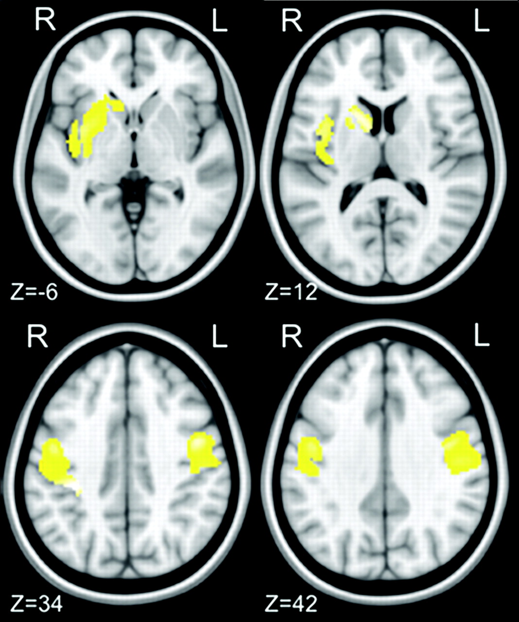
- Fig 1.
Images demonstrating focal decrease in GM volume between 19 patients with CD and 28 healthy controls. Clusters of decreased GM volume (in yellow) are overlapped with the T1-weighted mean image. Four axial sections passing at MNI z-coordinates of −6, 12, 34, 42 are shown. Clusters significant at P < .05 (2-sample t test; FDR corrected) are observed in the left caudate head and putamen and in the premotor and primary sensory motor cortices bilaterally. Images are displayed according to neurologic convention.
- Fig 2.
Images demonstrating longitudinal changes in GM volume in 12 patients with CD during 5 years of follow-up. Clusters of decreased GM volume (in yellow) are overlapped with the T1-weighted mean image. Two axial sections passing at MNI z-coordinates of 70 and 76 are shown. Clusters significant at P < .05 (paired t test, FDR corrected) are observed in the left primary sensory motor cortex. Images are displayed according to neurologic convention.


