Advertisement

Main menu

  • Home
  • Content
    • Current Issue
    • Accepted Manuscripts
    • Article Preview
    • Past Issue Archive
    • Video Articles
    • AJNR Case Collection
    • Case of the Week Archive
    • Case of the Month Archive
    • Classic Case Archive
  • Special Collections
    • AJNR Awards
    • Low-Field MRI
    • Alzheimer Disease
    • ASNR Foundation Special Collection
    • Photon-Counting CT
    • View All
  • Multimedia
    • AJNR Podcasts
    • AJNR SCANtastic
    • Trainee Corner
    • MRI Safety Corner
    • Imaging Protocols
  • For Authors
    • Submit a Manuscript
    • Submit a Video Article
    • Submit an eLetter to the Editor/Response
    • Manuscript Submission Guidelines
    • Statistical Tips
    • Fast Publishing of Accepted Manuscripts
    • Graphical Abstract Preparation
    • Imaging Protocol Submission
    • Author Policies
  • About Us
    • About AJNR
    • Editorial Board
    • Editorial Board Alumni
  • More
    • Become a Reviewer/Academy of Reviewers
    • Subscribers
    • Permissions
    • Alerts
    • Feedback
    • Advertisers
    • ASNR Home

User menu

  • Alerts
  • Log in

Search

  • Advanced search
American Journal of Neuroradiology

American Journal of Neuroradiology

ASHNR American Society of Functional Neuroradiology ASHNR American Society of Pediatric Neuroradiology ASSR
  • Alerts
  • Log in

Advanced Search

  • Home
  • Content
    • Current Issue
    • Accepted Manuscripts
    • Article Preview
    • Past Issue Archive
    • Video Articles
    • AJNR Case Collection
    • Case of the Week Archive
    • Case of the Month Archive
    • Classic Case Archive
  • Special Collections
    • AJNR Awards
    • Low-Field MRI
    • Alzheimer Disease
    • ASNR Foundation Special Collection
    • Photon-Counting CT
    • View All
  • Multimedia
    • AJNR Podcasts
    • AJNR SCANtastic
    • Trainee Corner
    • MRI Safety Corner
    • Imaging Protocols
  • For Authors
    • Submit a Manuscript
    • Submit a Video Article
    • Submit an eLetter to the Editor/Response
    • Manuscript Submission Guidelines
    • Statistical Tips
    • Fast Publishing of Accepted Manuscripts
    • Graphical Abstract Preparation
    • Imaging Protocol Submission
    • Author Policies
  • About Us
    • About AJNR
    • Editorial Board
    • Editorial Board Alumni
  • More
    • Become a Reviewer/Academy of Reviewers
    • Subscribers
    • Permissions
    • Alerts
    • Feedback
    • Advertisers
    • ASNR Home
  • Follow AJNR on Twitter
  • Visit AJNR on Facebook
  • Follow AJNR on Instagram
  • Join AJNR on LinkedIn
  • RSS Feeds

AJNR Awards, New Junior Editors, and more. Read the latest AJNR updates

LetterLetter

Atypical Meningeal Involvement in Wegener Granulomatosis: MR Imaging Survey of a Particular Case

N. Tasali, R. Cubuk, L. Celik and B. Canbora
American Journal of Neuroradiology September 2009, 30 (8) E116; DOI: https://doi.org/10.3174/ajnr.A1645
N. Tasali
  • Find this author on Google Scholar
  • Find this author on PubMed
  • Search for this author on this site
R. Cubuk
  • Find this author on Google Scholar
  • Find this author on PubMed
  • Search for this author on this site
L. Celik
  • Find this author on Google Scholar
  • Find this author on PubMed
  • Search for this author on this site
B. Canbora
  • Find this author on Google Scholar
  • Find this author on PubMed
  • Search for this author on this site
  • Article
  • Figures & Data
  • Info & Metrics
  • Responses
  • References
  • PDF
Loading

Wegener granulomatosis (WG) is a rare multisystem inflammatory disorder characterized by necrotizing granulomas in the respiratory tract with or without focal necrotizing glomerulonephritis and a systemic vasculitis. Meningeal involvement, which may be diffuse or focal, is rarely represented in 4% to 11% of cases.1 We present a patient with WG having meningeal lesions in an unusual location (intraventricular) with consecutive cranial MR imaging scans.

A 33-year-old woman who was diagnosed earlier with WG and was undergoing treatment for approximately 2 months began to experience some neurologic symptoms. Contrast-enhanced MR imaging scans revealed 2 nodular enhancing extra-axial lesions on the posterior wall of the fourth ventricle, which appeared to be slightly dilated. The diameter of the largest lesion was 13 mm; in addition, there was linear meningeal enhancement close to the lesions (Fig. 1). After 20 days of steroid treatment, control MR imaging examination demonstrated regression in both nodular-enhancing lesions; the largest was 10 mm in diameter (Fig 2).

Fig 1.
  • Download figure
  • Open in new tab
  • Download powerpoint
Fig 1.

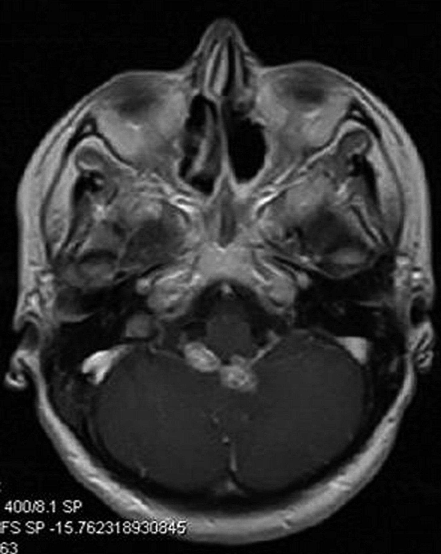
Axial T1-weighted contrast-enhanced MR image reveals 2 nodular-enhancing extra-axial lesions on the posterior wall of the fourth ventricle, which appears to be slightly dilated. The diameter of the largest lesion is 13 mm; in addition, there is linear meningeal enhancement close to the lesions.

Fig 2.
  • Download figure
  • Open in new tab
  • Download powerpoint
Fig 2.

Control MR imaging examination, which was performed after 20 days of treatment, demonstrates regression in both nodular-enhancing lesions (the largest was 10 mm in diameter) in an axial T1-weighted contrast-enhanced sequence.

Three major mechanisms causing central nervous system disease (CNS) in WG have been implicated: contiguous invasion of the granuloma from extracranial sites, CNS vasculitis, and remote intracranial granuloma. Meningeal involvement, which usually presents with diffuse or focal forms of dural thickening and enhancement, was previously reported in a small number (< 100) of cases in the literature.2 In the case of our patient, the meningeal lesions were located at the posterior wall of the fourth ventricle and the adjacent parenchyma. Therefore, it is predicated that these lesions did not derive from the dura, which is known to be the first affected meningeal layer throughout the contiguous invasion process. Taking all of these findings into consideration, we determined that the lesions in our patient may have developed secondary to leptomeningeal vasculitis. Leptomeningeal angiitis or isolated angiitis of the CNS is a rare entity, and, to our knowledge, only small case series have been reported.3 The primary cause of this entity, which is also termed primary angiitis, is unknown; however, the secondary form is known to appear as part of a systemic autoimmune disease.

In conclusion, the focal nodular type of meningeal involvement in WG may appear on the ventricle walls in association with parenchymal changes. The leptomeningeal vasculitic process seems to be the most probable mechanism of intraventricular secondary or granulomatous leptomeningeal angiitis vs meningeal involvement. We believe that an MR imaging survey of our patient will contribute to the limited imaging literature available on WG with meningeal involvement. On the other hand, it must be maintained that, as in the case of our patient, nodular-enhancing regions after administration of contrast material may mimic a brain tumor or primary lymphoma on neuroimaging.4

References

  1. ↵
    1. Cruz DN,
    2. Segal AS
    . A patient with Wegener's granulomatosis presenting with a subarachnoid hemorrhage: case report and review of CNS disease associated with Wegener's granulomatosis. Am J Nephrol 1997; 17: 181– 86
    CrossRefPubMed
  2. ↵
    1. Murphy JM,
    2. Gomez-Anson B,
    3. Gillard JH,
    4. et al
    . Wegener granulomatosis: MR imaging findings in brain and meninges. Radiology 1999; 213: 794– 99
    PubMed
  3. ↵
    1. Rollnik JD,
    2. Brandis A,
    3. Dehghani K,
    4. et al
    . [Primary angiitis of CNS (PACNS)]. Nervenarzt 2001; 72: 798– 801
    CrossRefPubMed
  4. ↵
    1. Hocaoglu C,
    2. Tan M
    . Isolated angiitis of the central nervous system: a case presented with atypical psychiatric symptoms. Prog Neuropsychopharmacol Biol Psychiatry 2005; 29: 627– 31
    CrossRefPubMed
  • Copyright © American Society of Neuroradiology
PreviousNext
Back to top

In this issue

American Journal of Neuroradiology: 30 (8)
American Journal of Neuroradiology
Vol. 30, Issue 8
1 Sep 2009
  • Table of Contents
  • Index by author
Advertisement
Print
Download PDF
Email Article

Thank you for your interest in spreading the word on American Journal of Neuroradiology.

NOTE: We only request your email address so that the person you are recommending the page to knows that you wanted them to see it, and that it is not junk mail. We do not capture any email address.

Enter multiple addresses on separate lines or separate them with commas.
Atypical Meningeal Involvement in Wegener Granulomatosis: MR Imaging Survey of a Particular Case
(Your Name) has sent you a message from American Journal of Neuroradiology
(Your Name) thought you would like to see the American Journal of Neuroradiology web site.
CAPTCHA
This question is for testing whether or not you are a human visitor and to prevent automated spam submissions.
Cite this article
N. Tasali, R. Cubuk, L. Celik, B. Canbora
Atypical Meningeal Involvement in Wegener Granulomatosis: MR Imaging Survey of a Particular Case
American Journal of Neuroradiology Sep 2009, 30 (8) E116; DOI: 10.3174/ajnr.A1645

Citation Manager Formats

  • BibTeX
  • Bookends
  • EasyBib
  • EndNote (tagged)
  • EndNote 8 (xml)
  • Medlars
  • Mendeley
  • Papers
  • RefWorks Tagged
  • Ref Manager
  • RIS
  • Zotero
0 Responses
Respond to this article
Share
Bookmark this article
Atypical Meningeal Involvement in Wegener Granulomatosis: MR Imaging Survey of a Particular Case
N. Tasali, R. Cubuk, L. Celik, B. Canbora
American Journal of Neuroradiology Sep 2009, 30 (8) E116; DOI: 10.3174/ajnr.A1645
del.icio.us logo Twitter logo Facebook logo Mendeley logo
  • Tweet Widget
  • Facebook Like
  • Google Plus One
Purchase

Jump to section

  • Article
    • References
  • Figures & Data
  • Info & Metrics
  • Responses
  • References
  • PDF

Related Articles

  • No related articles found.
  • PubMed
  • Google Scholar

Cited By...

  • No citing articles found.
  • Crossref
  • Google Scholar

This article has not yet been cited by articles in journals that are participating in Crossref Cited-by Linking.

More in this TOC Section

  • Letter to the Editor regarding “Automated Volumetric Software in Dementia: Help or Hindrance to the Neuroradiologist?”
  • Reply:
  • Brain AVM’s Nidus: What if We Hadn’t Understood Anything?
Show more LETTERS

Similar Articles

Advertisement

Indexed Content

  • Current Issue
  • Accepted Manuscripts
  • Article Preview
  • Past Issues
  • Editorials
  • Editor's Choice
  • Fellows' Journal Club
  • Letters to the Editor
  • Video Articles

Cases

  • Case Collection
  • Archive - Case of the Week
  • Archive - Case of the Month
  • Archive - Classic Case

More from AJNR

  • Trainee Corner
  • Imaging Protocols
  • MRI Safety Corner
  • Book Reviews

Multimedia

  • AJNR Podcasts
  • AJNR Scantastics

Resources

  • Turnaround Time
  • Submit a Manuscript
  • Submit a Video Article
  • Submit an eLetter to the Editor/Response
  • Manuscript Submission Guidelines
  • Statistical Tips
  • Fast Publishing of Accepted Manuscripts
  • Graphical Abstract Preparation
  • Imaging Protocol Submission
  • Evidence-Based Medicine Level Guide
  • Publishing Checklists
  • Author Policies
  • Become a Reviewer/Academy of Reviewers
  • News and Updates

About Us

  • About AJNR
  • Editorial Board
  • Editorial Board Alumni
  • Alerts
  • Permissions
  • Not an AJNR Subscriber? Join Now
  • Advertise with Us
  • Librarian Resources
  • Feedback
  • Terms and Conditions
  • AJNR Editorial Board Alumni

American Society of Neuroradiology

  • Not an ASNR Member? Join Now

© 2025 by the American Society of Neuroradiology All rights, including for text and data mining, AI training, and similar technologies, are reserved.
Print ISSN: 0195-6108 Online ISSN: 1936-959X

Powered by HighWire