
Article Figures & Data
Figures
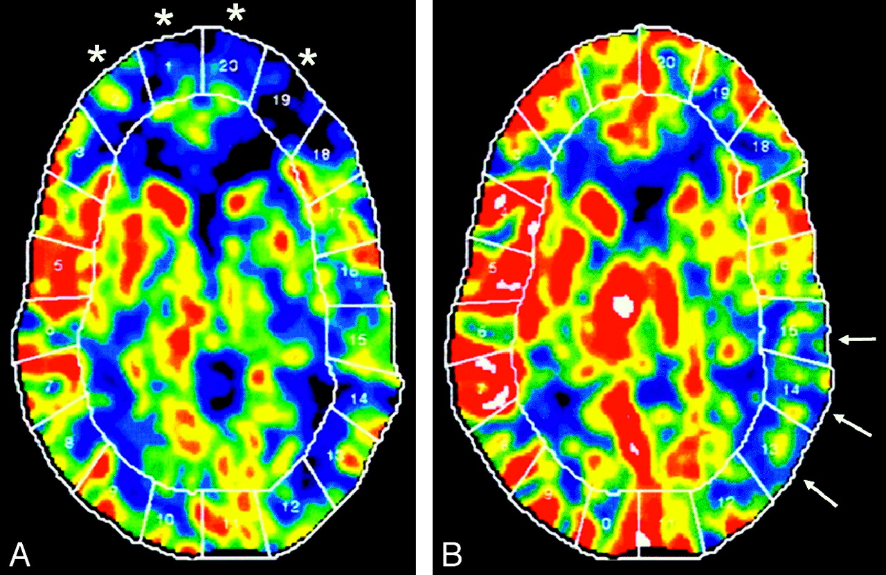
- Fig 1.
Xe-CT CBF maps in a patient with Moyamoya disease. A, Baseline. B, After ACZ administration. Baseline scan (A) shows reduced CBF in the bilateral ACA and anterior watershed areas (areas 1, 2, 19, and 20, asterisk). After ACZ, there is a robust increase in the CBF, indicating a normal cerebral reserve in these territories. There is reduced baseline flow with decreased augmentation of CBF after ACZ, indicating poor cerebral reserve in the left posterior MCA and the left posterior watershed territories (areas 13–15, arrows).
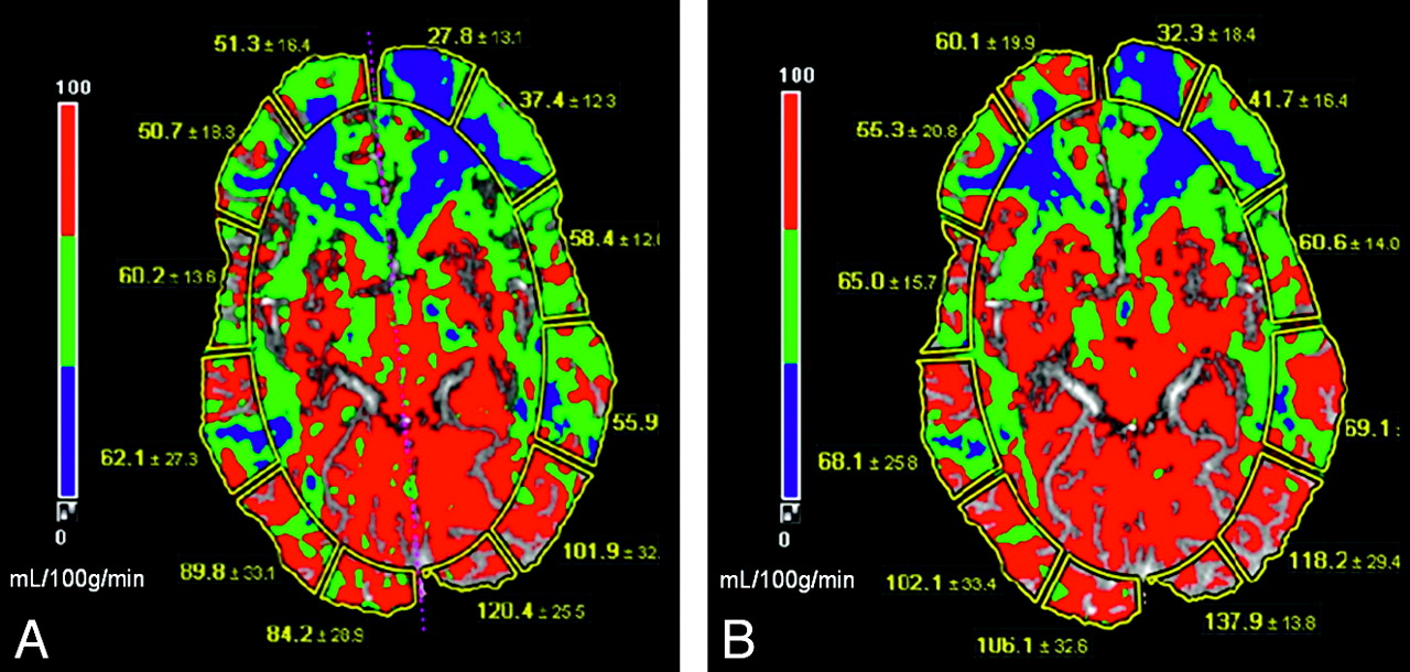
- Fig 2.
A 34-year-old man with severe headache and blurry vision was diagnosed with Moyamoya disease. CT perfusion maps. A, Baseline. B, After ACZ administration. CBF (measured in mL/100 g/min) is diminished in bilateral ACA, ACA-MCA watershed, and the MCA territories (depicted as blue to green). These areas show a very suboptimal increase in CBF after ACZ administration and thus exhibit limited CVR. Note the increased CBF secondary to vasodilatory capacity in the PCA and MCA-PCA distribution after ACZ administration.
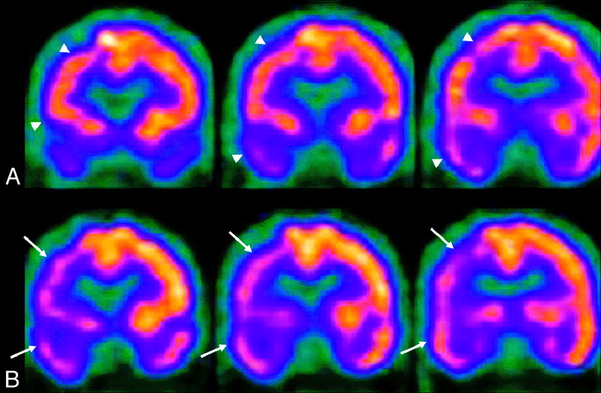
- Fig 3.
SPECT perfusion study of a 64-year-old man with high-grade right M1 and A1 segment stenosis. A, Baseline. B, After ACZ administration. There is decreased uptake and perfusion (arrows) involving the right frontal, parietal, and temporal lobes on the ACZ study (B), which resolve during baseline conditions (arrowheads).
- Fig 4.
CTP. Chronic left internal carotid artery occlusion. A, Baseline. B, After ACZ administration. There is significant hypoperfusion in the left hemisphere at baseline with decreased CBF and increased TTP (A, short arrows). After administration of ACZ (B), CBF decreases throughout the left hemisphere, resulting in negative calculated CVR percentages. The left-sided TTP increases even further in post-ACZ flow maps as seen by the accentuated asymmetry (B, long arrows). Note the normal increase in the right-sided CBF after the vasodilatory stimulus of ACZ (B, arrowheads).
- Fig 5.
CT perfusion maps in a 51-year-old patient presenting with right-sided hemiparesis who was diagnosed with Moyamoya disease, demonstrating bilateral supraclinoid internal carotid occlusion. A, Baseline. B, After ACZ administration. The baseline pre-ACZ PCT (A) demonstrates the typical pattern of Moyamoya disease with decreased CBF and increased MTT and TTP in the bilateral anterior and middle cerebral distributions (arrows). After ACZ challenge (B), the CBF in the anterior circulation decreases consistent with steal phenomenon (B, CBF, arrows). The CBF map demonstrates a normal expected increase in the PCA territories. There is further prolongation of the MTT and TTP in both ACA and MCA distributions (arrowheads, B), consistent with worsening of cerebral hemodynamics after ACZ, and type III physiology. The patient successfully underwent left-sided ECIC bypass surgery.
- Fig 6.
CT perfusion flow maps in a 56-year-old patient presenting with multiple transient ischemic episodes and diagnosed with Moyamoya disease with bilateral carotid occlusion on digital subtraction angiography before (A) and 12 months after (B) bilateral ECIC bypass surgery. The preoperative baseline maps (A) show a typical pattern of Moyamoya disease with decreased CBF and increased MTT in the anterior circulation. After bilateral ECIC bypass (STA-MCA, B), there is an increase in the CBF in the ACA and MCA territories with minimal decrease in the MTT. The improvement is more pronounced in the MCA territory because of the proximity of the graft. Quantitatively, there is improvement in the CVR in these distributions. After bypass, the patient's ischemic symptoms resolved.


In this issue


Jump to section
- Article
- Abstract
- Pathophysiology of Chronic Cerebrovascular Disease
- Cerebrovascular Reactivity
- ACZ
- Imaging Techniques
- Xe-CT
- PCT
- MR Perfusion
- ASL MR Perfusion
- PET
- SPECT
- Clinical Applications of the ACZ Challenge
- Moyamoya Disease
- Pre- and Postoperative Evaluation of Extracranial-Intracranial Bypass for Flow Augmentation
- Carotid Balloon Occlusion
- Hyperperfusion Syndrome
- Conclusions
- Acknowledgments
- References
- Figures & Data
- Info & Metrics
- Responses
- References
Related Articles
Cited By...
- AER-270 and TGN-020 are not aquaporin-4 water channel blockers
- Modern endovascular management of chronic total carotid artery occlusion: technical results and procedural challenges
- The Significance and Limited Influence of Cerebrovascular Reactivity on Age and Sex Effects in Task- and Resting-State Brain Activity
- High Intravascular Signal Arterial Transit Time Artifacts Have Negligible Effects on Cerebral Blood Flow and Cerebrovascular Reserve Capacity Measurement Using Single Postlabel Delay Arterial Spin-Labeling in Patients with Moyamoya Disease
- Evaluating off-label uses of acetazolamide
- Comparison of Blood Oxygenation Level-Dependent fMRI and Provocative DSC Perfusion MR Imaging for Monitoring Cerebrovascular Reserve in Intracranial Chronic Cerebrovascular Disease
- Can Arterial Spin-Labeling with Multiple Postlabeling Delays Predict Cerebrovascular Reserve?
- The Brain Thermal Response as a Potential Neuroimaging Biomarker of Cerebrovascular Impairment
- Acetazolamide potentiates the afferent drive to prefrontal cortex in vivo
- The Effects of Acetazolamide on the Evaluation of Cerebral Hemodynamics and Functional Connectivity Using Blood Oxygen Level-Dependent MR Imaging in Patients with Chronic Steno-Occlusive Disease of the Anterior Circulation
- Angiographic Correlates of Cerebral Hemodynamic Changes With Diamox Challenge Assessed by Quantitative Magnetic Resonance Angiography
- Neuroradiologic Correlates of Cognitive Impairment in Adult Moyamoya Disease