
Article Figures & Data
Figures
- Fig 1.
The midbrain as a region-of-interest on 4T SWI magnitude images shown in sagittal and coronal sections, with and without section locators. Arrows 1, 2, and 3 point to the red nucleus, the subthalamic nucleus, and the substantia nigra, respectively.
- Fig 2.
A comparison of 1 region of the mesencephalon for different sequences and field strengths with Duvernoy's stained cadaver brain results.16 A, T1 at 1.5T; B, T1 at 4T; C, T2 at 1.5T; D, T2 at 4T; E, Duvernoy's india ink-stained image; F, SWI magnitude at 1.5T; G, SWI HP-filtered phase at 1.5T; H, SWI magnitude at 4T; I, SWI HP-filtered phase at 4T; J, Duvernoy's india ink-stained image. Images E and J reprinted with permission from Duvernoy HM. Human Brain Stem Vessels. 2nd ed. Berlin, Germany: Springer-Verlag; 1999;206–13.
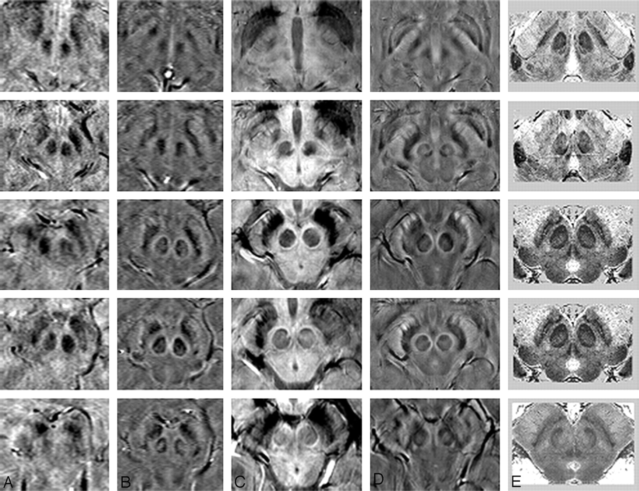
- Fig 3.
A comparison of adjacent sections in SWI magnitude and HP-filtered-phase images at 1.5T and 4T with Duvernoy's india ink-stained results.16 A, Magnitude SWI at 1.5T; B, HP-filtered phase at 1.5T; C, Magnitude SWI at 4T; D, HP-filtered phase at 4T; E, Duvernoy's results. Only 3 unique images from Duvernoy are shown here because his section thickness is 3 mm. The third and fourth images from the top are identical. Images in column E reprinted with permission from Duvernoy HM. Human Brain Stem Vessels. 2nd ed. Berlin, Germany: Springer-Verlag; 1999;206–13.
- Fig 4.
A comparison of SWI HP-filtered-phase images (B and D) with Duvernoy's india ink–stained results (A and C).16 Anterior (A and B) and posterior (C and D) mesencephalic structures are indicated. Arrow 1 indicates the crus cerebri; 2a, substantia nigra, pars reticulata; 2b, substantia nigra, pars compacta; 3, capsule of the red nucleus; 4a, red nucleus (vascularized region); 4b, red nucleus (nonvascularized region); 5, fascicula nigrale; 6, medial geniculate body; 7, superior colliculus; 8, intermediate area. Images A and C reprinted with permission from Duvernoy HM. Human Brain Stem Vessels. 2nd ed. Berlin, Germany: Springer-Verlag; 1999;206–13.


Tables
Sequence Red Nucleus CNR Substantia Nigra CNR Medial Geniculate CNR IA/RN IA/RN capsule RN/RN capsule IA/SNc CC/SNr SNc/SNr CMG/LMG CMG/IA T2 tse 4.13 – – 6.19† 4.97† – – – T2 tse SD 1.23 – – 1.44† 1.48† – – – SWI 5.97 1.43 4.54 5.58 2.84 3.74 1.38 0.45 SWI SD 1.78 0.73 1.62 0.6 0.75 1.46 1.47 0.35 RN indicates red nucleus; SNc, substantia nigra, pars compacta; SNr, substantia nigra, pars reticulata; IA, intermediate area between the RN and SN; CC, crus cerebri; LMG, lateral aspect of the medial geniculate body; CMG, central aspect of medial geniculate body; tse, turbo spin-echo; CNR, contrast-to-noise ratio.
* The first row for each sequence represents the mean in a given region of interest and the second row represents the SD in the same region of interest. HP-filtered-phase data were used for SWI CNR measurements; no contrast for the given regions of interest was observed in the gradient-echo T1 data. Empty data boxes show that CNRs between certain structures could not be calculated because the structures could not be detected.
† Because SNc and SNr were not separable for T2, CNR is recorded here for IA/SN and CC/SN.
Sequence Red Nucleus CNR Substantia Nigra CNR Medial Geniculate CNR IA/RN IA/RN capsule RN/RN capsule IA/SNc CC/SNr SNc/SNr CMG/LMG CMG/IA T2 tse 3.36 – – 4.53† 6.27† – – – T2 tse SD 0.77 – – 0.48† 1.64† – – – SWI 6.98 1.13 5.85 8.75 5.19 3.56 4.57 1.64 SWI SD 2.60 1.34 2.23 1.67 1.51 1.08 2.49 0.89 * The first row for each sequence represents the mean in a given region of interest and the second row represents the SD in the same region of interest. HP-filtered-phase data were used for SWI CNR measurements; no contrast for the given regions of interest was observed in the gradient-echo T1 data. Empty data boxes show that CNRs between certain structures could not be calculated because the structures could not be detected.
† Because SNc and SNr were not separable for T2, CNR is recorded here for IA/SN and CC/SN.
In this issue


Jump to section
Related Articles
Cited By...
- Multimodal anatomical mapping of subcortical regions in Marmoset monkeys using high-resolution MRI and matched histology with multiple stains
- Direct In Vivo MRI Discrimination of Brain Stem Nuclei and Pathways
- 3T MRI Whole-Brain Microscopy Discrimination of Subcortical Anatomy, Part 2: Basal Forebrain
- 3T MRI Whole-Brain Microscopy Discrimination of Subcortical Anatomy, Part 1: Brain Stem
- Comparison of 3D FLAIR, 2D FLAIR, and 2D T2-Weighted MR Imaging of Brain Stem Anatomy
- Using High-Resolution MR Imaging at 7T to Evaluate the Anatomy of the Midbrain Dopaminergic System
- Genetic Variation in FGF20 Modulates Hippocampal Biology
- Localization of the Subthalamic Nucleus: Optimization with Susceptibility-Weighted Phase MR Imaging