
Article Figures & Data
Figures
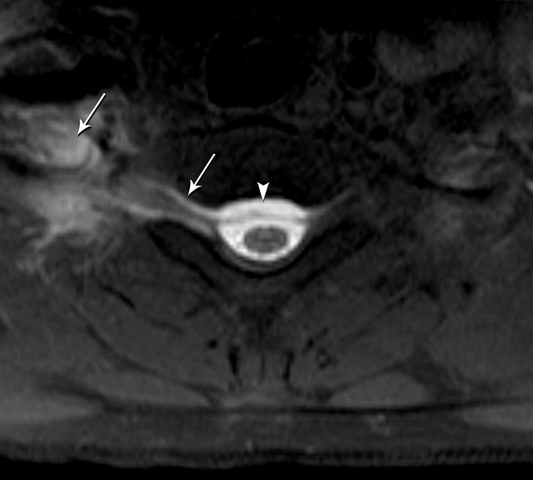
- Fig 1.
Axial T1-weighted fat-saturated MR image shows a right-sided CSF leak at the cervicothoracic junction extending into the right paraspinal soft tissue (arrows) and epidural collection (arrowhead).
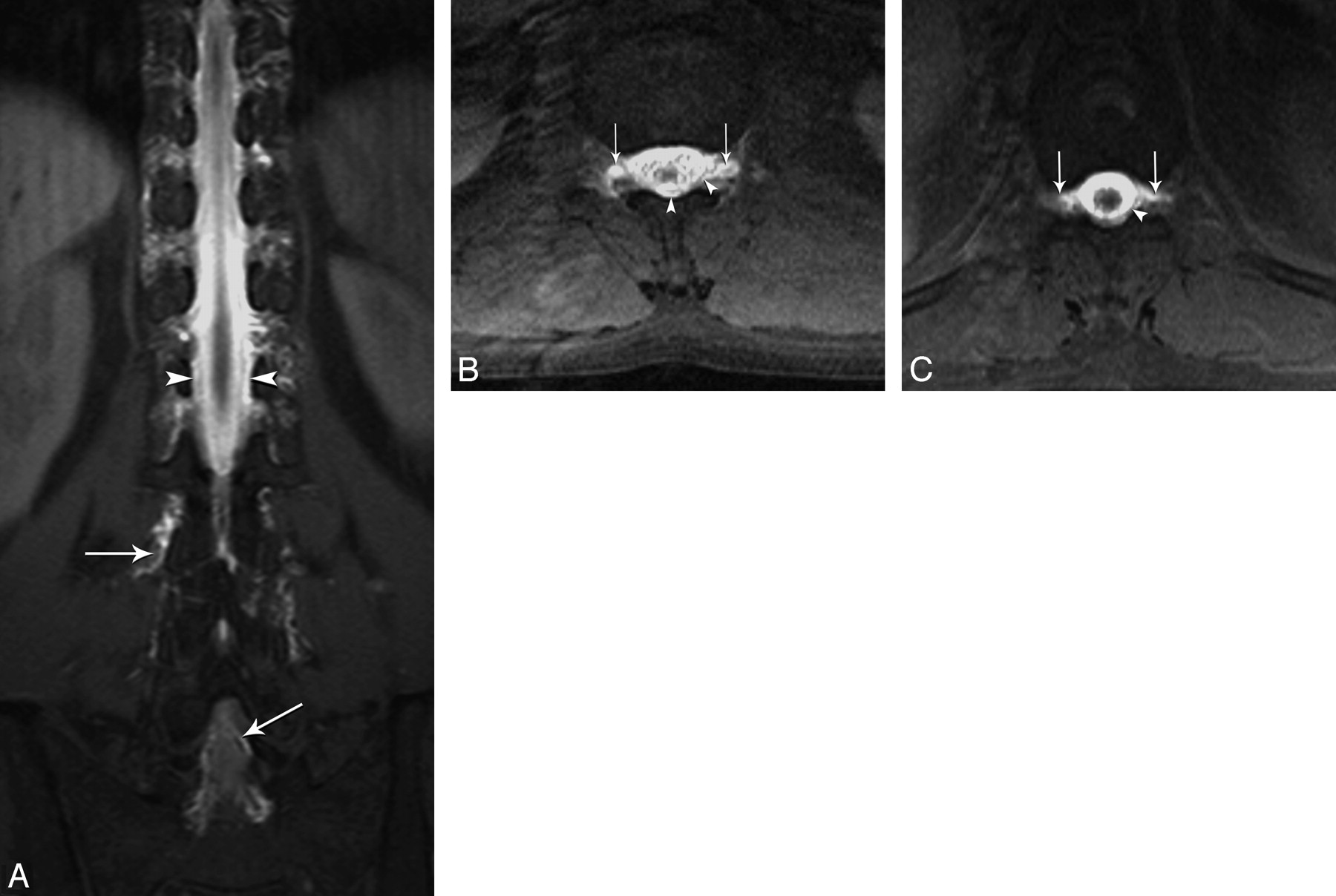
- Fig 2.
Coronal (A) and axial (B and C) T1-weighted fat-saturated MR images reveal diffuse epidural (arrowhead) and paravertebral contrast accumulation (arrows) at the thoracolumbar area.
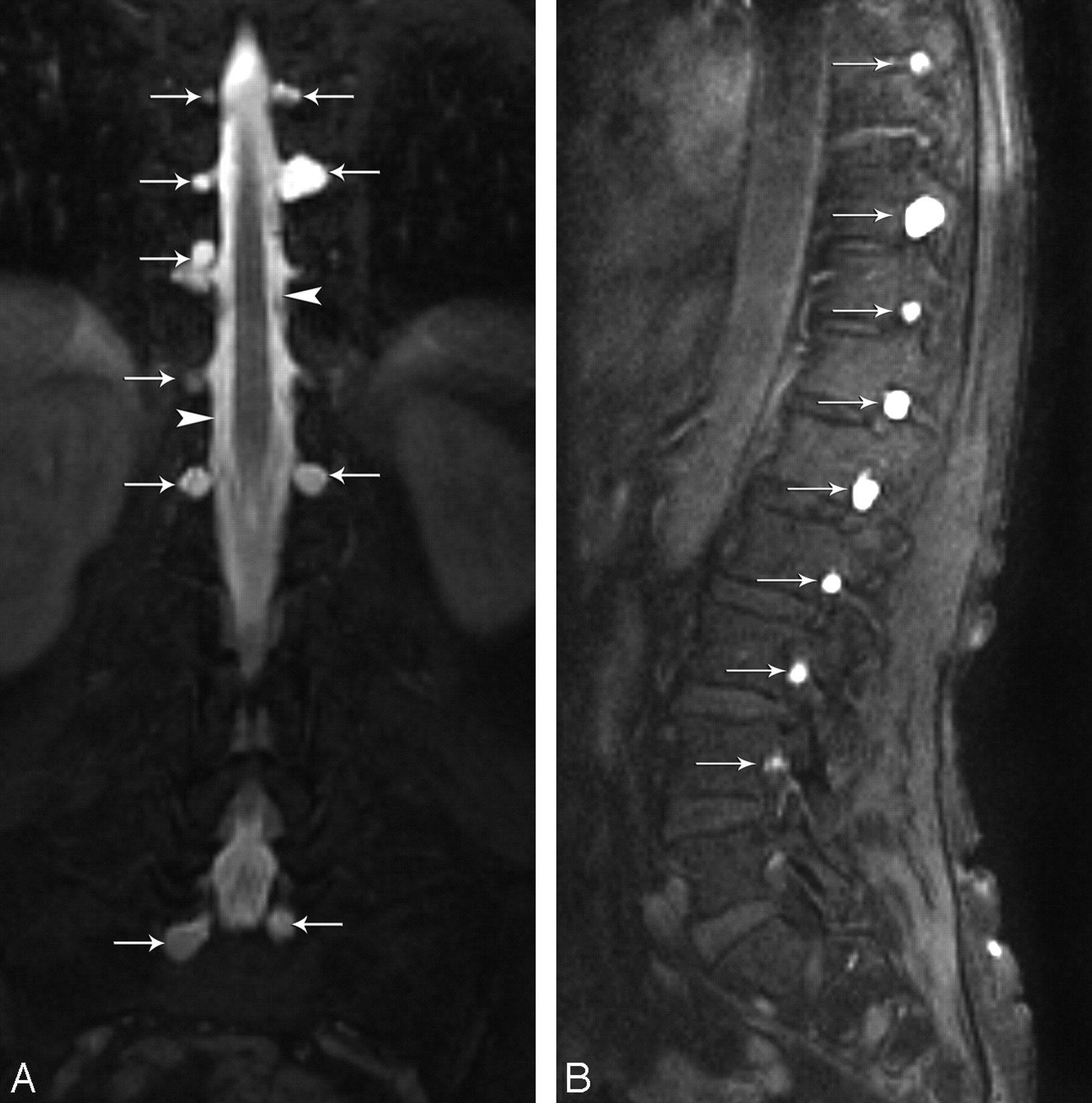
- Fig 3.
Coronal and sagittal T1-weighted fat-saturated MR images show multiple bilateral meningeal diverticula (arrows) at the thoracic and lumbar level.
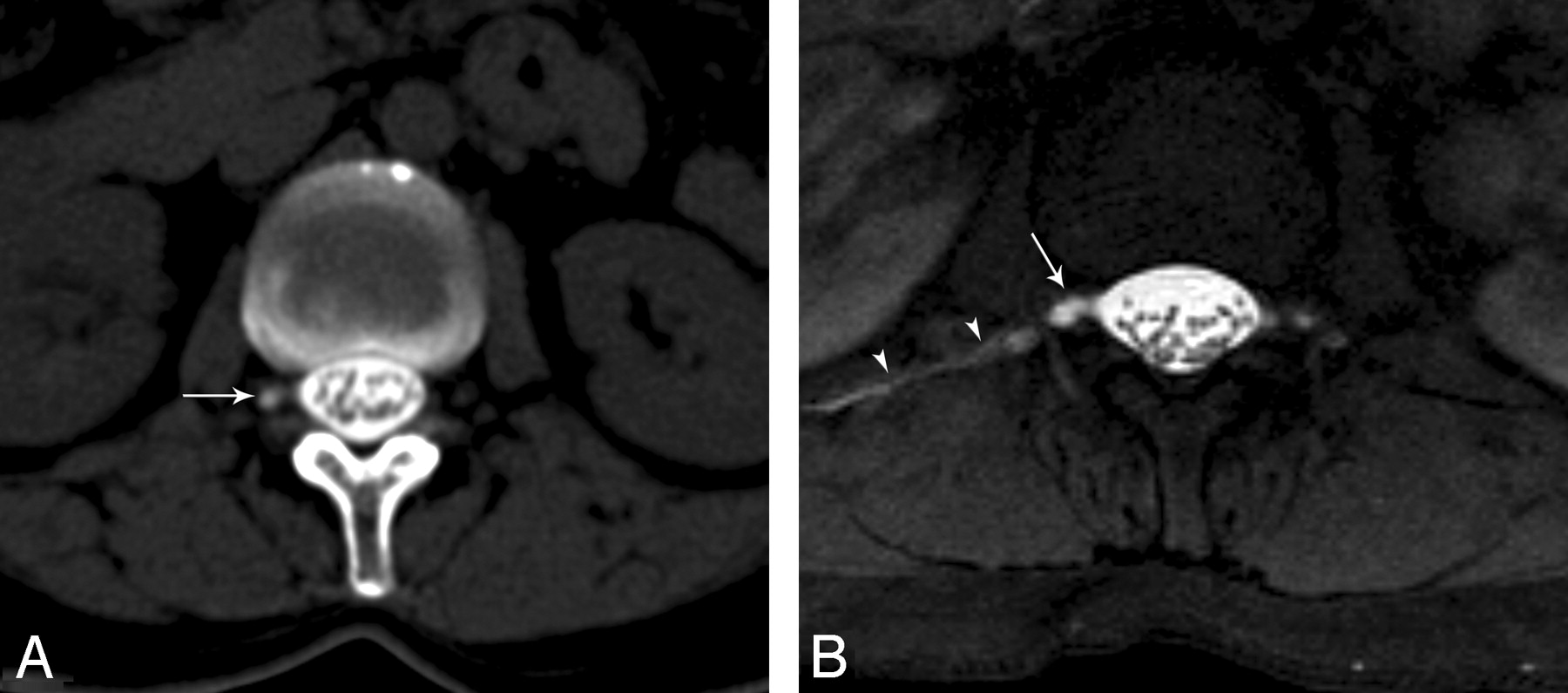
- Fig 4.
A, Axial CT cisternography image shows right meningeal diverticulum (arrow) without CSF leak at the level of L1. B, Axial T1-weighted fat-saturated MR image reveals both a CSF leak on the right side (arrowheads) and a right meningeal diverticulum (arrow).


Tables
- Table 1:
The diagnostic criteria for SIH proposed by Headache3 Classification Subcommittee of the International Headache Society
Diffuse and/or dull headache that worsens within 15 minutes after sitting or standing, with at least 1 of the following and fulfilling criterion:
Neck stiffness
Tinnitus
Hyperacusia
Photophobia
Nausea
At least 1 of the following:
Evidence of low CSF pressure on MR imaging (eg, pachymeningeal enhancement)
Evidence of CSF leakage on conventional myelography, CT myelography, or cisternography
CSF opening pressure <60 mm H2O in sitting position
No history of dural puncture or other cause of CSF fistula
Headache resolves within 72 hours after epidural blood patching
Patient No. Cranial MR Imaging Spinal MR Imaging CSF Pressure CT Myelography or RC Dural Puncture or Other Causes Response to EBP (Immediate Relief*) 1 − + N − − + 2 + − ↓ − − + 3 + − 0 − − + 4 + − 0 − − + 5 + − ↓ − − + 6 + − ↓ − − + 7 + − ↓ − − + 8 + − ↓ − − + 9 + + ↓ − − + 10 + − ↓ − − + 11 + − ↓ − − + 12 − + N + − + 13 + − ↓ − − + 14 + − ↓ − − + 15 + − 0 − − + 16 + − ↓ − − + 17 + + ↓ + − + 18 + + ↓ − − + 19 + − ↓ − − + Note:—SIH indicates spontaneous intracranial hypotension; RC, radionuclide cisternography; EBP, epidural blood patch; N, normal; ↓, low; +, yes; −, no.
* Immediate relief means that the patient symptoms resolve due to the pressure effect of EBP. This does not address complete cure of the disease.
Patient No. CSF Leak Exact Location of CSF Leak No. of the Dural Tear Location of CSF Leak Epidural Collection Paravertebral Collection Meningeal Diverticulum Associated Findings 1 + + Single Right T12 + + − − 2 − − − − − − + − 3 + Diffuse ? Bilateral T11-12 and L1-2 + + − − 4 + Diffuse ? Bilateral T10-11-12 and L1-2-3 + + + − 5 + + Single Left T1 + + − − 6 + + Single C6 anteriorly + + + Cervical osteophyte and hernia 7 + + Multiple Bilateral C7 and T1 + + + − 8 + + Single Right T10 + + − − 9 + + Single Right T2 + + − − 10 + + Single Left L2 − + + − 11 + + Multiple Bilateral C7 and T5 + + + Thoracic osteophyte and hernia 12 + + Single Right L1 − + + − 13 + + Single Left T4 + + + − 14 + + Multiple Bilateral T4-T5 − + + − 15 + + Single Bilateral T7 + + − − 16 + Diffuse ? Bilateral T11-T12 and Left T9-T10 + + − Prominent epidural veins 17 − − − − − − + − 18 + + Multiple Bilateral T2-T3 and Bilateral T11-T12 + + − − Note:—SIH indicates spontaneous intracranial hypotension; C, cervical; T, thoracic; L, lumbar; ?, not detected due to diffuse CSF leakage; +, yes; −, no.
In this issue


Jump to section
Related Articles
Cited By...
- Spine MRI in Spontaneous Intracranial Hypotension for CSF Leak Detection: Nonsuperiority of Intrathecal Gadolinium to Heavily T2-Weighted Fat-Saturated Sequences
- Fatal gadolinium-induced encephalopathy following accidental intrathecal administration: a case report and a comprehensive evidence-based review
- MR Myelography for Identification of Spinal CSF Leak in Spontaneous Intracranial Hypotension
- Intrathecal Gadolinium-Enhanced MR Cisternography: A Comprehensive Review
- CT Myelography for the Planning and Guidance of Targeted Epidural Blood Patches in Patients with Persistent Spinal CSF Leakage
- The Role of MR Myelography with Intrathecal Gadolinium in Localization of Spinal CSF Leaks in Patients with Spontaneous Intracranial Hypotension
- MRI with intrathecal gadolinium to detect a CSF leak: a prospective open-label cohort study
- Intrathecal Gadolinium-Enhanced MR Cisternography in the Evaluation of CSF Leakage
- Dural Tears in Spinal Burst Fractures: Predictable MR Imaging Findings