Advertisement

Main menu

  • Home
  • Content
    • Current Issue
    • Accepted Manuscripts
    • Article Preview
    • Past Issue Archive
    • Video Articles
    • AJNR Case Collection
    • Case of the Week Archive
    • Case of the Month Archive
    • Classic Case Archive
  • Special Collections
    • AJNR Awards
    • Low-Field MRI
    • Alzheimer Disease
    • ASNR Foundation Special Collection
    • Photon-Counting CT
    • View All
  • Multimedia
    • AJNR Podcasts
    • AJNR SCANtastic
    • Trainee Corner
    • MRI Safety Corner
    • Imaging Protocols
  • For Authors
    • Submit a Manuscript
    • Submit a Video Article
    • Submit an eLetter to the Editor/Response
    • Manuscript Submission Guidelines
    • Statistical Tips
    • Fast Publishing of Accepted Manuscripts
    • Graphical Abstract Preparation
    • Imaging Protocol Submission
    • Author Policies
  • About Us
    • About AJNR
    • Editorial Board
    • Editorial Board Alumni
  • More
    • Become a Reviewer/Academy of Reviewers
    • Subscribers
    • Permissions
    • Alerts
    • Feedback
    • Advertisers
    • ASNR Home

User menu

  • Alerts
  • Log in

Search

  • Advanced search
American Journal of Neuroradiology

American Journal of Neuroradiology

ASHNR American Society of Functional Neuroradiology ASHNR American Society of Pediatric Neuroradiology ASSR
  • Alerts
  • Log in

Advanced Search

  • Home
  • Content
    • Current Issue
    • Accepted Manuscripts
    • Article Preview
    • Past Issue Archive
    • Video Articles
    • AJNR Case Collection
    • Case of the Week Archive
    • Case of the Month Archive
    • Classic Case Archive
  • Special Collections
    • AJNR Awards
    • Low-Field MRI
    • Alzheimer Disease
    • ASNR Foundation Special Collection
    • Photon-Counting CT
    • View All
  • Multimedia
    • AJNR Podcasts
    • AJNR SCANtastic
    • Trainee Corner
    • MRI Safety Corner
    • Imaging Protocols
  • For Authors
    • Submit a Manuscript
    • Submit a Video Article
    • Submit an eLetter to the Editor/Response
    • Manuscript Submission Guidelines
    • Statistical Tips
    • Fast Publishing of Accepted Manuscripts
    • Graphical Abstract Preparation
    • Imaging Protocol Submission
    • Author Policies
  • About Us
    • About AJNR
    • Editorial Board
    • Editorial Board Alumni
  • More
    • Become a Reviewer/Academy of Reviewers
    • Subscribers
    • Permissions
    • Alerts
    • Feedback
    • Advertisers
    • ASNR Home
  • Follow AJNR on Twitter
  • Visit AJNR on Facebook
  • Follow AJNR on Instagram
  • Join AJNR on LinkedIn
  • RSS Feeds

AJNR Awards, New Junior Editors, and more. Read the latest AJNR updates

Research ArticleFunctional

Nonlinear Changes in Brain Activity During Continuous Word Repetition: An Event-Related Multiparametric Functional MR Imaging Study

R.E. Hagenbeek, S.A.R.B. Rombouts, D.J. Veltman, J.W. Van Strien, M.P. Witter, P. Scheltens and F. Barkhof
American Journal of Neuroradiology October 2007, 28 (9) 1715-1721; DOI: https://doi.org/10.3174/ajnr.A0632
R.E. Hagenbeek
  • Find this author on Google Scholar
  • Find this author on PubMed
  • Search for this author on this site
S.A.R.B. Rombouts
  • Find this author on Google Scholar
  • Find this author on PubMed
  • Search for this author on this site
D.J. Veltman
  • Find this author on Google Scholar
  • Find this author on PubMed
  • Search for this author on this site
J.W. Van Strien
  • Find this author on Google Scholar
  • Find this author on PubMed
  • Search for this author on this site
M.P. Witter
  • Find this author on Google Scholar
  • Find this author on PubMed
  • Search for this author on this site
P. Scheltens
  • Find this author on Google Scholar
  • Find this author on PubMed
  • Search for this author on this site
F. Barkhof
  • Find this author on Google Scholar
  • Find this author on PubMed
  • Search for this author on this site
  • Article
  • Figures & Data
  • Info & Metrics
  • Responses
  • References
  • PDF
Loading

Article Figures & Data

Figures

  • Fig 1.
    • Download figure
    • Open in new tab
    • Download powerpoint
    Fig 1.

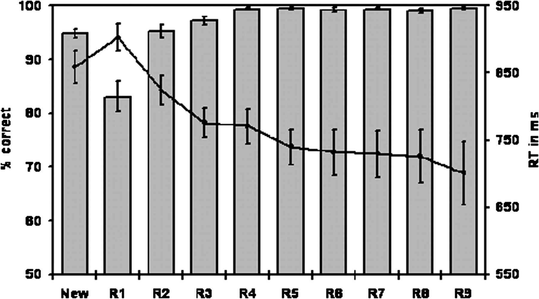
    Mean percentages of correct responses (bar charts) and mean RTs in milliseconds (line chart) of 15 participants to new words (identified as a new word) and repeated words (R1-R9). Error bars depict the standard error of the means.

  • Fig 2.
    • Download figure
    • Open in new tab
    • Download powerpoint
    Fig 2.

    Brain areas showing positive activity for encoding (new words) versus baseline, by using fixed-effects analyses. Positive activity is found in the left inferior frontal gyrus, bilateral middle frontal gyrus, nucleus lentiformis, left motor cortex, and anterior cingulate cortex. Z-values of 3.0–4.7 correspond with changing brain activation colors from red to yellow, respectively. The right side of the brain on the image corresponds with the left hemisphere, and vice versa.

  • Fig 3.
    • Download figure
    • Open in new tab
    • Download powerpoint
    Fig 3.

    Correct responses to repeated words, each occurring with the same amplitude, reveal, in a fixed-effects analysis, activation in the right inferior, middle, and superior frontal gyrus; anterior cingulate cortex; right posterior central sulcus; and left precentral sulcus. Z-values of 3.0–5.8 correspond with changing brain activation colors from red to yellow, respectively. The right side of the brain on the image corresponds with the left hemisphere, and vice versa.

  • Fig 4.
    • Download figure
    • Open in new tab
    • Download powerpoint
    Fig 4.

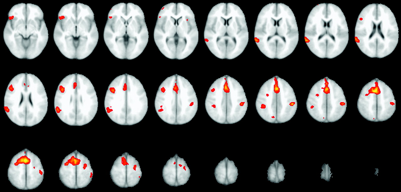
    Brain areas showing a linear decrease in activity as a function of word recognition, by using fixed-effects analyses. Linear changes in brain activation are found in prefrontal, thalamus, anterior cingulate cortex, and parietal regions. Z-values of 3.0–5.1 correspond with changing brain activation colors from red to yellow, respectively. The right side of the brain on the image corresponds with the left hemisphere, and vice versa.

  • Fig 5.
    • Download figure
    • Open in new tab
    • Download powerpoint
    Fig 5.

    A, Brain areas negatively associated with group average RT, shown in the top row, found to be the anterior cingulate cortex. Changing brain activation colors from red to yellow correspond with Z-values of 3.0–4.3, respectively. B, Both center rows reveal brain areas positively associated with group average RT. Regions involved were found to be the posterior cingulate cortex and left middle frontal gyrus. Changing brain activation colors from red to yellow correspond with Z-values of 3.0–4.3, respectively. C, The bottom row shows that the anterior cingulate cortex was also negatively associated with individual RT. Brain regions involved were calculated by using fixed-effects analysis (P < .001, uncorrected). Changing brain activation colors from red to yellow correspond with Z-values of 3.0–3.8 for the bottom row. The right side of the brain on the image corresponds with the left hemisphere, and vice versa.

  • Fig 6.
    • Download figure
    • Open in new tab
    • Download powerpoint
    Fig 6.

    Plotted Z-scores for each regressor in the most significant brain areas for constant activation and activation following group RT. For the RIFG there is a significant fit to the experimental data for the model “brain activation with constant amplitude as function of word repetition.” No significant fit was found for linear or nonlinear regressors. For the PCC, we found a significant fit for the model “positive nonlinear brain activation following group RT,” whereas no significant fit was found for constant, linear, or nonlinear brain activation following individual RT. RIFG indicates right inferior frontal gyrus; indiv, individual.

PreviousNext
Back to top

In this issue

American Journal of Neuroradiology: 28 (9)
American Journal of Neuroradiology
Vol. 28, Issue 9
October 2007
  • Table of Contents
  • Index by author
Advertisement
Print
Download PDF
Email Article

Thank you for your interest in spreading the word on American Journal of Neuroradiology.

NOTE: We only request your email address so that the person you are recommending the page to knows that you wanted them to see it, and that it is not junk mail. We do not capture any email address.

Enter multiple addresses on separate lines or separate them with commas.
Nonlinear Changes in Brain Activity During Continuous Word Repetition: An Event-Related Multiparametric Functional MR Imaging Study
(Your Name) has sent you a message from American Journal of Neuroradiology
(Your Name) thought you would like to see the American Journal of Neuroradiology web site.
CAPTCHA
This question is for testing whether or not you are a human visitor and to prevent automated spam submissions.
Cite this article
R.E. Hagenbeek, S.A.R.B. Rombouts, D.J. Veltman, J.W. Van Strien, M.P. Witter, P. Scheltens, F. Barkhof
Nonlinear Changes in Brain Activity During Continuous Word Repetition: An Event-Related Multiparametric Functional MR Imaging Study
American Journal of Neuroradiology Oct 2007, 28 (9) 1715-1721; DOI: 10.3174/ajnr.A0632

Citation Manager Formats

  • BibTeX
  • Bookends
  • EasyBib
  • EndNote (tagged)
  • EndNote 8 (xml)
  • Medlars
  • Mendeley
  • Papers
  • RefWorks Tagged
  • Ref Manager
  • RIS
  • Zotero
0 Responses
Respond to this article
Share
Bookmark this article
Nonlinear Changes in Brain Activity During Continuous Word Repetition: An Event-Related Multiparametric Functional MR Imaging Study
R.E. Hagenbeek, S.A.R.B. Rombouts, D.J. Veltman, J.W. Van Strien, M.P. Witter, P. Scheltens, F. Barkhof
American Journal of Neuroradiology Oct 2007, 28 (9) 1715-1721; DOI: 10.3174/ajnr.A0632
del.icio.us logo Twitter logo Facebook logo Mendeley logo
  • Tweet Widget
  • Facebook Like
  • Google Plus One
Purchase

Jump to section

  • Article
    • Abstract
    • Methods
    • Results
    • Discussion
    • Conclusion
    • References
  • Figures & Data
  • Info & Metrics
  • Responses
  • References
  • PDF

Related Articles

  • No related articles found.
  • PubMed
  • Google Scholar

Cited By...

  • No citing articles found.
  • Crossref
  • Google Scholar

This article has not yet been cited by articles in journals that are participating in Crossref Cited-by Linking.

More in this TOC Section

  • Kurtosis and Epileptogenic Tubers: A Pilot Study
  • Glutaric Aciduria Type 1: DK vs. Conventional MRI
  • Choroid Plexus Calcification&Microglial Activation
Show more Functional

Similar Articles

Advertisement

Indexed Content

  • Current Issue
  • Accepted Manuscripts
  • Article Preview
  • Past Issues
  • Editorials
  • Editor's Choice
  • Fellows' Journal Club
  • Letters to the Editor
  • Video Articles

Cases

  • Case Collection
  • Archive - Case of the Week
  • Archive - Case of the Month
  • Archive - Classic Case

More from AJNR

  • Trainee Corner
  • Imaging Protocols
  • MRI Safety Corner
  • Book Reviews

Multimedia

  • AJNR Podcasts
  • AJNR Scantastics

Resources

  • Turnaround Time
  • Submit a Manuscript
  • Submit a Video Article
  • Submit an eLetter to the Editor/Response
  • Manuscript Submission Guidelines
  • Statistical Tips
  • Fast Publishing of Accepted Manuscripts
  • Graphical Abstract Preparation
  • Imaging Protocol Submission
  • Evidence-Based Medicine Level Guide
  • Publishing Checklists
  • Author Policies
  • Become a Reviewer/Academy of Reviewers
  • News and Updates

About Us

  • About AJNR
  • Editorial Board
  • Editorial Board Alumni
  • Alerts
  • Permissions
  • Not an AJNR Subscriber? Join Now
  • Advertise with Us
  • Librarian Resources
  • Feedback
  • Terms and Conditions
  • AJNR Editorial Board Alumni

American Society of Neuroradiology

  • Not an ASNR Member? Join Now

© 2025 by the American Society of Neuroradiology All rights, including for text and data mining, AI training, and similar technologies, are reserved.
Print ISSN: 0195-6108 Online ISSN: 1936-959X

Powered by HighWire