Advertisement

Main menu

  • Home
  • Content
    • Current Issue
    • Accepted Manuscripts
    • Article Preview
    • Past Issue Archive
    • Video Articles
    • AJNR Case Collection
    • Case of the Week Archive
    • Case of the Month Archive
    • Classic Case Archive
  • Special Collections
    • AJNR Awards
    • Low-Field MRI
    • Alzheimer Disease
    • ASNR Foundation Special Collection
    • Photon-Counting CT
    • View All
  • Multimedia
    • AJNR Podcasts
    • AJNR SCANtastic
    • Trainee Corner
    • MRI Safety Corner
    • Imaging Protocols
  • For Authors
    • Submit a Manuscript
    • Submit a Video Article
    • Submit an eLetter to the Editor/Response
    • Manuscript Submission Guidelines
    • Statistical Tips
    • Fast Publishing of Accepted Manuscripts
    • Graphical Abstract Preparation
    • Imaging Protocol Submission
    • Author Policies
  • About Us
    • About AJNR
    • Editorial Board
    • Editorial Board Alumni
  • More
    • Become a Reviewer/Academy of Reviewers
    • Subscribers
    • Permissions
    • Alerts
    • Feedback
    • Advertisers
    • ASNR Home

User menu

  • Alerts
  • Log in

Search

  • Advanced search
American Journal of Neuroradiology

American Journal of Neuroradiology

ASHNR American Society of Functional Neuroradiology ASHNR American Society of Pediatric Neuroradiology ASSR
  • Alerts
  • Log in

Advanced Search

  • Home
  • Content
    • Current Issue
    • Accepted Manuscripts
    • Article Preview
    • Past Issue Archive
    • Video Articles
    • AJNR Case Collection
    • Case of the Week Archive
    • Case of the Month Archive
    • Classic Case Archive
  • Special Collections
    • AJNR Awards
    • Low-Field MRI
    • Alzheimer Disease
    • ASNR Foundation Special Collection
    • Photon-Counting CT
    • View All
  • Multimedia
    • AJNR Podcasts
    • AJNR SCANtastic
    • Trainee Corner
    • MRI Safety Corner
    • Imaging Protocols
  • For Authors
    • Submit a Manuscript
    • Submit a Video Article
    • Submit an eLetter to the Editor/Response
    • Manuscript Submission Guidelines
    • Statistical Tips
    • Fast Publishing of Accepted Manuscripts
    • Graphical Abstract Preparation
    • Imaging Protocol Submission
    • Author Policies
  • About Us
    • About AJNR
    • Editorial Board
    • Editorial Board Alumni
  • More
    • Become a Reviewer/Academy of Reviewers
    • Subscribers
    • Permissions
    • Alerts
    • Feedback
    • Advertisers
    • ASNR Home
  • Follow AJNR on Twitter
  • Visit AJNR on Facebook
  • Follow AJNR on Instagram
  • Join AJNR on LinkedIn
  • RSS Feeds

AJNR Awards, New Junior Editors, and more. Read the latest AJNR updates

Research ArticleBrain

New Morphologic Variants of the Hand Motor Cortex as Seen with MR Imaging in a Large Study Population

M. Caulo, C. Briganti, P.A. Mattei, B. Perfetti, A. Ferretti, G.L. Romani, A. Tartaro and C. Colosimo
American Journal of Neuroradiology September 2007, 28 (8) 1480-1485; DOI: https://doi.org/10.3174/ajnr.A0597
M. Caulo
  • Find this author on Google Scholar
  • Find this author on PubMed
  • Search for this author on this site
C. Briganti
  • Find this author on Google Scholar
  • Find this author on PubMed
  • Search for this author on this site
P.A. Mattei
  • Find this author on Google Scholar
  • Find this author on PubMed
  • Search for this author on this site
B. Perfetti
  • Find this author on Google Scholar
  • Find this author on PubMed
  • Search for this author on this site
A. Ferretti
  • Find this author on Google Scholar
  • Find this author on PubMed
  • Search for this author on this site
G.L. Romani
  • Find this author on Google Scholar
  • Find this author on PubMed
  • Search for this author on this site
A. Tartaro
  • Find this author on Google Scholar
  • Find this author on PubMed
  • Search for this author on this site
C. Colosimo
  • Find this author on Google Scholar
  • Find this author on PubMed
  • Search for this author on this site
  • Article
  • Figures & Data
  • Info & Metrics
  • Responses
  • References
  • PDF
Loading

Article Figures & Data

Figures

  • Tables
  • Fig 1.
    • Download figure
    • Open in new tab
    • Download powerpoint
    Fig 1.

    Schematic representation of the criteria used for morphologic classification and corresponding MR image of a typical example. The position of the third fissure, which segments the knob, modifying its appearance from an omega to an epsilon in the lateral, central, or medial part of the hand knob indicates a medially asymmetric epsilon, epsilon, and laterally asymmetric epsilon, respectively. To distinguish an omega from a null, the height of the knob must be greater than the thickness of the precentral gyrus measured at the base of the knob. If the height is smaller than the thickness, the HMC is classified as null. Multiplanar reformatted MR imaging axial sections were obtained from left hemispheres 50 ± 2 mm above the Talairach ACPC plane. The red area in the MR images highlights the morphologic variant.
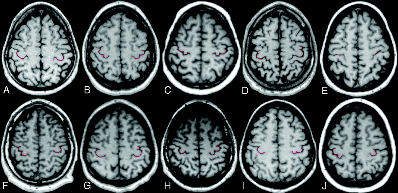
  • Fig 2.
    • Download figure
    • Open in new tab
    • Download powerpoint
    Fig 2.

    Multiplanar reformatted axial sections obtained 50 ± 2 mm above the Talairach anterior AC-PC plane. Images are shown using the right-left radiologic convention. The following are representative examples of the different combinations of HMC morphologic variants (red highlight) in the 2 hemispheres: A, Omega-omega. B, Epsilon-epsilon. C, Medially asymmetric epsilon-medially asymmetric epsilon. D, Laterally asymmetric epsilon-laterally asymmetric epsilon. E, Null-null. F, Epsilon-omega. G, Omega-laterally asymmetric epsilon. H, Omega-epsilon. I, Omega-medially asymmetric epsilon. J, Medially asymmetric epsilon-omega. The first 5 indicate concordant combinations, and the latter 5, the most common disconcordant combinations.

Tables

  • Figures
    • View popup
    Table 1:

    HMC morphologic variants by gender

    OmegaMedially Asymmetric EpsilonEpsilonLaterally Asymmetric EpsilonNullTotal
    No. (%)No. (%)No. (%)No. (%)No. (%)No.
    Overall
        Left hemisphere201 (78.2)7 (2.7)31 (12.1)14 (5.4)4 (1.6)257
        Right hemisphere201 (78.2)8 (3.1)21 (8.2)22 (8.6)5 (1.9)257
        Total402 (78.2)15 (2.9)52 (10.1)36 (7.0)9 (1.8)514
    Men
        Left hemisphere105 (73.4)4 (2.8)23 (16.1)10 (7.0)1 (0.7)143
        Right hemisphere114 (79.7)2 (1.4)16 (11.2)10 (7.0)1 (0.7)143
        Total*219 (76.6)6 (2.1)39 (13.6)20 (7.0)2 (0.7)286
    Women
        Left hemisphere96 (84.2)3 (2.6)8 (7.0)4 (3.5)3 (2.6)114
        Right hemisphere87 (76.3)6 (5.3)5 (4.4)12 (10.5)4 (3.5)114
        Total†183 (80.3)9 (3.9)13 (5.7)16 (7.0)7 (3.1)228
    • * χ2 = 13.675 with 4 df; P = .008.

    • † χ2 = 7.933 with 1 df; P = .005.

    • View popup
    Table 2:

    Correlation of morphologic variants of the HMC for hemispheres in men and women

    LeftRightTotal
    OmegaMedially Asymmetric EpsilonEpsilonLaterally Asymmetric EpsilonNull
    Men*
        Omega88 (61.5)4 (2.8)18 (12.6)3 (2.1)1 (0.7)114 (79.7)
        Medially asymmetric epsilon2 (1.4)0 (0.0)0 (0.0)0 (0.0)0 (0.0)2 (1.4)
        Epsilon8 (5.6)0 (0.0)4 (2.8)4 (2.8)0 (0.0)16 (11.2)
        Laterally asymmetric epsilon7 (4.9)0 (0.0)1 (0.7)2 (1.4)0 (0.0)10 (7.0)
        Null0 (0.0)0 (0.0)0 (0.0)1 (0.7)0 (0.0)1 (0.7)
        Total105 (73.4)4 (2.8)23 (16.1)10 (7.0)1 (0.7)143
    Women†
        Omega76 (66.7)1 (0.9)6 (5.3)2 (1.8)2 (1.8)87 (76.5)
        Medially asymmetric epsilon5 (4.4)1 (0.9)0 (0.0)0 (0.0)0 (0.0)6 (5.3)
        Epsilon3 (2.6)0 (0.0)1 (0.9)1 (0.9)0 (0.0)5 (4.4)
        Laterally asymmetric epsilon9 (7.6)1 (0.9)1 (0.9)1 (0.9)0 (0.0)12 (10.3)
        Null3 (2.6)0 (0.0)0 (0.0)0 (0.0)1 (0.9)4 (3.5)
        Total96 (83.9)3 (2.7)8 (7.1)4 (3.6)3 (2.7)114
    • Note:—Concordant hemispheres are in bold typeset.

    • * Kappa test: z = 2.09, P < .05 (not statistically significant).

    • † Kappa test: z = 3.03, P < .01.

PreviousNext
Back to top

In this issue

American Journal of Neuroradiology: 28 (8)
American Journal of Neuroradiology
Vol. 28, Issue 8
September 2007
  • Table of Contents
  • Index by author
Advertisement
Print
Download PDF
Email Article

Thank you for your interest in spreading the word on American Journal of Neuroradiology.

NOTE: We only request your email address so that the person you are recommending the page to knows that you wanted them to see it, and that it is not junk mail. We do not capture any email address.

Enter multiple addresses on separate lines or separate them with commas.
New Morphologic Variants of the Hand Motor Cortex as Seen with MR Imaging in a Large Study Population
(Your Name) has sent you a message from American Journal of Neuroradiology
(Your Name) thought you would like to see the American Journal of Neuroradiology web site.
CAPTCHA
This question is for testing whether or not you are a human visitor and to prevent automated spam submissions.
Cite this article
M. Caulo, C. Briganti, P.A. Mattei, B. Perfetti, A. Ferretti, G.L. Romani, A. Tartaro, C. Colosimo
New Morphologic Variants of the Hand Motor Cortex as Seen with MR Imaging in a Large Study Population
American Journal of Neuroradiology Sep 2007, 28 (8) 1480-1485; DOI: 10.3174/ajnr.A0597

Citation Manager Formats

  • BibTeX
  • Bookends
  • EasyBib
  • EndNote (tagged)
  • EndNote 8 (xml)
  • Medlars
  • Mendeley
  • Papers
  • RefWorks Tagged
  • Ref Manager
  • RIS
  • Zotero
0 Responses
Respond to this article
Share
Bookmark this article
New Morphologic Variants of the Hand Motor Cortex as Seen with MR Imaging in a Large Study Population
M. Caulo, C. Briganti, P.A. Mattei, B. Perfetti, A. Ferretti, G.L. Romani, A. Tartaro, C. Colosimo
American Journal of Neuroradiology Sep 2007, 28 (8) 1480-1485; DOI: 10.3174/ajnr.A0597
del.icio.us logo Twitter logo Facebook logo Mendeley logo
  • Tweet Widget
  • Facebook Like
  • Google Plus One
Purchase

Jump to section

  • Article
    • Abstract
    • Materials and Methods
    • Results
    • Discussion
    • Conclusions
    • Acknowledgments
    • Footnotes
    • References
  • Figures & Data
  • Info & Metrics
  • Responses
  • References
  • PDF

Related Articles

  • No related articles found.
  • PubMed
  • Google Scholar

Cited By...

  • Functional Magnetic Resonance Spectroscopy of Prolonged Motor Activation using Conventional and Spectral GLM Analyses
  • A roadmap for implanting microelectrode arrays to evoke tactile sensations through intracortical microstimulation
  • Increased cortical inhibition immediately following brief motor memory reactivation supports reconsolidation and overnight offline learning gains
  • Early excitatory and inhibitory modifications in the motor cortex following skill learning support motor memory consolidation and cortical plasticity overnight
  • Morphologic Variants of the Hand Motor Cortex in Developing Brains from Neonates through Childhood Assessed by MR Imaging
  • White matter variability, cognition, and disorders: a systematic review
  • Reduced asymmetry of the hand knob area and decreased sensorimotor u-fiber connectivity in middle-aged adults with autism
  • Laminar perfusion imaging with zoomed arterial spin labeling at 7 Tesla
  • Crossref
  • Google Scholar

This article has not yet been cited by articles in journals that are participating in Crossref Cited-by Linking.

More in this TOC Section

  • Optimal MRI Sequence for Identifying Occlusion Location in Acute Stroke: Which Value of Time-Resolved Contrast-Enhanced MRA?
  • Evaluating the Effects of White Matter Multiple Sclerosis Lesions on the Volume Estimation of 6 Brain Tissue Segmentation Methods
  • Quiet PROPELLER MRI Techniques Match the Quality of Conventional PROPELLER Brain Imaging Techniques
Show more Brain

Similar Articles

Advertisement

Indexed Content

  • Current Issue
  • Accepted Manuscripts
  • Article Preview
  • Past Issues
  • Editorials
  • Editor's Choice
  • Fellows' Journal Club
  • Letters to the Editor
  • Video Articles

Cases

  • Case Collection
  • Archive - Case of the Week
  • Archive - Case of the Month
  • Archive - Classic Case

More from AJNR

  • Trainee Corner
  • Imaging Protocols
  • MRI Safety Corner
  • Book Reviews

Multimedia

  • AJNR Podcasts
  • AJNR Scantastics

Resources

  • Turnaround Time
  • Submit a Manuscript
  • Submit a Video Article
  • Submit an eLetter to the Editor/Response
  • Manuscript Submission Guidelines
  • Statistical Tips
  • Fast Publishing of Accepted Manuscripts
  • Graphical Abstract Preparation
  • Imaging Protocol Submission
  • Evidence-Based Medicine Level Guide
  • Publishing Checklists
  • Author Policies
  • Become a Reviewer/Academy of Reviewers
  • News and Updates

About Us

  • About AJNR
  • Editorial Board
  • Editorial Board Alumni
  • Alerts
  • Permissions
  • Not an AJNR Subscriber? Join Now
  • Advertise with Us
  • Librarian Resources
  • Feedback
  • Terms and Conditions
  • AJNR Editorial Board Alumni

American Society of Neuroradiology

  • Not an ASNR Member? Join Now

© 2025 by the American Society of Neuroradiology All rights, including for text and data mining, AI training, and similar technologies, are reserved.
Print ISSN: 0195-6108 Online ISSN: 1936-959X

Powered by HighWire